Introduction
Aspilia africana (Pers.) C. D. Adams popularly known as wild sunflower is a valuable medicinal plant which has been used for many years to treat different diseases across various African communities1. The plant belongs to family Asteraceae and it is distributed across tropical African countries including Nigeria, Ethiopia, Uganda, Central African Republic, Congo, and Senegal2. The diseases that A. africana has been used to treat traditionally include wounds, malaria, measles, osteoporosis, gonorrhea, gastrointestinal disorders such as stomach ache, diabetes mellitus, tuberculosis, and rheumatic pains3,4. Accordingly, pharmacological studies have shown that phytochemicals of A. africana either singly or in combination exhibited various bioactivities namely wound healing, anti-inflammatory, antimicrobial, antidiabetic, gastroprotective, anticancer, antioxidant, and antimalarial effects5. Apart from its important medicinal uses, A. africana is also utilized as forage for livestock, such as chicken, goats, sheep, cattle, and rabbits, and it is reported to be one of the most browsed plants by these animals in Africa1,6. The expanding therapeutic and other uses due to explosion of human population, climate change, and environmental pollution are causing depletion of wild A. africana population6thus requiring propagation of this valuable plant. However, conventional plant propagation is associated with many challenges including low viability and occurrence of seed borne diseases7. Indeed, Okello et al.8 reported very low percent seed germination (between 11.67% and 15.67%) of A. africana in various soil types.
Micropropagation serves as a solution to these challenges. Regarding A. africana in vitro propagation, direct and indirect in vitro regeneration methods via organogenesis have been established9,10. As an alternative production technique, somatic embryogenesis is a powerful biotechnological tool for clonal regeneration, germplasm conservation, synthetic seed production, cryopreservation, and genetic improvement11,12. Somatic embryogenesis occurs if a somatic cell dedifferentiates to a totipotent embryonic stem cell, which can become an embryo in vitro13. Direct somatic embryogenesis and indirect somatic embryogenesis are the two ways of inducing somatic embryogenesis14. During direct somatic embryogenesis, somatic embryos are induced directly from explants under specific conditions without an intermediate callus phase, while indirect somatic embryogenesis happens through an intermediate callus phase11,12. Somatic and zygotic embryos undergo comparable developmental stages of globular, heart, torpedo, and cotyledonary in dicots such as A. africana, or globular, scutellar, and coleoptilar in monocots11,15.
In plant tissue cultures, regeneration through somatic embryogenesis could present several advantages over organogenesis, including the attainability of single cell origin and possible automation of the massive production of embryos in bioreactors and ex vitro planting as synthetic seeds16. Additionally, the bipolar nature of embryos enables direct growth into plantlets, eliminating need for the rooting stage required for plant regeneration through organogenesis, thereby making regeneration via somatic embryogenesis quicker compared to organogenesis11. Further, single epidermal cell origins for embryos could prevent chimeras, allowing preference of somatic embryogenesis for plant transformation17.
Somatic embryogenesis has been induced in various species of plants namely Digitalis lanata Ehrh, Camellia oleifera Abel, and Castanea mollissima Blume among others18,19,20. However, to the best of our knowledge, reports on somatic embryogenesis in A. africana are not available in literature to date. In this view therefore, this study aimed to establish an efficient and scalable method for direct somatic embryogenesis in A. africana using leaf explants. We evaluated the effects of exogenous PGRs, phytosulfokine-α (PSK), AMP, and NAD on induction, development, and maturation of somatic embryos. The ploidy level of regenerated plantlets was measured using flow cytometry to confirm their genetic stability. Furthermore, SPAD and the FluorPen FP110 series were used to test and compare chlorophyll pigment content and photosynthetic rates, respectively, of somatic and zygotic embryo derived plants. The proposed method could be useful for mass clonal regeneration, conservation, synthetic seed production, cryopreservation, and genetic improvement of A. africana. Additionally, this system would provide suitable model for investigating molecular, biochemical, and physiological events, which occur at the induction and development of embryogenesis in A. africana.
Materials and methods
Plant material and Preparation of explants
With permission from local authorities, A. africana seeds (mature and dry) were collected and provided by Natural Chemotherapeutics Research Institute (NCRI) from about 50 healthy plants growing at Pece, Gulu, Uganda in East Africa. On arrival at Korea Institute of Oriental Medicine (KIOM), Republic of Korea, the seeds were stored in a room at temperature 25 ± 1 °C until the time of experiment. A voucher specimen (number KYM-KIOM-2021-1) was deposited by Dr. Sungyu Yang at the Korean Herbarium of Standard Herbal Resources (Index Herbarium code: KIOM) at KIOM, Herbal Medicine Resources Research Center, Republic of Korea. To raise seedlings for explant sources in aseptic conditions, seeds of A. africana were germinated in vitro by adopting the method of Okello et al.1 with slight modification. Briefly, the seeds were initially washed for 2 min under running tap water and taken to a laminar flow cabinet. Next, the seeds were again washed using autoclaved double-distilled water, surface sterilized for 1 min with 70% (v/v) ethanol, and rinsed thrice with autoclaved double-distilled water. This was followed by surface sterilizing with 2% (v/v) sodium hypochlorite for 3 min and rinsing thrice using autoclaved double-distilled water. The seeds were allowed to dry between sterile filter papers and afterwards inoculated on MS medium with vitamins augmented with gibberellin (GA; 0.5 mg/L) and transferred to culture room. After growth for four weeks, the in vitro germinated seedlings were cultured in MS medium containing vitamins, supplemented with BAP (1.0 mg/L)2. For constant supply of plant materials, shoots of seedlings raised in vitro were sub-cultured in the same medium after every four weeks. Leaves obtained from in vitro shoots served as explants.
Embryo initiation and proliferation
Effects of PGRs on somatic embryogenesis of A. africana
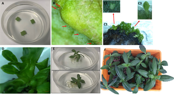
Effects of PGRs on somatic embryogenesis in A. africana was evaluated in two phased experiments using leaf explants of size approximately 1 cm x 1 cm (obtained from 4 weeks old healthy plantlets growing in vitro) (Fig. 1A). In the first experiment, effects of four different cytokinins, BAP, thidiazuron (TDZ), kinetin (Kn), isopentenyl adenine (2iP), at varying concentrations (0.1, 0.5, 1.0, 2.0, and 3.0 mg/L) were evaluated separately. MS medium containing vitamins was supplemented with the cytokinins (BAP, TDZ, Kn, 2iP), 30 g/L sucrose, and 3 g/L gelrite; pH of the medium was adjusted to 5.7–5.8 and autoclaved for 20 min at 121 °C, then poured in crystal-grade polystyrene petri dishes (100 × 20 mm) to 50 mL level. Leaf explants were placed (adaxial surface in contact with medium) on the solidified medium and each petri dish contained three explants with eight replicates (total 24 explants per treatment). All cultures were incubated at 16 h photoperiod (33.73 µmol/(m2/s) light intensity)) and 80% r.h. Unless otherwise mentioned, culture conditions and other medium components in all subsequent evaluations were identical to those described at this induction stage. After four weeks of culture, frequency (%) of explants that developed somatic embryos and the number of somatic embryos per explant were captured (Fig. 1B). The cytokinin that showed optimal result (1.0 mg/L BAP) was used in the next experiment.
In the second experiment, leaf explants were cultured in MS medium containing 1.0 mg/L BAP combined separately with three different auxins, indole-3-acetic acid (IAA), indole-3-butyric acid (IBA), and NAA at concentrations of 0.1, 0.5, 1.0, 2.0, and 3.0 mg/L. Similarly, each petri dish contained three explants and this was replicated eight times (total 24 explants per treatment). Frequency (%) of explants that developed somatic embryos and the number of somatic embryos per explant were registered after four weeks of culture (Fig. 1B). Treatment with optimal result (still 1.0 mg/L BAP alone) was employed in further experiment.

Direct somatic embryogenesis in A. africana from leaf explants. (A) Leaf explants on induction medium. (B) Induction and development of globular shape embryos. (C1) development of heart and torpedo embryos. (C2) heart shape embryo. (C3) Torpedo shape embryo. (D) Proliferated cotyledonary embryos. (E) Well developed plantlets. (F) Plantlets growing in containers having horticultural soil mixed with perlite (2:1). Bars: (b-d) = 1 mm.
Effects of combining PGR with PSK, AMP, and NAD on somatic embryogenesis of A. africana
Leaf explants were cultured in MS medium containing 1.0 mg/L BAP fortified independently with PSK, AMP, and NAD at various concentrations, PSK (8.449 × 10⁻⁴, 8.449 × 10⁻³, 8.449 × 10⁻², and 8.449 × 10⁻¹ mg/L), AMP (3.472 × 10⁻⁴, 3.472 × 10⁻³, 3.472 × 10⁻², and 3.472 × 10⁻¹ mg/L), and NAD (6.634 × 10⁻⁴, 6.634 × 10⁻³, 6.634 × 10⁻², and 6.634 × 10⁻¹ mg/L). Each petri dish consisted of three explants with eight replicates (total 24 explants per treatment). Notably, globular embryos developed earlier on the surface of leaf explants in media having both PGR and any of PSK, AMP, and NAD (twenty-four days), in contrast with medium devoid of PSK, AMP, and NAD (four weeks or 28 days). After twenty-four days of culture, frequency (%) of explants that developed somatic embryos and the number of somatic embryos per explant were captured (Fig. 1B). Leaf explants with embryos at twenty-four days of culture were used to evaluate differentiation and maturation in subsequent experiment.
Embryo differentiation and maturation
Effects of ABA on A. africana somatic embryo differentiation and maturation
Leaf explants with embryos at twenty-four days of culture from initiation medium were randomly transferred to MS medium augmented with ABA at different concentrations of 0.1, 0.5, 1.0, 2.0, 3.0, and 5.0 mg/L for differentiation and maturation of somatic embryos. Accordingly, ABA was added when medium temperature was about 60 °C after autoclaving. MS medium devoid of ABA served as control. Each petri dish contained a single explant with 15 replicates (a total of 15 explants per treatment). After ten days of culture in maturation medium, number of somatic embryos per leaf explant at various stages viz. globular, heart, torpedo, and cotyledonary was counted and registered (Fig. 1C1, C2, C3, and D). Treatment with best result, in this case, 0.5 mg/L ABA was used in the following evaluation.
Effects of combination of ABA with PSK, AMP, and NAD on A. africana somatic embryo differentiation and maturation
To observe influence of combining ABA with PSK, AMP, and NAD on differentiation and maturation of somatic embryos, leaf explants with embryos at twenty-four days of culture from initiation medium were randomly shifted to MS medium having 0.5 mg/L ABA supplemented separately with PSK (8.449 × 10⁻⁴, 8.449 × 10⁻³, 8.449 × 10⁻², and 8.449 × 10⁻¹ mg/L), AMP (3.472 × 10⁻⁴, 3.472 × 10⁻³, 3.472 × 10⁻², and 3.472 × 10⁻¹ mg/L), and NAD (6.634 × 10⁻⁴, 6.634 × 10⁻³, 6.634 × 10⁻², and 6.634 × 10⁻¹ mg/L). MS medium fortified with 0.5 mg/L ABA alone served as control. Every petri dish had a single explant with 15 replicates (total 15 explants per treatment). Number of somatic embryos per leaf explant at various stages (globular, heart, torpedo, and cotyledonary) was counted and recorded, after ten days of culture in maturation medium (Fig. 1C1, C2, C3, and D). Treatment that showed best result (0.5 mg/L ABA + 6.634 × 10⁻² mg/L NAD) was used in the next assessment.
Effects of osmotic stress, ABA, and NAD on A. africana somatic embryo differentiation and maturation
To examine the role of ABA and NAD coupled with osmotic stress in maturation of A. africana somatic embryos, MS medium fortified with 0.5 mg/L ABA and 6.634 × 10⁻² mg/L NAD was combined with either 3 g/L or 9 g/L gelrite (total two treatments). Leaf explants with embryos at twenty-four days of culture from initiation medium were randomly transferred to respective culture medium. Each petri dish contained a single explant with 15 replicates (total 15 explants per treatment). After ten days of culture in maturation medium, number of somatic embryos per leaf explant at various stages (globular, heart, torpedo, and cotyledonary) were counted and noted.
Embryo pre-germination treatments, germination and conversion into plantlets, and acclimatization
Suitable medium for embryo germination was determined by culturing cotyledonary stage somatic embryos on embryo germination media of full/half/quarter strength semi solid MS fortified with 0.5 mg/L GA and 0.1 mg/L NAA. After three weeks, germination frequency (percent of germination) was determined as the percent of somatic embryos showing emergence of shoot out of total somatic embryos inoculated. The best medium for germination (Half strength MS medium fortified with 0.5 mg/L GA and 0.1 mg/L NAA) was used in further evaluation.
Regarding pre-germination treatments, mature somatic embryos (cotyledonary and torpedo stages) were obtained from maturation medium and subjected to two conditions (1) stratification treatment at 4O C for 5 days while on germination medium and (2) direct placement of embryos onto germination medium and kept in culture room (this served as control).
Embryos from stratification treatment were kept in the dark for 5 days before being shifted to light in a 16-h photoperiod to germinate. After 3 weeks of growth, the germinated embryos were shifted to same germination medium for additional 4 weeks. Germination frequency (percent of germination) as the percent of somatic embryos showing emergence of shoot out of total somatic embryos inoculated, and percent of plantlets as percent of germinated somatic embryos with shoot and root out of total germinated somatic embryos with shoot inoculated, were determined after 3 weeks and 7 weeks of growth, respectively.
Upon sufficient growth (Fig. 1E), the plantlets were removed from culture containers, medium adhered to the roots washed with water, and planted in sterile horticultural soil mixed with perlite (2:1) contained in plastic containers placed in a greenhouse (Fig. 1F). Plantlets were covered with a transparent polythene bag which were removed after fourteen days. The plantlets were watered two times a week, and rate of survival was recorded after seven weeks of growth.
Morphological and histological observations
Observation of embryos at various stages of development were performed under a stereo-zoom binocular microscope (SMZ745T, China) and a digital camera (Nikon, China) was used to photograph images (Fig. 1B, C1, C2, C3, and D).
Histological analysis of A. africana somatic embryos was performed at various developmental stages (globular, heart, torpedo, and cotyledonary) at 21–40 days after inoculation following the method of Okello et al.10 with slight modification. Briefly, fresh leaf explant tissues with somatic embryos at various stages of development were immediately fixed in 70% ethanol for at least 24 h. After, the tissues were sequentially dehydrated in ethanol 50, 70, 80, 90, 95, 98, and 100% (for 30 min each) at 25 ± 1 °C. The dehydrated tissues were successively cleared in ethanol-xylene mixtures at different ratios (ethanol : xylene) 3:1, 1:1, and 1:3, and twice in xylene (for 30 min each) at 25 ± 1 °C. Further clearing was done in xylene-paraffin mixtures at different ratios (xylene : paraffin) 2:1, 1:2, and twice in paraffin (for 1 h in each) at 60 ± 1 °C. Then, the tissues were embedded in paraffin wax overnight and 12-µm sections were sliced using a microtome. Tissue sections were dewaxed by sequentially dipping for 5 min in the following: xylene (twice), ethanol-xylene mixture (1:1), ethanol 100% (twice), 95%, 70%, and 50%. Next, the sections were stained by successively dipping in the following: safranine 1% (1 h), water, ethanol 50%, 70%, 95% (3 min each), fast-green (3 min), ethanol 100% (twice and 1 min each time), and xylene (thrice and 15 min each time). After mounting with Balsam medium, images of stained tissues were scanned and observed and captured using digital slide scanner (3DHistech Pannoramic Desk II DW; 3DHistech Kft., Budapest, Hungary) and CaseViewer software (version 2.4.0; 3DHistech Kft., Budapest, Hungary), respectively.
Genetic stability assessment by flow cytometry
Flow cytometry was used to determine the genetic stability of direct somatic embryo A. africana plants following the method of Choi et al.21 with slight modifications. With a known 2 C DNA content of 2.50 pg, the leaves of Glycine max cv. Polanka served as an internal standard. Briefly, randomly picked leaf tissues (approximately 0.5 cm2 from somatic embryo A. africana plants and zygotic embryo A. africana plants were independently co-chopped together with leaf tissue of internal standard in a plastic petri dish containing 500 µL of nuclei extraction buffer (CyStain Ultraviolet Precise P Nuclei Extraction Buffer; Sysmex Partec, Germany). Prior to filtering the contents through 50 μm mesh (CellTrics, Sysmex Partec, Germany) into tubes, they were incubated for 30–90 s. To each filtrate, staining solution (2000 µL) consisting of staining buffer, propidium iodide, and RNAse A was added, and the mixtures were incubated for 60 min at 25 °C while protecting from light. Measurement of the fluorescence intensity of isolated nuclei was performed using a flow cytometer (BD FACSCalibur, USA). The ploidy of somatic embryo A. africana and zygotic embryo A. africana plants was determined through comparing their relative fluorescence intensity with that of the internal standard, based on the formula below:
2 C DNA of A. africana = (:2.50:pg:frac{::Intensity:of:test}{Intensity:of:standard:}).
Where intensity is the fluorescence intensity of isolated nuclei.
Chlorophyll content measurement
The leaf chlorophyll content of somatic embryo A. africana plants (five weeks after acclimatization) and zygotic embryo A. africana (about 2 months old) growing in the same greenhouse was measured and compared using SPAD chlorophyll meter (SPAD-502 Plus, Konica Minolta, Inc., Japan). Before clumping the SPAD chlorophyll meter onto A. africana leaves to get readings of chlorophyll content, it was calibrated. Per plant, six leaves were chosen randomly from lower-stem region (a pair), mid-stem region (a pair), and apical region (a pair) and registered chlorophyll content values from four points of each leaf (Fig. 2A and B). Average chlorophyll content value of the six leaves represented chlorophyll content of an individual plant. Chlorophyll contents of same plants (10 somatic embryo A. africana plants and 10 zygotic embryo A. africana plants) were recorded weekly across seven weeks (Fig. 2C).

Chlorophyll content measurement of zygotic embryo plant leaves and direct somatic embryo plant leaves of A. africana. (A) Measuring chlorophyll content of leaf using SPAD meter (B) Specific points on a leaf taken to represent the average chlorophyll content of an individual leaf. (C) Chlorophyll contents of zygotic embryo A. africana plants compared with that of the direct somatic embryo A. africana plants across 7 weeks.
Chlorophyll fluorescence measurement
FluorPen FP110 (Drásov 470, 664 24 Drásov, Czech Republic) was employed to capture chlorophyll fluorescence of the plants. Prior to recording chlorophyll fluorescence, plant leaves were dark-adapted with leaf clip gaskets for about 1 h. Chlorophyll fluorescence measurements were taken weekly for seven weeks from six randomly selected leaves per zygotic embryo A. africana plant (total 10 plants) and somatic embryo A. africana plants (total 10 plants). To get Fv/Fm values, more than 4,000 µmol m−2 s−1 saturating flash was used as per the FluorPen FP110 OJIP protocol. In the dark-adapted state, minimal chlorophyll fluorescence intensity (Fo) was measured when photosystem II (PSII) reaction center was open, maximal chlorophyll fluorescence intensity (Fm) was measured during application of saturation light pulse, and variable chlorophyll fluorescence (Fv) was recorded when minimal non-photochemical processes occurred.
Statistical analysis
Completely randomized design was applied to the experiments performed. Embryogenesis initiation and proliferation stage consisted of three experiments, and during all, each treatment petri dish contained three explants with eight replicates (total 24 explants per treatment). In the first experiment, effects of four different cytokinins (BAP, TDZ, Kn, and 2iP) at five varying concentrations (total 21 treatments) were evaluated. In the second experiment, 1.0 mg/L BAP was combined separately with three different auxins (IAA, IBA, and NAA) at five varying concentrations (total 16 treatments). In the third experiment, 1.0 mg/L BAP was fortified independently with three molecules (PSK, AMP, and NAD) at four various concentrations (total 13 treatments).
The embryo differentiation and maturation stage comprised three experiments, and during all, each treatment petri dish contained a single explant with 15 replicates (total 15 explants per treatment). During the first experiment, effect of ABA at six different concentrations (total 7 treatments) was investigated. In the second experiment, 0.5 mg/L ABA was combined with three molecules (PSK, AMP, and NAD) at four various concentrations (total 13 treatments). In the third experiment, 0.5 mg/L ABA + 6.634 × 10⁻² mg/L NAD was combined with either 3 g/L or 9 g/L gelrite (total 2 treatments).
During embryo germination and conversion into plantlets stage, two experiments were conducted, and during both experiments, each treatment had 100 embryos. In the first experiment, embryo germination was evaluated in MS medium at three different strengths (full/half/quarter) supplemented with 0.5 mg/L GA and 0.1 mg/L NAA (total 4 treatments). The second experiment involved two embryo germination treatments of stratification at 4o C before transfer to culture room and direct placement in culture room. Unless otherwise stated, all experimental data were analyzed by one-way analysis of variance (ANOVA) followed by Tukey’s post hoc tests using GraphPad Prism v 10.1.1. Means were considered significantly different at P < 0.05.
Results
Embryo initiation and proliferation
Effects of PGRs on somatic embryogenesis of A. africana
Except Kn, all cytokinins induced somatic embryo formation from leaf explants, although with varying degree of response (Fig. 3A and B). Initially, the number of somatic embryos increased with increasing concentration of cytokinins before decreasing at high concentrations (Fig. 3A). Accordingly, swelling and formation of embryogenic clumps on leaf explants occurred in the first and second week, respectively. After four weeks, globular embryos developed on the surface of leaf explants. In the initial test, among cytokinins, MS medium augmented with 1.0 mg/L BAP showed the best results with 5.92 ± 0.29 somatic embryos per explant and 100% response (Fig. 3A and B). These values of BAP (1.0 mg/L) did not markedly differ from those of TDZ (2.0 mg/L) (Fig. 3A and B). The lowest values were observed in MS medium devoid of cytokinins and those supplemented with Kn at all concentrations, which did not form somatic embryos (i.e., 0.0 somatic embryos and 0.0% frequency of explants that developed somatic embryos) (Fig. 3A and B).

Effects of Cytokinins at different concentrations on number of somatic embryos formed and percent of explants that developed somatic embryos via direct somatic embryogenesis from leaf explants of A. africana. (A) Number of somatic embryos formed. (B) Percent of explants that developed somatic embryos. The medium used was MS, and all concentrations are in mg/L. Values represent the Mean ± SE of 24 explants. Same letters are not significantly different by Tukey’s test and p ≤ 0.05.
In the successive experiment, excluding MS medium fortified with 1.0 mg/L BAP + 1.0 mg/L IBA, combination of BAP (1.0 mg/L) with different auxins resulted in decreased formation of somatic embryos in all concentrations (Fig. 4A and B). MS medium supplemented with 1.0 mg/L BAP + 1.0 mg/L IBA generated the highest number of somatic embryos per explant (6.33 ± 1.35) and this did not differ significantly (p < 0.05) from those of MS medium supplemented with BAP (1.0 mg/L), BAP (1.0 mg/L) + IBA (2.0 mg/L), BAP (1.0 mg/L) + IBA (3.0 mg/L), and BAP (1.0 mg/L) + IAA (3.0 mg/L) (Fig. 4A and B). Without significant (p < 0.05) variation from the values of MS medium fortified with BAP (1.0 mg/L) + IBA (2.0 mg/L), BAP (1.0 mg/L) + NAA (3.0 mg/L), BAP (1.0 mg/L) + IAA (3.0 mg/L), and BAP (1.0 mg/L) + IBA (0.5 mg/L), the highest percentage of explants that developed somatic embryos (91.67% ± 5.46%) was recorded in MS medium fortified with BAP (1.0 mg/L), BAP (1.0 mg/L) + IBA (1.0 mg/L), and BAP (1.0 mg/L) + IBA (3.0 mg/L) treatments (Fig. 4A and B). However, the lowest number of somatic embryos formed (0.0) and frequency of explants that developed somatic embryos (0.0%) were registered in MS medium augmented with BAP 1.0 (mg/L) + NAA (0.1 mg/L). 1.0 mg/L BAP alone with the second highest number of somatic embryos (5.75 ± 0.48 and no marked difference from that of first, 1.0 mg/L BAP + 1.0 mg/L IBA) and joint highest percentage response of explants (91.67% ± 5.46%) was taken as the optimal PGR treatment for inducing direct somatic embryogenesis, since some calluses were observed in 1.0 mg/L BAP + 1.0 mg/L IBA treatment. Therefore, 1.0 mg/L BAP was used for further evaluation.

Effects of PGR combination at different concentrations on number of somatic embryos formed and percent of explants that developed somatic embryos via direct somatic embryogenesis from leaf explants of A. africana. (A) Number of somatic embryos formed. (B) Percent of explants that developed somatic embryos. All media (MS) contained 1.0 BAP, and all concentrations are in mg/L. Values represent the Mean ± SE of 24 explants. Same letters are not significantly different by Tukey’s test and p ≤ 0.05.
Effects of combining PGR with PSK, AMP, and NAD on somatic embryogenesis of A. africana
Combination of 1.0 mg/L BAP with PSK, AMP, and NAD, except AMP (3.472 × 10⁻³, 3.472 × 10⁻², and 3.472 × 10⁻¹ mg/L) reduced both number of somatic embryos and frequency of embryogenesis (Fig. 5A and B). MS medium fortified with BAP (1.0 mg/L) plus AMP (3.472 × 10⁻⁴ mg/L) yielded the highest mean somatic embryo number (9.50 ± 0.29), and this was markedly higher (p < 0.05) than the mean somatic embryo number of other treatments (apart from 1.0 mg/L BAP + 3.472 × 10⁻³ mg/L AMP) (Fig. 5A). Lowest mean somatic embryo number of 4.0 ± 1.19 was recorded in MS medium augmented with BAP (1.0 mg/L) plus NAD (6.634 × 10⁻³ mg/L) (Fig. 5A). Similarly, MS augmented with BAP (1.0 mg/L) combined with AMP (3.472 × 10⁻², 3.472 × 10⁻³ mg/L) produced maximum percentage of explants that developed somatic embryos (100%), while BAP (1.0 mg/L) combined with NAD (6.634 × 10⁻³ mg/L) showed the lowest percentage of explants that developed somatic embryos (33.33%) (Fig. 5B). As stated above, globular embryos developed earlier on the surface of leaf explants in media containing both PGR and any of PSK, AMP, and NAD (twenty-four days) compared to medium devoid of PSK, AMP, and NAD (four weeks or twenty-eight days). Thus, 1.0 mg/L BAP + 3.472 × 10⁻² mg/L AMP optimally induced somatic embryogenesis in A. africana from leaf explants compared to other treatments in the present study.

Effects of combination of Cytokinin and PSK, AMP, and NAD at various concentrations on number of somatic embryos formed and percent of explants that developed somatic embryos via direct somatic embryogenesis from leaf explants of A. africana. (A) Number of somatic embryos formed. (B) Percent of explants that developed somatic embryos. All media (MS) contained 1.0 BAP, and all concentrations are in mg/L. Values represent the Mean ± SE of 24 explants. Same letters are not significantly different by Tukey’s test and p ≤ 0.05.
Embryo differentiation and maturation
Effects of ABA on A. africana somatic embryo differentiation and maturation
Differentiation and development of various somatic embryo stages of globular, heart, torpedo, and cotyledonary occurred when leaf explants with embryos at twenty-four days of culture from initiation medium were transferred to MS medium augmented with ABA at different concentrations (0.1, 0.5, 1.0, 2.0, 3.0, and 5.0 mg/L) (Fig. 6A, B, C, and D). Maximum mean cotyledonary stage embryo number (1.67 ± 0.16) was registered in MS medium augmented with 0.5 mg/L ABA, and this markedly differed (p < 0.05) from that in all other treatments (Fig. 6D). Based on this result, 0.5 mg/L ABA was employed in further assessments.

Effects of different concentrations of abscisic acid (ABA) on differentiation and maturation of somatic embryos from leaf explants of A. africana. (A) Globular shape embryos. (B) Heart shape embryos. (C) Torpedo shape embryos. (D) Cotyledonary shape embryos. Same letters are not significantly different by Tukey’s test and p ≤ 0.05.
Effects of combination of ABA with PSK, AMP, and NAD on A. africana somatic embryo differentiation and maturation
ABA (0.5 mg/L) combined with PSK, AMP, and NAD at concentrations PSK (8.449 × 10⁻⁴, 8.449 × 10⁻³, 8.449 × 10⁻², and 8.449 × 10⁻¹ mg/L), AMP (3.472 × 10⁻⁴, 3.472 × 10⁻³, 3.472 × 10⁻², and 3.472 × 10⁻¹ mg/L), and NAD (6.634 × 10⁻⁴, 6.634 × 10⁻³, 6.634 × 10⁻², and 6.634 × 10⁻¹ mg/L) differently influenced differentiation and development of somatic embryos at various stages (globular, heart, torpedo, and cotyledonary) on leaf explants containing embryos transferred from initiation medium (Fig. 7A, B, C, and D). The initial globular, heart, torpedo, and cotyledonary embryo stages formed at 23, 25, 26, and 28 days, respectively. MS medium fortified with 0.5 mg/L ABA + 6.634 × 10⁻² mg/L NAD showed the highest number of cotyledonary stage somatic embryos per explant (3.33 ± 0.33), which was markedly different (p < 0.05) from control (0.5 mg/L ABA alone) and most other treatments (Fig. 7D). The lowest mean cotyledonary stage somatic embryo number of 0.80 ± 0.22 was observed in MS medium fortified with 0.5 mg/L ABA + 8.449 × 10⁻¹ mg/L PSK (Fig. 7D).

Effects of combination of abscisic acid (ABA) and PSK, AMP, and NAD at various concentrations on differentiation and maturation of somatic embryos from leaf explants of A. africana. (A) Globular shape embryos. (B) Heart shape embryos. (C) Torpedo shape embryos. (C) Cotyledonary shape embryos. All media (MS) contained 0.5 ABA, and all concentrations are in mg/L. Same letters are not significantly different by Tukey’s test and p ≤ 0.05.
Effects of osmotic stress, ABA, and NAD on A. africana somatic embryo differentiation and maturation
Osmotic stress showed increased differentiation and development of various stages of somatic embryos on leaf explants having embryos transferred from initiation medium (Table 1). Maturation of somatic embryos was markedly higher (p < 0.05) in osmotic medium (MS + ABA 0.5 mg/L + NAD 6.634 × 10⁻² mg/L + Gelrite 9 g/L) with 4.73 ± 0.41 number of cotyledonary embryos per explant than in non-osmotic medium (MS + ABA 0.5 mg/L + NAD 6.634 × 10⁻² mg/L + Gelrite 3 g/L), which yielded 3.20 ± 0.35 number of cotyledonary embryos per explant (Table 1).
Embryo pre-germination treatments, germination and conversion into plantlets, and acclimatization
In addition to reducing the strength of MS medium, its supplementation with 0.5 mg/L GA and 0.1 mg/L NAA increased germination of cotyledonary stage somatic embryos (Fig. 8). The cotyledonary somatic embryos elongated and developed shoot after three weeks in germination media. Half strength MS medium fortified with 0.5 mg/L GA and 0.1 mg/L NAA produced the highest germination percent (31 ± 1.73%), and this was markedly (p < 0.05) different from the rest of the treatments, except in quarter strength MS medium augmented with 0.5 mg/L GA and 0.1 mg/L NAA (Fig. 8). The lowest percent of germination (14 ± 1.15%) was observed in PGR free MS medium.

Germination of somatic embryos in different medium compositions. Same letters are not significantly different by Tukey’s test and p ≤ 0.05.
Generally, stratification at 4o C treatment influenced the germination of somatic embryos (Fig. 9 and Table 2). Stratification at 4o C resulted into better embryo germination (60.00%) and conversion to plantlets (26.67%) compared to direct culture in medium (Table 2).

Effect of stratification at 4o C on germination of A. africana somatic embryos and their conversion to plantlets. (A) Cotyledonary somatic embryos cultured in germination medium before stratification at 4o C. (B) Somatic embryo derived plantlets growing in germination medium two weeks after stratification at 4o C.
After five weeks of acclimatization, the survival rate of somatic embryo derived A. africana plants was 75.00% (Table 3). The plantlets were morphologically similar to zygotic embryo derived A. africana plants.
Morphological and histological observations
Histological analysis showed the regeneration pathway of somatic embryogenesis in A. africana. Longitudinal sections of embryonic structures revealed that accelerated cell divisions begin from the leaf explant surfaces and cut edges (Fig. 10A). Within 2 weeks of culture, formation of cell aggregates occurred, developing into globular embryos (Fig. 10A). The globular somatic embryos majorly comprised small compact meristematic cells, having dense cytoplasm, notable nuclei, and a layer of protoderm on their surface (Fig. 10A). From globular embryos, subsequent stages of embryos developed asynchronously, including heart, torpedo, and cotyledonary stages, after 4 weeks of culture (Fig. 10B, C, D). It was observed that the cotyledonary stage embryos had a closed vascular system, thus no vascular bundle connection with mother tissue (Fig. 10D).

Histological observation of direct somatic embryogenesis in A. africana from leaf explants. (A) Histological section of globular somatic embryo. (B) Histological section of heart stage somatic embryo. (C) Histological section of torpedo stage somatic embryo. (D) Histological section of cotyledonary somatic embryo. cp.: cotyledon primordia, mp: mother plant, se: somatic embryo, su: suspensor, vb: vascular bundle. Bars: (b, d) = 0.100 mm, (a, c) = 0.200 mm.
Genetic stability assessment by flow cytometry
As per flow cytometry histograms, the ploidy levels of somatic and zygotic embryo A. africana plants were identical (Fig. 11A and B). The 2 C DNA contents of zygotic and somatic embryo A. africana plants were 6.33 pg and 6.25 pg, respectively (Fig. 11A and B). The 2 C DNA contents of zygotic (6.33 pg) and somatic (6.25 pg) embryo A. africana plants corresponded to the genome size (2 C) of 6,190 Mbp and 6,113 Mbp, respectively (where 1 pg is ~ 978 Mbp, Doležel et al.22.

Flow cytometry histograms of fluorescent nuclei isolated from A. africana. (A) 2 C-DNA content from zygotic embryo A. africana plants. (B) 2 C-DNA content from direct somatic embryo A. africana plant.
Chlorophyll content measurement
Initially (in the first week), the chlorophyll contents of zygotic embryo A. africana plants (35.06 ± 0.30) were comparatively higher than that of somatic embryo A. africana plants (29.5 ± 0.35) (Fig. 2C). During the first two weeks, increase in chlorophyll contents of somatic embryo A. africana plants was relatively higher than that in zygotic embryo A. africana plants, and this led to comparable contents by the third week (Fig. 2C). From week three to seven, minimal increase in chlorophyll contents was registered for both zygotic embryo A. africana plants (from 40.01 ± 0.54 to 43.75 ± 0.35) and somatic embryo A. africana plants (from 39.55 ± 0.39 to 43.26 ± 0.46) (Fig. 2C). Notably, from third week onwards, there was no significant difference in chlorophyll contents of zygotic embryo A. africana plants and somatic embryo A. africana plants (Fig. 2C).
Chlorophyll fluorescence measurement
Measurement of the Fv/Fm values in dark-adapted state, which is an indicator of potential maximum quantum efficiency of PSII, revealed the photosynthetic rates of zygotic embryo A. africana plants and somatic embryo A. africana plants. Across seven weeks, the mean Fv/Fm values of zygotic embryo A. africana plants and somatic embryo A. africana plants ranged from 0.78 ± 0.01 to 0.83 ± 0.01 and from 0.76 ± 0.0 to 0.83 ± 0.02, respectively (Fig. 12). Accordingly, Fv/Fm values of zygotic embryo A. africana plants and somatic embryo A. africana plants across the last four weeks were similar (Fig. 12).

Comparison of mean Fv/Fm ratio of zygotic embryo A. africana plants compared with that of the direct somatic embryo A. africana plants across 7 weeks.
Discussion
Generally, somatic embryogenesis is induced when plant tissues are exposed to the right stimulus, in most cases PGRs23,24. PGR requirement for optimal somatic embryogenesis induction varies among plant species and even genotypes18,20 thus, types, concentrations, combinations, and ratios of exogenous cytokinins and/or auxins play a crucial role. Importance of cytokinins and auxins in triggering embryogenic response could possibly be attributed to their involvement in cell cycle regulation and cell division25.
Among cytokinins used in this study, 1.0 mg/L BAP optimally induced somatic embryogenesis from A. africana leaf explants (number of somatic embryos per explant and percentage response of explants being 5.75 ± 0.48 and 91.67% ± 5.46%, respectively). In most species, auxins have been used for inducing somatic embryogenesis. However, in the present study, cytokinins effectively induced direct somatic embryogenesis. Noteworthy is that combination of cytokinins and auxins did not show synergistic effects on initiation and proliferation of somatic embryos. Superiority of BAP in inducing somatic embryogenesis could probability be due to its metabolic stability in in vitro cultures and ability to initiate cell division26,27. This finding is in agreement with other reports that also recorded highest number of somatic embryos in media containing BAP, including in Curcuma longa L28. and Metabriggsia ovalifolia W. T. Wang29.
Exogeneous metabolites, small molecules, and peptides such as PSK, AMP, and NAD not only similarly regulate plant growth like cytokinins and auxins but also regulate other processes including reproduction, bioenergetic, biotic and abiotic responses, essential compounds for nucleic acid synthesis, and other components of cell metabolism30,31. Some researchers have used these metabolites, molecules, and peptides including PSK, AMP, and NAD to supplement culture media and promote somatic embryogenesis in different plant species32,33,34,35.
In this study, for induction and proliferation of somatic embryos, supplementation of MS medium containing 1.0 mg/L BAP with 3.472 × 10⁻² mg/L AMP generated the biggest number of somatic embryos per explant (9.50 ± 0.29) and percent of explants that developed somatic embryos (100%), coupled with earlier formation of embryos than control. The observed result could possibly be because adenosine, a component of AMP is reported to be a major substrate for purine salvage, which is highly associated with growth and development of embryos30. Additionally, during early phase of somatic embryogenesis, active utilization of purines may be necessary to enhance endogenous nucleotide pool, needed to maintain rapid proliferation and cell division of embryogenic tissue in the presence of BAP or/and 2, 4-D30. This finding suggests that AMP is potentially a major metabolite that confers embryogenic competence during A. africana somatic embryogenesis. Similarly, Lee et al.34 alluded to the enhancement of somatic embryo development in Peucedanum japonicum Thunb. by AMP.
Maturation is a critical stage of somatic embryogenesis since accumulation of storage materials occurs at this point36,37. In many plant species, presence of exogenous ABA at various concentrations has been reported to promote maturation of somatic embryos38,39. In the current study, maximum number of cotyledonary stage embryos (1.67 ± 0.16) was registered in 0.5 mg/L ABA. Ability of ABA to enhance embryo maturation may potentially be attributed to its capacity to induce key metabolic changes that facilitate storage of reserves, thus increasing contents of fatty acids and storage proteins in somatic embryos40,41. Furthermore, ABA has been reported to improve quality of somatic embryos through increasing their desiccation tolerance and prevention of precocious germination42. Our result implies that exogenous application of ABA at low concentration (0.5 mg/L) favours maturation of A. africana somatic embryos. Consistent with the present finding, exogenous ABA favoured maturation of somatic embryos in other species such as Pinus elliottii Engelm43. Eucalyptus camaldulensis Dehnh44. and Copiapoa tenuissima Ritt. forma monstruosa36.
Further supplementation of maturation medium (MS containing 0.5 mg/L ABA) with 6.634 × 10⁻² mg/L NAD yielded the highest number of cotyledonary embryos per explant (3.33 ± 0.33). Effectiveness of NAD in improving maturation of A. africana somatic embryos could likely be explained by the fact that it is a core component of reduced nicotinamide adenine dinucleotide (phosphate)-oxidised nicotinamide adenine dinucleotide (phosphate) [NAD(P)+/NAD(P)H], an important redox couple in plant cells45. Accordingly, somatic embryogenesis is known to be influenced by redox environment of the culture medium, and oxidized environment favours completion of embryogenesis through actions such as improving functionality of shoot apical meristems (SAMs)46. Relatedly, Kintzios et al.33 also showed that exogeneous application of nicotinic acid, a key constituent of NAD + and NADP+, strongly supported embryogenesis in Rosa hybrids L. leaf explants.
Another important factor that is reported to influence maturation of somatic embryos is osmotic water potential of the culture medium47. Indeed, in our study, maturation of somatic embryos was enhanced in osmotic stress medium (4.73 ± 0.41 cotyledonary embryos per explant) compared to non-osmotic medium (3.20 ± 0.35 cotyledonary embryos per explant). The increased level of maturation of somatic embryos in osmotic stress medium is likely because this condition allows proper development of root apical meristem (RAM) and SAM47. This implies that somatic embryos matured under osmotic stress potentially have high conversion rate to plantlets due to well-developed RAM and SAM48. This could explain successful development of plantlets from A. africana somatic embryos obtained from leaf explants recorded in this study. Similarly, Valencia-Lozano et al.47 recorded more efficiency in maturation and conversion to plantlets of somatic embryos of Coffee arabica var. Typica under osmotic stress and PGR presence in medium.
Accordingly, media strength markedly influenced germination of A. africana somatic embryos with highest percent of germination observed in half strength MS medium (31 ± 1.73%). The reason may be that variation in strength of culture medium is also known to directly affect osmotic potential49. Indeed, half strength MS medium appreciably supported germination of somatic embryos in other species such as Hypoxis hemerocallidea Fisch., C.A. Mey. & Avé-Lall49 and Silybum marianum L50. Additionally, augmentation of germination medium with 0.5 mg/L GA and 0.1 mg/L NAA optimally supported germination of somatic embryos in A. africana. This supplementation was based on previous findings of Okello et al.1 and Okello et al.8 ho revealed that 1.44 × 10⁻³ M (0.5 mg/L) GA and 0.1 mg/L NAA strongly enhanced in vitro seed germination and growth of A. africana, respectively. Positive effects of GA on somatic embryo germination have been recorded in various plant species28,51.
Cold treatment has commonly been applied to break dormancy and improve seed germination52,53. Indeed, in this study, subjecting the embryos to cold treatment at 4o C improved germination and conversion to plantlets, and this may be because cold mimics stratification54. Our finding is consistent with that of Fernández-Guijarro et al.52 and Deng & Cornu55 who reported enhanced somatic embryo germination after cold treatment in Quercus suber L and Juglans regia L, respectively.
Due to the fact that in vitro regenerated plants can sometimes show abnormal anatomies, morphologies, and physiologies, their acclimatization is important because it facilitates survival of the plantlets ex vitro26. In our study, the survival rate of somatic embryo derived A. africana plantlets was 75.00% after acclimatization, suggesting appropriate adaptation of the plantlets to greenhouse condition. Several other workers also registered more than 70.00% survival rate of somatic embryo plantlets after acclimatization including in Viola canescens Wall. Ex. Roxb56 Tolumnia Louise Elmore ‘Elsa’57 and Hemides musindicus58.
Histological observation is a key tool in proving the morphological differentiation and structure development of somatic embryos20. Isolation of vascular bundle from the mother tissue has been used to distinguish somatic embryogenesis from organogenesis20 and in this study, cotyledonary stage embryos had a closed vascular system, indicating reliability of somatic embryogenesis. Histological analysis also revealed that embryonic cells aggregation directly formed somatic embryos, confirming direct somatic embryogenesis in A. africana. Anatomical evidences of somatic embryogenesis exhibited sequential cellular differentiation and stages of development in A. africana, similar to zygotic embryogenesis (in dicots), characterized by developmental stages of globular, torpedo, and cotyledonary stages11.
Flow cytometry has become a primary method used for measuring content of plant nuclear DNA such as genome size and its related parameters like ploidy level59. The wide application of this technique is attributed to associated merits, including being rapid, convenience of sample preparation, and ability to accurately detect small differences in DNA content60. Several researchers have employed flow cytometry in determining stability of plant genome size/ploidy level, following in vitro regeneration as tissue culture might lead to somaclonal variation18,61,62. During somatic embryogenesis, somaclonal variation is reported to be influenced by many factors, including PGRs such as BAP, light conditions, wounding, and it is most common when callus phase is involved63. For example, Catalano et al.63 revealed that alteration of ploidy levels (from diploid to tetraploid) in 9.3% of somatic embryo derived Vitis vinifera occurred when pistil explants were cultured in medium containing BAP (4.4 µM) and beta-naphthoxyacetic acid (10 µM). In this study, there was no marked variation between the genome size/ploidy level of zygotic embryo derived A. africana plants (6.33 pg/6,190 Mbp) and somatic embryo derived A. africana plants (6.25 pg/6,113 Mbp), meaning genetic integrity of the regenerated plants was maintained. This result corresponds to that reported for Juglans regia64 Cucumis melo L65. and Brassica juncea L62.
Chlorophyll is a key photosynthetic pigment that affects photosynthetic capacity, health, and growth of plants26. SPAD meter has been commonly used for measuring chlorophyll content because it is non-destructive, quick, and its result is consistent with those of other destructive chlorophyll measuring methods66,67. The first week of measurement in the current study revealed that chlorophyll contents in zygotic embryo A. africana plants (35.06 ± 0.30) were higher than those in somatic embryo A. africana plants (29.5 ± 0.35). This may probably be due to indisposed chloroplasts of in vitro raised plants, decreased phytochemical activity of leaves during initial periods of acclimatization, characteristic lower chlorophyll content in younger leaves compared to older leaves as somatic embryo A. africana plants were younger26,68. Previously, lower chlorophyll contents in in vitro regenerated plants were reported for some plants, for instance Tecoma stans L69. and Apios americana Medik26. In the initial two weeks, there was relatively higher rate of increase in chlorophyll contents of somatic embryo A. africana plants compared to that in zygotic embryo A. africana plants, resulting into comparable chlorophyll contents by the third week. The observed higher rates of chlorophyll content increase in somatic embryo A. africana plants could likely be attributed to increased: supply of nutrients such as magnesium and potassium (necessary for chlorophyll synthesis) from soil, phytochemical activity, and age of leaves2. From week three to seven, with minimal increase in chlorophyll contents, there was no significant difference in chlorophyll contents of zygotic embryo A. africana plants and somatic embryo A. africana plants. The minimal increase in chlorophyll contents at this stage might be due to the fact that mature leaves tend to have more stable content of chlorophyll than young leaves67. Lack of significant difference in chlorophyll contents of zygotic embryo A. africana plants and somatic embryo A. africana plants revealed similarity of photosynthetic rates in both plant groups across the last four weeks. Okello et al.2 reported comparable trend for in vitro A. africana regenerated via organogenesis and mother plants.
The quantum yield efficiency of PSII (Fv/Fm) is a sensitive chlorophyll parameter that is commonly used to show photosynthetic performance of plants70. In this study, the initially lower Fv/Fm values (2 weeks) expressed in somatic embryo A. africana plants may possibly be attributed to poorly differentiated and immature photosynthetic tissues26. Fv/Fm values increased in somatic embryo A. africana plants and became similar to those of zygotic embryo A. africana plants in the third week; this could be because as acclimatization progresses, further development of tissues and leaf mesophyll differentiation occur, making the plants more adapted for photosynthesis71. Fv/Fm values of zygotic embryo A. africana plants and somatic embryo A. africana plants across the last four weeks were similar, suggesting similar rate of photosynthesis between them. Moreover, the Fv/Fm values of both plant groups were within range for plants growing under no stress (0.75 to 0.85)72 meaning their growth conditions were favourable and devoid of stress. This finding is consistent with that of Okello et al.2 who observed related Fv/Fm values between in vitro A. africana regenerated via organogenesis and donor plants. In other species, similar Fv/Fm values were also recorded and these include Prunus africana (Hook f.) Kalkman67 Paphiopedilum insigne (Wall. ex Lindl.) Pfitzer73 and Guadua chacoensis (Rojas) Londoño & P. M. Peterson74.
Conclusions
For the first time, micropropagation method via direct somatic embryogenesis in A. africana from leaf explants was established. MS medium supplemented with 1.0 mg/L BAP and 3.472 × 10⁻² mg/L AMP optimally induced direct somatic embryogenesis in leaf explants (100% response and 9.50 ± 0.29 somatic embryos per explant). Differentiation and maturation of somatic embryos was enhanced in MS medium augmented with 0.5 mg/L ABA, 6.634 × 10⁻² mg/L NAD, and 9 g/L gelrite (6.27 ± 0.36 globular, 3.40 ± 0.35 heart, 2.60 ± 0.51 torpedo, and 4.73 ± 0.41 cotyledonary). Half strength MS medium containing 0.5 mg/L GA and 0.1 mg/L NAA optimally supported germination (31 ± 1.73%) of cotyledonary somatic embryos. Pre-germination treatment of stratification at 4o C further stimulated somatic embryo germination (60.00%) and their conversion into plantlets (26.67%). Histological observations and flow cytometric analysis confirmed different somatic embryo stages and stability in genome size of regenerated plants, respectively. Moreover, chlorophyll contents and photosynthetic rates were similar between zygotic and somatic embryo derived plants. However, the major limitations registered in this study are the asynchronous formation of embryos and relatively low conversion of somatic embryos to plantlets (26.67%), requiring improvement. These bottlenecks could be addressed by conducting molecular studies (such as genomics and transcriptomics) to understand molecular basis of somatic embryogenesis in A. africana, which would possibly enable identification of molecular markers and manipulation of key factors, leading to improved formation and development of somatic embryos at the same time (synchronously) as well as conversion to plants. Additionally, we also recommend exploring other methods reported to improve conversion of somatic embryos to plantlets in other plant species such as the use of temporary immersion bioreactors in coffee (Coffea canephora and Coffea arabica)23. Overall, the established method in this study may be used for mass clonal regeneration, conservation, synthetic seed production, cryopreservation, and genetic improvement of A. africana. Additionally, this system could be utilized for investigating molecular, biochemical, and physiological events, that take place during embryogenesis in A. africana.
Data availability
The datasets used and/or analysed during the current study are available from the corresponding author on reasonable request.
References
-
Okello, D. et al. Influence of various temperatures, seed priming treatments and durations on germination and growth of the medicinal plant aspilia Africana. Sci. Rep. 12, 14180 (2022).
-
Okello, D. et al. An in vitro propagation of aspilia Africana (Pers.) CD adams, and evaluation of its anatomy and physiology of acclimatized plants. Frontiers Plant. Science 12, 704896 (2021).
-
Komakech, R., Matsabisa, M. G. & Kang, Y. The wound healing potential of Aspilia africana (Pers.) CD Adams (Asteraceae). Evidence-based complementary and alternative medicine: eCAM (2019). (2019).
-
Okello, D., Lee, J. & Kang, Y. Ethnopharmacological potential of Aspilia africana for the treatment of inflammatory diseases. Evidence-Based Complementary and Alternative Medicine (2020). (2020).
-
Ogbuehi, G. U. I. & Echeme, J. B. O. Chemical constituents of methanol leaf extract of aspilia Africana cd Adams by GC MS. Int. J. Adv. Res. Chem. Sci. 5, 21–29 (2018).
-
Gang, R., Okello, D., Ban, Y. & Kang, Y. A systematic review of aspilia Africana (Pers.) CD Adams traditional medicinal uses, phytoconstituents, bioactivities, and toxicities. Pharmacol Res 212, 107590 (2025).
-
Gang, R. et al. In vitro propagation of Codonopsis pilosula (Franch.) nannf. Using apical shoot segments and phytochemical assessments of the maternal and regenerated plants. BMC Plant. Biol. 23, 1–16 (2023).
-
Okello, D. et al. Effects of commercial soils on germination, early growth, and chlorophyll content of aspilia africana, a medicinal plant. J. Plant. Biotechnol. 48, 115–122 (2021).
-
Okello, D. et al. Indirect in vitro regeneration of the medicinal plant, aspilia africana, and histological assessment at different developmental stages. Front. Plant Sci. 12, 797721 (2021).
-
Okello, D. et al. An in vitro propagation of aspilia Africana (Pers.) CD adams, and evaluation of its anatomy and physiology of acclimatized plants. Front. Plant Sci. 12, 704896 (2021).
-
Guan, Y., Li, S. G., Fan, X. F. & Su, Z. H. Application of somatic embryogenesis in Woody plants. Front. Plant Sci. 7, 938 (2016).
-
Martínez, M. T. & Corredoira, E. Recent advances in plant somatic embryogenesis: where we stand and where to go?? Int. J. Mol. Sci. 25, 8912 (2024).
-
Ikeuchi, M. et al. PRC2 represses dedifferentiation of mature somatic cells in Arabidopsis. Nat. Plants. 1, 1–7 (2015).
-
Yang, X. & Zhang, X. Regulation of somatic embryogenesis in higher plants. Crit. Reviews Plant. Sci. 29, 36–57 (2010).
-
Winkelmann, T. Somatic versus zygotic embryogenesis: learning from seeds. In Vitro Embryogenesis High. Plants 1359, 25–46 (2016).
-
Giri, C., Shyamkumar, B. & Anjaneyulu, C. Progress in tissue culture, genetic transformation and applications of biotechnology to trees: an overview. Trees 18, 115–135 (2004).
-
Normah, M. & Rohani, E. & Mohamed-Hussein, Z. Somatic embryogenesis in higher plants. Malaysian Appl. Biology 42, 2 (2013).
-
Bhusare, B., John, C., Bhatt, V. & Nikam, T. Induction of somatic embryogenesis in leaf and root explants of digitalis Lanata ehrh.: direct and indirect method. S Afr. J. Bot. 130, 356–365 (2020).
-
Lu, D. et al. Establishment of a somatic embryo regeneration system and expression analysis of somatic embryogenesis-related genes in Chinese chestnut (Castanea mollissima Blume). Plant. Cell. Tissue Organ. Cult. (PCTOC). 130, 601–616 (2017).
-
Zhang, M. et al. Direct and indirect somatic embryogenesis induction in camellia Oleifera Abel. Front. Plant Sci. 12, 644389 (2021).
-
Choi, B., Yang, S., Song, J. H. & Jang, T. S. Karyotype and genome size variation in Ajuga L.(Ajugoideae–Lamiaceae). Nord J. Bot 37, 5 (2019).
-
Doležel, J., Greilhuber, J. & Suda, J. Estimation of nuclear DNA content in plants using flow cytometry. Nat. Protoc. 2, 2233–2244 (2007).
-
Campos, N. A., Panis, B. & Carpentier, S. C. Somatic embryogenesis in coffee: the evolution of biotechnology and the integration of omics technologies offer great opportunities. Front. Plant Sci. 8, 282888 (2017).
-
Ossai, C. O., Balogun, M. O. & Maroya, N. G. Status and prospects of Yam somatic embryogenesis: a pathway for biotechnology applications. In Vitro Cell. & Dev. Biology-Plant 60, 1–11 (2024).
-
Jiménez, V. M. Involvement of plant hormones and plant growth regulators on in vitro somatic embryogenesis. Plant. Growth Regul. 47, 91–110 (2005).
-
Gang, R. et al. In vitro multiplication and phytochemical evaluation of Apios Americana medik for enhanced production of the staple food and tissues with versatile bioactivities. Sci. Hort. 331, 113130 (2024).
-
Vondrakova, Z. et al. Profiles of endogenous phytohormones over the course of Norway Spruce somatic embryogenesis. Front. Plant Sci. 9, 1283 (2018).
-
Soundar Raju, C., Aslam, A. & Shajahan, A. High-efficiency direct somatic embryogenesis and plant regeneration from leaf base explants of turmeric (Curcuma longa L). Plant. Cell. Tissue Organ. Cult. (PCTOC). 122, 79–87 (2015).
-
Ouyang, Y. et al. Somatic embryogenesis and enhanced shoot organogenesis in metabriggsia ovalifolia WT Wang. Sci. Rep. 6, 24662 (2016).
-
Ashihara, H., Stasolla, C., Loukanina, N. & Thorpe, T. A. Purine metabolism during white Spruce somatic embryo development: salvage of adenine, adenosine, and inosine. Plant. Sci. 160, 647–657 (2001).
-
Sauter, M. Phytosulfokine peptide signalling. J. Exp. Bot. 66, 5161–5169 (2015).
-
Igasaki, T. et al. Phytosulfokine stimulates somatic embryogenesis in cryptomeria Japonica. Plant Cell Physiol. 44, 1412–1416 (2003).
-
Kintzios, S., Drossopoulos, J. & Lymperopoulos, C. Effect of vitamins and inorganic micronutrients on callus growth and somatic embryogenesis from young mature leaves of Rose. J. Plant. Nutr. 23, 1407–1420 (2000).
-
Lee, H. G. et al. Adenosine monophosphate enhances callus regeneration competence for de Novo plant organogenesis. Mol. Plant. 16, 1867–1870 (2023).
-
Ochatt, S. et al. Phytosulfokine-alpha, an enhancer of in vitro regeneration competence in recalcitrant legumes. Plant. Cell. Tissue Organ. Cult. (PCTOC). 135, 189–201. https://doi.org/10.1007/s11240-018-1455-0 (2018).
-
Lema-Rumińska, J., Goncerzewicz, K. & Gabriel, M. Influence of abscisic acid and sucrose on somatic embryogenesis in cactus Copiapoa tenuissima Ritt. forma mostruosa. The Scientific World Journal (2013). (2013).
-
Kong, E. Y. Y. et al. Improvement of somatic embryo maturation and shoot formation in somatic embryogenesis of Asian coconut varieties. Sci. Hort. 343, 114069 (2025).
-
Vale, E. M., Reis, R. S., Passamani, L. Z., Santa-Catarina, C. & Silveira, V. Morphological analyses and variation in carbohydrate content during the maturation of somatic embryos of carica Papaya. Physiol. Mol. Biol. Plants. 24, 295–305 (2018).
-
Ali, N. B. et al. Secondary somatic embryogenesis in Cork oak: influence of plant growth regulators. For. Sci. Technol. 19, 78–88 (2023).
-
Acanda, Y., Martínez, Ó., Prado, M. J., González, M. V. & Rey, M. Changes in abscisic acid metabolism in relation to the maturation of grapevine (Vitis vinifera L., cv. Mencía) somatic embryos. BMC Plant. Biol. 20, 1–16 (2020).
-
Rai, M. K. et al. The role of abscisic acid in plant tissue culture: a review of recent progress. Plant. Cell. Tissue Organ. Cult. (PCTOC). 106, 179–190 (2011).
-
Walther, M., Wagner, I., Raschke, J., Zoglauer, K. & Rupps, A. Abscisic acid induces somatic embryogenesis and enables the capture of high-value genotypes in Douglas fir (Pseudotsuga menziesii [MIRB.] Franco). Plant Cell, Tissue and Organ Culture (PCTOC), 1–15 (2021).
-
Yang, F., Xia, X. R., Ke, X., Ye, J. & Zhu, L. -h. Somatic embryogenesis in Slash pine (Pinus Elliottii Engelm): improving initiation of embryogenic tissues and maturation of somatic embryos. Plant. Cell. Tissue Organ. Cult. (PCTOC). 143, 159–171 (2020).
-
Prakash, M. & Gurumurthi, K. Effects of type of explant and age, plant growth regulators and medium strength on somatic embryogenesis and plant regeneration in Eucalyptus camaldulensis. Plant. Cell. Tissue Organ. Cult. (PCTOC). 100, 13–20 (2010).
-
Rose, R. J. et al. In Somatic Embryogenesis and Gene Expression146–156 (Narosa Publishing House New Delhi, 2013).
-
Belmonte, M. F., Donald, G., Reid, D. M., Yeung, E. C. & Stasolla, C. Alterations of the glutathione redox state improve apical meristem structure and somatic embryo quality in white Spruce (Picea glauca). J. Exp. Bot. 56, 2355–2364 (2005).
-
Valencia-Lozano, E., Ibarra, J. E., Herrera-Ubaldo, H., De Folter, S. & Cabrera-Ponce, J. L. Osmotic stress-induced somatic embryo maturation of coffee coffea Arabica L., shoot and root apical meristems development and robustness. Sci. Rep. 11, 9661 (2021).
-
Evans, M. M. & Barton, M. K. Genetics of angiosperm shoot apical meristem development. Annu. Rev. Plant. Biol. 48, 673–701 (1997).
-
Kumar, A., Jnanesha, A. & Cultivation Utilization and role of medicinal plants in tradition medicine in Deccan Eco-climate. Int. J. Agricultural Sci. 8, 98–103 (2017).
-
Khan, M. A. et al. Temporal variations in metabolite profiles at different growth phases during somatic embryogenesis of Silybum Marianum L. Plant. Cell. Tissue Organ. Cult. (PCTOC). 120, 127–139 (2015).
-
Kumar, V., Moyo, M. & Van Staden, J. Somatic embryogenesis of pelargonium Sidoides DC. Plant. Cell. Tissue Organ. Cult. (PCTOC). 121, 571–577 (2015).
-
Fernández-Guijarro, B., Celestino, C. & Toribio, M. Influence of external factors on secondary embryogenesis and germination in somatic embryos from leaves of Quercus suber. Plant. Cell. Tiss Org. Cult. 41, 99–106 (1995).
-
Stoian-Dod, R. L. et al. Seed germination within genus rosa: the complexity of the process and influencing factors. Horticulturae 9, 914 (2023).
-
Liao, Y. & Amerson, H. Slash pine (Pinus Elliottii Engelm.) somatic embryogenesis II. Maturation of somatic embryos and plant regeneration. New Forest. 10, 165–182 (1995).
-
Deng, M. D. & Cornu, D. Maturation and germination of walnut somatic embryos. Plant. Cell. Tiss Org. Cult. 28, 195–202 (1992).
-
Khajuria, A. K., Hano, C. & Bisht, N. S. Somatic embryogenesis and plant regeneration in viola canescens wall. Ex. Roxb.: an endangered Himalayan herb. Plants 10, 761 (2021).
-
Shen, H. J., Chen, J. T., Chung, H. H. & Chang, W. C. Plant regeneration via direct somatic embryogenesis from leaf explants of Tolumnia Louise Elmore ‘elsa’. Bot. Stud. 59, 1–7 (2018).
-
Yadav, V., Ahmad, Z., Shahzad, A. & Upadhyay, A. Advancing hemidesmus indicus propagation and conservation: somatic embryogenesis, histology, metabolite assessment and genetic stability. S Afr. J. Bot. 168, 394–405 (2024).
-
Loureiro, J. et al. In Plant Cytogenetics and Cytogenomics: Methods and Protocols25–64 (Springer, 2023).
-
Sliwinska, E. et al. Application-based guidelines for best practices in plant flow cytometry. Cytometry Part. A. 101, 749–781 (2022).
-
de Sousa, P. C. A., Souza, Nogueira, S. S. S., de Araújo Silva-Cardoso, G. F., Scherwinski-Pereira, J. E. & I. M. & Indirect somatic embryogenesis of Piper hispidinervum L. and evaluation of the regenerated plants by flow cytometry. J. Genetic Eng. Biotechnol. 20, 40 (2022).
-
Faisal, M., Abdel-Salam, E. M., Alatar, A. A. & Qahtan, A. A. Induction of somatic embryogenesis in brassica juncea L. and analysis of regenerants using ISSR-PCR and flow cytometer. Saudi J. Biol. Sci. 28, 1147–1153 (2021).
-
Catalano, C. et al. Autotetraploid emergence via somatic embryogenesis in vitis vinifera induces marked morphological changes in shoots, mature leaves, and stomata. Cells 10, 1336 (2021).
-
Sadat-Hosseini, M., Vahdati, K. & Leslie, C. A. Germination of Persian walnut somatic embryos and evaluation of their genetic stability by ISSR fingerprinting and flow cytometry. HortScience 54, 1576–1580 (2019).
-
Raji, M. R. et al. Somatic embryogenesis of muskmelon (Cucumis Melo L.) and genetic stability assessment of regenerants using flow cytometry and ISSR markers. Protoplasma 255, 873–883 (2018).
-
Brown, L. A., Williams, O. & Dash, J. Calibration and characterisation of four chlorophyll meters and transmittance spectroscopy for non-destructive Estimation of forest leaf chlorophyll concentration. Agric. For. Meteorol. 323, 109059 (2022).
-
Komakech, R. et al. A micropropagation protocol for the endangered medicinal tree Prunus Africana (Hook f.) kalkman: genetic fidelity and physiological parameter assessment. Front. Plant Sci. 11, 548003 (2020).
-
Suryani, C. L. S., Wahyuningsih, T. D., Supriyadi, S. & Santoso, U. The potential of mature Pandan leaves as a source of chlorophyll for natural food colorants. Jurnal Teknologi Dan. Industri Pangan. 31, 127–137 (2020).
-
Hussain, S. A., Anis, M. & Alatar, A. A. Efficient in vitro regeneration system for Tecoma stans L., using shoot tip and assessment of genetic fidelity among regenerants. Proc. Natl. Acad. Sci. India Sect. B: Biol. Sci. 90, 171–178 (2020).
-
Pollastrini, M., Brüggeman, W., Fotelli, M. & Bussotti, F. Downregulation of PSI regulates photosynthesis in early successional tree species. Evidence from a field survey across European forests. J. Photochem. Photobiology. 12, 100145 (2022).
-
Shekhawat, M. S. & Manokari, M. Micromorphological and anatomical evaluation of in vitro and field transferred plants of coccinia indica. Agricultural Res. 7, 135–144 (2018).
-
Roux, D. et al. Ecophysiological and phytochemical characterization of wild populations of Inula Montana L.(Asteraceae) in southeastern France. Flora 236, 67–75 (2017).
-
Poniewozik, M., Parzymies, M., Szot, P. & Rubinowska, K. Paphiopedilum insigne morphological and physiological features during in vitro rooting and ex vitro acclimatization depending on the types of auxin and substrate. Plants 10, 582 (2021).
-
Ornellas, T. S., Marchetti, C. K., Oliveira, G. H., Fritsche, Y. & Guerra, M. P. Micropropagation of Guadua chacoensis (Rojas) Londoño & PM Peterson. Pesquisa Agropecuária Tropical 49, e55450 (2019).
Funding
This research was funded by the Development of Sustainable Application for Standard Herbal Resources (KSN1823320), Development of Innovative Technologies for the Future Value of Herbal Medicine Resources (KSN2511030) Korea Institute of Oriental Medicine, through the Ministry of Science and ICT, Republic of Korea.
Ethics declarations
Competing interests
The authors declare no competing interests.
Additional information
Publisher’s note
Springer Nature remains neutral with regard to jurisdictional claims in published maps and institutional affiliations.
Rights and permissions
Open Access This article is licensed under a Creative Commons Attribution-NonCommercial-NoDerivatives 4.0 International License, which permits any non-commercial use, sharing, distribution and reproduction in any medium or format, as long as you give appropriate credit to the original author(s) and the source, provide a link to the Creative Commons licence, and indicate if you modified the licensed material. You do not have permission under this licence to share adapted material derived from this article or parts of it. The images or other third party material in this article are included in the article’s Creative Commons licence, unless indicated otherwise in a credit line to the material. If material is not included in the article’s Creative Commons licence and your intended use is not permitted by statutory regulation or exceeds the permitted use, you will need to obtain permission directly from the copyright holder. To view a copy of this licence, visit http://creativecommons.org/licenses/by-nc-nd/4.0/.
About this article

Cite this article
Gang, R., Yang, S., Happy, K. et al. Direct somatic embryogenesis induction in Aspilia Africana (Pers.) C. D. Adams, and assessment of genetic homogeneity and physiology of regenerants. Sci Rep 15, 27791 (2025). https://doi.org/10.1038/s41598-025-13476-4
-
Received:
-
Accepted:
-
Published:
-
DOI: https://doi.org/10.1038/s41598-025-13476-4