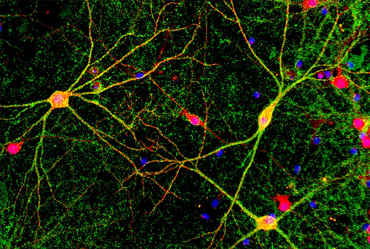
Stem cells anchor the development of human organs and other structures, and, in some cases, can also perform repairs. The intensive interest in these developmental masterminds relates directly to their ability, under the right conditions, to develop into any tissue in vitro, securing their prime position as research tools as well as in therapeutic applications.
One of the ways researchers use stem cells is for the development of organoids–3D self-assembled structures that mimic the function of human organs. Organoid models are relevant in many areas of research and drug development, but are particularly much-needed, useful models for neural and brain research. Significant progress has been made on cancer therapeutics, but individuals suffering from degenerative neurological disorders like Alzheimer’s and Parkinson’s diseases still desperately need options to slow or even reverse the disorders.
Neural and brain organoids hold great promise to further understand brain development and function, model neural diseases, and test potential therapeutic approaches. Possibly, in certain circumstances, they might also be grown as surrogate sources for transplantation.
But, as always, when trying to replicate in vivo conditions in an in vitro model, challenges abound. For stem cells to be induced to follow a certain path of development, in vivo environments must be replicated as closely as possible.
This has led to the advancement of electroporation instruments, specific growth factors, and other exciting technologies that allow scientists to perform single-cell biopsies that can follow RNA translation from the same cell over time, as well as new microelectrode array (MEA) technology specifically designed for cells that use electrical potential to communicate. And these emerging technologies are only the front edge of an expected avalanche of innovative evolutions.
Tailoring products
Capable of nearly unlimited cell renewal, pluripotent stem cells (PSCs) have the capacity to differentiate into nearly every human cell type. As such, PSCs have been foundational tools in understanding human development and, more recently, as attractive platforms for cell therapy and regenerative medicine applications.
According to David Kuninger, PhD, director of cell biology R&D at Thermo Fisher Scientific, PSCs have typically been cultured in a monolayer, but the estimated commercial needs of PSC-derived cell types have led to the development of scaled-up 3D-suspension culture media and methodologies. In a generalized workflow, PSCs are expanded and characterized for genetic stability and pluripotency. These cells can then be taken into, typically, a multi-step differentiation process, driving cell commitment to specific lineage and downstream cell types in ways that mimic development in vivo.
“The Neon NxT Electroporation system uses advanced technology to transform transfection efficiency, particularly in PSCs, by employing finely tuned electrical pulses to maximize cell permeability, while minimizing cytotoxicity,” said Kuninger. Tailored to PSCs’ delicate nature, customizable pulse parameters result in high viability.
The system’s proprietary open-design specialized electroporation chamber ensures uniform electric field distribution, enhancing the consistency of nucleic acid uptake. Additionally, unique tip technology provides precise delivery and high transfection efficiency. A user-friendly interface allows for adaptation to a wide range of cell types and experimental conditions.
CultureCEPT Supplement, a chemically-defined cell culture supplement, has been shown to increase cell survival and viability during handling and processing steps. Based on discoveries made at the National Center for Advancing Translational Sciences, the supplement contains the molecules chroman 1, emricasan, polyamines, and trans-ISRIB, which collectively reduce cellular stress and prevent DNA damage.
Other research has demonstrated that CultureCEPT Supplement enhances normal expression patterns of cytoskeletal and cell-membrane associated proteins, prevents bleb formation, increases expression of glutathione and procaspase 3, and supports normal cell cycles. Kuninger noted that the “formula significantly improves outcomes in cell culture workflows, including long-term passaging, cryopreservation, and organoid formation.”
Refining microenvironments
Laminins are essential proteins both in vivo and in vitro, according to Malin Kele, PhD, director of business development at BioLamina. “Over the past 15 years, the use of biorelevant laminin isoforms in protocols has contributed to improved reproducibility, quality, and functionality of cellular models and enabled clinical translation.”
The family of laminin proteins plays a key role in organizing tissue-specific extracellular matrices (ECM), acting as reservoirs for growth factors, and directly engaging cell surface receptors to regulate vital cellular processes such as adhesion, identity, differentiation, survival, and migration. “In mammals, there are 16 distinct laminin isoforms. The proper combination of these isoforms is essential for healthy tissue function,” explained Kele.
Tissue engineering, for modeling and cell replacement therapy, requires protocols resulting in high specificity, functionality, and reproducibility. Stage-specific media and biorelevant substrates facilitate mimicking development in a dish. Isoform-specific, recombinant full-length human laminin cell culture matrices (Biolaminin®) allow for precise control of the cellular niche at all protocol stages.

“We are the sole producer of full-length human recombinant laminins,” said Kele. “We currently offer a toolbox of 11 different isoform-specific, chemically defined, animal origin-free laminin proteins for various applications, from R&D to clinical translation.”
In general, PSCs and adult stem cells thrive on Biolaminin 521. However, differentiation protocols often require the addition of other isoforms to accelerate the process. An example is iPSC-derived dopamine neurons, where the supporting laminin shifts from the early-stage laminin 521 to laminin 111 during differentiation.
Another product, Biosilk®, a non-immunogenic and biodegradable, recombinant fragment of the spider silk protein, forms a robust, fibrous network that mimics the structure of natural ECMs and allows free movement of nutrients and patterning factors. Biosilk 3D networks can be coated with Biolaminin to generate tissue-specific 3D environments. For example, Biosilk functionalized with Biolaminin 111 formed the base for long-term and reproducible tissue-like ventral midbrain organoids.

Kele believes that, in the future, cell culture substrates will play an increasingly important role beyond upstream processing in supporting cell survival, integration, and function at cell therapy delivery sites, enhancing their efficacy and durability in clinical applications.
Tracking single cells
Dynamic, non-destructive transcriptomic profiling is needed to decode cell state transitions in development, aging, and disease in the neuronal research field.
The FluidFM® OMNIUM platform performs live-cell cytoplasmic biopsies using a proprietary hollow cantilever with a nanoscale tip, combining atomic force microscopy precision with microfluidics. The process begins with target cell selection using the integrated microscope and software to position the FluidFM Nanosyringe precisely at the desired location on the cell. Then, with controlled force, the syringe gently penetrates the cell membrane and extracts a small volume of cytoplasm using negative pressure. The full transcriptome can be retrieved from this pl-sized cytoplasmic liquid biopsy.
“The cell remains viable throughout and after the procedure,” said Dario Ossola, PhD, head of applications and product manager of instruments at Cytosurge. “The tiny biopsy contains sufficient RNA to retrieve the full transcriptome of the cell and is about 10 to 50% of a typical cell’s total cytoplasm.” A Nature article describes how the target cell is minimally perturbed, even at a transcriptional level, in a sequencing technology called Live-seq.1
“Live-seq’s time-resolved, single-cell expression tracking is a new lens for developmental biology,” Ossola emphasized. The workflow tracks the same cell’s transcriptional changes over time to generate longitudinally-paired molecular and phenotypical data that intrinsically contains cause-and-effect information.
The FluidFM OMNIUM platform handles the targeting, extraction, and initial sample deposition into a buffer droplet containing RNA stabilization components. Then the workflow uses the highly sensitive Luthor HD kit, integrated through a partnership with Lexogen, for an ultra-low input RNA library preparation protocol. Libraries are sequenced conventionally, followed by specialized bioinformatic analysis.

“Neural development is a very interesting use case,” said Ossola. While 3D models are increasingly adopted, the neuroscience community still relies on 2D cell cultures for better experimental control. Indeed, MEAs and live-cell imaging techniques are greatly contributing to understanding how these cells differentiate and communicate.
FluidFM complements these established techniques by enabling temporal profiling of individual neural stem cells as they differentiate, providing direct insight into the molecular mechanisms driving fate decisions as well as investigation of responses to external stimuli. The platform also has significant potential in cancer heterogeneity studies.
Monitoring cellular communication
Stem cell-derived organoids mirror the complex spatial organization found in organs. Typically, they are grown in suspension to maintain their 3D structure and viability, and, thus, their translational capability. For example, iPSC-derived brain organoids offer a human-relevant model system to study brain development and function as well as dysfunction in neurological disorders.
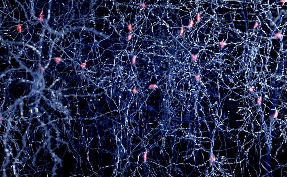
Neurons communicate via electrical signals, and monitoring this activity in organoids is crucial for assessing neural circuit activity and drug effects. The challenge is attaching tiny microelectrodes to a 3D moving target. Most organoids cultured under continuous movement settle and deform on solid surfaces.
“A mesh microelectrode array (MEA) has advantages over solid surface MEAs when it comes to brain and neural organoids,” said Peter Jones, PhD, group leader of biomedical micro and nano engineering at the NMI Natural and Medical Sciences Institute in Reutlingen, Germany. “With a mesh MEA the organoids are not constrained, so do not deform and can function normally.”
“Our Mesh MEA chip is a new way to obtain real-time recordings over time from any organoids that contain cells that use electrical signals to communicate,” said Sven Schönecker, PhD, global product manager at MultiChannel Systems. “Interestingly, the cells grow along the mesh and use it as a type of scaffold.” As the organoids grow, they internalize the mesh MEA to enable simultaneous recording from different locations within the tissue. The internalization amount depends on the application.

The Mesh MEA chip consists of 60 electrodes on a 12µm-thin polyimide mesh. The Mesh MEA chip is compatible with the small footprint MEA2100-Mini System, a miniaturized in vitro recording system, for continuous, undisturbed recordings and stimulation of samples in an incubator or on a lab bench.
In addition, incubator-compatible mini Mesh MEA systems are available for monitoring individual organoids. “You can also merge organoids and use two or three organoids on the same chip to get different brain regions close together,” said Schönecker.
Mesh MEAs will continue to evolve. For instance, a mesh with multiple 3D layers would allow evaluating larger circuits and more regions of a brain organoid. In addition, NMI and MultiChannel Systems are considering a simpler multi-well high-throughput system and a multi-disciplinary approach that could, for example, merge electrophysiological recordings and live imaging as well as integration with organ-on-a-chip devices.
Reference
- Chen W, Guillaume-Gentil O, Rainer PY et al. Live-seq enables temporal transcriptomic recording of single cells. Nature. 2022; 608:733-740. https://doi.org/10.1038/s41586-022-05046-9
The post Reading the Masterminds of the Human Body appeared first on GEN – Genetic Engineering and Biotechnology News.


