Introduction
Bone tissue engineering presents a promising alternative to conventional grafting techniques for the treatment of critical-sized bone defects. However, the clinical success of such approaches remains highly dependent on the design and functionality of the scaffold1. Recent scaffold designs increasingly prioritize biomimetic strategies that replicate the composition, structure, and functional properties of the native bone extracellular matrix (ECM)2. This paradigm shift recognizes that scaffolds must provide both physical support and bioactive cues to guide cellular behavior and mineralization3.
Although previous studies have made significant contributions to mimicking the extracellular matrix (ECM), challenges in fully replicating its native complexity remain4. These efforts should not be viewed as failures but rather as essential steps in a continuous process of advancement5,6. Building on these foundations, our research demonstrates notable improvements in achieving a more accurate and functional ECM structure, regular nanoscale structures, appropriate biochemical signaling, and dynamic mechanical responsiveness similar to that of bone. Furthermore, contributing of antibiotics could improve bone healing in infected defects7. Overall, scaffolds are essential for bone regeneration by providing a framework for new tissue growth and facilitating the repair and regeneration of damaged bone tissue8.
Piezoelectricity, the ability of certain crystal structures to generate an electric charge when subjected to mechanical stress, offers notable advantages for scaffold design9. Incorporating piezoelectric materials into bone scaffolds enables the generation of localized electric fields during mechanical loading, modulating bone cell activities and fostering bone formation, which is critical for tissue regeneration10,11,12. During electrospinning, the strong electric field orients the dipoles in the piezoelectric polymer chains, enhancing the formation of the electroactive β-phase. Therefore, combining electrospinning with piezoelectric materials enables the fabrication of scaffolds that closely mimic bone extracellular matrix (ECM) architecture13,14.
Polyvinylidene fluoride (PVDF) is widely utilized in bone regeneration due to its piezoelectric properties, biocompatibility, and capacity to enhance osteoblast adhesion and collagen formation15,16. PVDF exhibits many crystalline phases, with the β-phase being characterized by its polarity and demonstrating both ferroelectric and piezoelectric capabilities17,18. Many studies have tried to integrate barium titanate (BaTiO₃; BTO) particles into piezoelectric polymeric matrices, such as PVDF19. Adding nanoparticles such as BTO, ZnO, and others to PVDF-based nanocomposite can enhance their piezoelectric properties, making it suitable for sensors20, actuators21, and energy harvesting applications22. Furthermore, BTO has been researched for its biocompatibility and bioactivity, which are desirable characteristics for specific medical devices and implants23.
The hydrophobic nature of PVDF restricts cell attachment and growth24. Surface treatment techniques like chemical vapor deposition and plasma treatment are effective methods to enhance the hydrophilicity of inherently hydrophobic polyvinylidene fluoride (PVDF) nanofibers25. The modified nanofibers displayed improved adherence to cells and significant changes in surface wettability, making them more suitable for biomedical applications26. PVDF nanofiber scaffolds are widely used for their bioactive and biocompatible properties27.
Bacterial contamination remains a major challenge for medical implants and scaffolds. Emerging biomaterials can reduce bacterial growth and promote bone tissue regeneration due to their intrinsic bioactivity28. Biomaterials with antibacterial surfaces are essential for preventing biofilm formation and infection29. Nevertheless, it is essential to incorporate antibacterial properties when utilizing fluoropolymers in the medical and healthcare fields30. This is due to the documented resistance of PVDF surfaces to microbial colonization and the production of biofilms31.
Antibiotics are considered an effective antibacterial strategy for treating orthopedic implant-related infections32,33. Controlled and sustained release of antibiotics from the scaffold is crucial to maintaining therapeutic levels at the infection site over an extended period32,34. Vancomycin-laden scaffolds have osteogenic and antibacterial properties that can enhance bone healing in infected defects35. Using vancomycin in different scaffolds has been explored in various research studies, including bio-derived hydrogels, nanodiamond composite scaffolds, PLGA microspheres, poly(simvastatin) scaffolds, and extracellular matrix scaffolds crosslinked with vancomycin. These scaffolds can help prevent infection and support tissue regeneration in various medical applications36,37,38,39.
This study primarily investigates the fabrication of bioactive and biocompatible nanofiber scaffolds using PVDF/BTO nanocomposites. To our knowledge, no prior work has simultaneously combined piezoelectric stimulation, cold plasma treatment, and controlled antibiotic delivery in a single scaffold, addressing both mechanical and infectious challenges in bone defects. By integrating piezoelectric stimulation, cold plasma treatment, and drug delivery into a single scaffold, we address multiple challenges simultaneously, which existing solutions often fail to do. Piezoelectricity promotes osteogenesis by providing electrical cues, overcoming the limitations of passive scaffolds. Plasma treatment enhances cell attachment and integration, solving the issue of poor bioactivity in traditional materials. In this sense, drug delivery ensures localized infection control, addressing the critical challenge of implant-associated infections. To enhance the PVDF β phase, BTO filler was added in varying weight percentages through the high-voltage electrospinning method. Nanofibrous scaffolds loaded with vancomycin were tested for their antibacterial activity against Gram-positive and Gram-negative implant pathogens. Unlike many previous approaches that focus on either mechanical stimulation or antibacterial activity individually40,41, the scaffold developed in this study offers unique multiple functionalities by integrating piezoelectric properties, controlled antibiotic release, and cold plasma treatment. This innovative integration not only mimics the electrical microenvironment of native bone to stimulate osteogenesis but also addresses the critical challenge of implant-associated infections, positioning this scaffold as a comprehensive solution for bone tissue engineering.
Experimental and methods
Materials and samples fabrication
The main components used were PVDF powder (CH2=CF2, MW = 275,000 g/mol; Kaynar 761), BaTiO₃ nanoparticles42,43, and vancomycin hydrochloride powder (Sigma-Aldrich). N-dimethylformamide (C₃H₇NO; DMF, Merck) was used as the solvent to dissolve the PVDF/BTO mixture. Initially, 10 mL of DMF was ultrasonicated in a bath sonicate for 60 min to disperse barium titanate (BTO) ceramic nanoparticles at varying weight percentages (0, 5, 10, and 15%). Subsequently, 2 g of PVDF were added to the solution and stirred using a magnetic stirrer with an oil bath equipped with a proportional-integral-derivative (PID) controller. The solution was stirred at 60 °C for two hours until the PVDF polymer was fully dissolved.
Four concentrations of PVDF-BTO were determined for electrospinning. The samples contain PVDF, PVDF-5%BaTiO3 (PVDF/5BTO), PVDF-10% BaTiO3 (PVDF/10BTO), and PVDF-15% BaTiO3 (PVDF/15BTO). Electrospinning was performed at room temperature using a rotary collector electrospinning machine (Asia Nanostructure Technology Company, Iran) to produce uniform fibers. The polymeric solution was loaded into a syringe with a volume of approximately 10 mL, produced by Ava Company. The syringe was fitted with a metal needle gauge 38 mm in length and 21 gauges in diameter. Then, the solution was transferred to an infusion setup. Electrospinning was performed under the following conditions: collector speed of 800 rpm, distance of 12 cm between the needle and collector, applied voltage of 20 kV, and flow rate of 3 mL/h. The electrospinning process took approximately 60 min, and the metallic collectors were coated with aluminum foil (86 × 75 mm).
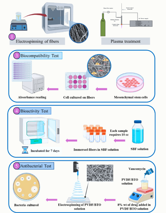
The electrospun scaffolds were treated with helium plasma on both sides. The following principles were followed while carrying out the treatment procedure: a Helium flow rate of 0.0012 kg/sec, an AC power source, and treatment at approximately 60 °C for 4 min. Plasma treatment and electrospinning processes are shown in Fig. 1, “Schematic was created with BioRender.com.”
To conduct the antibacterial test, barium titanate ceramic nanoparticles (10%, optimal percentage) with 0.2 gr of vancomycin and 2 gr of PVDF in DMF (10 ml) were solved to produce fibers with the specifications mentioned above.
Characterization
Morphology of fiber scaffolds
The morphology of the nanofibers was examined using scanning electron microscopy (SEM; EVO MA15, ZEISS, Germany) at an acceleration voltage range of 0.5–40 kV.
Mechanical properties of fiber scaffolds
An analysis was conducted on the mechanical properties of PVDF/BTO nanofiber scaffolds were analyzed to assess their stress response. The study was performed utilizing the NDY-150k universal machine with a testing speed of 12 mm/min. Three specimens of each nanofiber, with dimensions of 2 × 8 cm2, were tested to ascertain the mean values of Young’s modulus, ultimate tensile strength (UTS), and elongation at fracture.
Piezoelectric properties of fiber scaffolds
Samples were cut into 2 × 2 cm2 pieces and wrapped in aluminum foil for piezoelectric testing. Using a homemade piezo tester, The output voltage was measured using a custom-made piezoelectric tester under a force of 2.6 N and a frequency of 5 Hz, with detailed specifications provided elsewhere44. The force of 2.6 N and a frequency of 5 Hz were selected to provide standardized and repeatable testing conditions, ensuring the reliable evaluation of piezoelectric sensor performance.
Wettability of fiber scaffolds
Water contact angles (WCA) at ambient temperature were measured using an optical contact angle goniometer (Model CAG-1, NanoFMC, Iran). A droplet volume of 10 µl was consistently used for WCA measurements. The WCA was determined as the average of measurements taken at five distinct locations on each specimen.
Roughness of fiber scaffolds
Surface roughness was assessed at ambient temperature using a laser profilometer (Model LPM-D1, Fanavari-Kahroba Company, Iran). Specimens were cut into 1 × 1 cm2 segments and positioned under the profilometer laser. Roughness parameters were determined using Gwyddion software.
Cell culturing and cell migration of fiber scaffolds
Mesenchymal stem cells (MSCs) were derived from the Iran Biological Resource Center. The Dulbecco’s Modified Eagle Medium (DMEM-F12) was enriched with 10% (v/v) fetal bovine serum (FBS) from Gibco, USA, and 1% Pen-Strep. Cultures were maintained at 37 °C in a humidified atmosphere containing 5% CO2. In order to study cell adhesion and cell morphology, the nanofibrous mats were fabricated and then sterilized using gamma radiation at a dose rate of 25 kGy/h. Subsequently, the mats cut into discs of 2 cm diameter.
Following sterilization, MSCs were seeded onto the scaffolds at a density of 1 × 10⁴ cells per well in a 24-well plate. After 24 h of incubation, cells adhered to the scaffolds were fixed with 2.5% glutaraldehyde, followed by graded dehydration using ethanol concentrations ranging from 30 to 100%. Cellular morphology on the scaffolds was subsequently assessed by scanning electron microscopy (SEM). For the evaluation of cell viability, sterilized nanofiber mats were incubated in 3 mL of serum-free medium at 37 °C for 4 and 7 days to obtain material extracts. MSCs were seeded at 1.9 × 10⁴ cells per well in 96-well plates. After 24 h, the culture medium was replaced with the prepared extracts, and incubation continued for an additional 24 h. A culture medium without material extracts served as the control.
Cytocompatibility of fiber scaffolds
Cell viability was assessed using the MTT assay. Briefly, 3-[4,5-dimethylthiazol-2-yl]-2,5-diphenyltetrazolium bromide (MTT; Sigma, USA) was dissolved in phosphate-buffered saline (PBS) at a concentration of 0.5 mg/mL to prepare the stock solution. The MTT solution was added to each well at a 1:10 ratio with the culture medium. After 4 h of incubation at 37 °C, the culture medium was removed, and 100 µL of dimethyl sulfoxide (DMSO) was added to each well to dissolve the formazan crystals. Samples were then shaken at 37 °C for 15 min.
Subsequently, 100 µL from each well was transferred to a fresh 96-well plate, and absorbance was measured at 570 nm using a microplate reader (STAT FAX 2100, USA). The MTT assay was performed in triplicate for each sample, with tissue culture polystyrene (TPS) serving as the negative control. Results are reported as the mean ± standard deviation (SD).
Bioactivity of fiber scaffolds
Bioactivity analysis was conducted on nanofiber scaffolds by assessing calcium phosphate (CaP) deposition on their surfaces, following the ISO 23,317 standard for evaluating apatite-forming ability. Samples were placed in sterilized containers containing simulated body fluid (SBF). Samples measuring approximately 2 cm2 were immersed in SBF for 7 days. Post-immersion, the samples were analyzed using scanning electron microscopy (SEM) and inductively coupled plasma (ICP) spectroscopy to assess the extent of CaP deposition. The SBF solution was prepared according to the protocol outlined in references42,43.
Degradation of fiber scaffolds
Biodegradation studies were conducted following ASTM F1635-24, the Standard Test Method for in vitro Degradation Testing of Hydrolytically Degradable Polymer Resins and Fabricated Forms for Surgical Implants. Nanofiber samples measuring 2.5 cm2 were cut, weighed, and submerged in 10 mL of phosphate-buffered saline (PBS) at 37 °C for 1, 4, 7, 14, 28, and 56 days. At each time point, samples were rinsed with distilled water, air-dried at ambient temperature, and re-weighed to determine percentage weight loss due to degradation. Each experiment was performed in triplicate. The degradation rate was calculated using the following formula45:
$${text{Degradation}};{text{rate}};(% ) = left( {frac{{Wi – Wf}}{{Wi}}} right) times 100$$
(1)
Antibacterial activity of fiber scaffolds
The antibacterial efficacy of the PVDF/BTO nanofiber scaffold containing vancomycin against E. coli and S. aureus was assessed using the disk diffusion method. E. coli and S. aureus cultures were grown in Luria-Bertani broth for 18 h at 35 ± 2 °C, then diluted to a concentration of 1 × 10⁸ cells/ml with sterile water. A 40− l aliquot of the diluted bacterial suspension was spread-plated onto duplicate nutrient agar plates. Discs, 4 mm in diameter and weighing > 2 mg, were cut from the fibrous membrane and placed on the agar surface. The plates were incubated at 37 °C for 24 h, after which the zones of inhibition around the discs were measured.
Statistical analysis
All tests were carried out in triplicate, and outcomes were statistically assessed via SPSS software. The samples were analyzed using one-way analysis of variance (ANOVA) and Tukey’s post hoc test at p < 0.05.

Process of fabrication of PVDF/BTO nanofiber scaffolds.
Results and discussion
The PVDF/BTO bone scaffold nanofibers were fabricated utilizing the electrospinning technique, as shown in Fig. 2a. The morphology of the nanofibers was evaluated using SEM images shown in Fig. 2(b-e). Nanofibers were formed by applying a strong electric field between the syringe needle and the rotating drum collector, which overcame the surface tension of the droplets via electrostatic force.
The diameter distribution of electrospun PVDF nanofibers (Fig. 2b) ranged from 200 to 400 nm. Figure 2(c-e) shows that the diameter of fibers in PVDF/BTO ranges from 180 to 250 nm. Upon adding BTO nanoparticles to the PVDF solution, they were uniformly distributed throughout the polymer matrix and embedded within the fibers. It has been observed that an increase in the percentage of BTO leads to a slight decrease in fiber diameters. This occurs because BTO nanoparticles increase the electrical conductivity of the solution, leading to higher charge density on the ejected jet46,47. This amplifies the electrostatic repulsion forces, which dominate over surface tension and viscoelastic resistance, resulting in greater jet elongation and thinner fibers48. The PVDF/BTO solutions produced uniform fibers with minimal beading, achieving an average diameter of 180 nm for 15% BTO.
Nanofiber dimensions of the PVDF and PVDF/BTO scaffolds, calculated by the ImageJ program, showed the average diameters in histogram diagrams.
BTO nanoparticles tend to aggregate above 10% due to strong chemical interactions and weak van der Waals repulsion. As a result, the fiber tensile strength during the electrospinning process is affected by these nanoparticles. When the concentration of BTO particles increased to 15 wt%, a granular morphology appeared on the fiber surface (Fig. 2(e-2)).
An analysis was conducted on the mechanical characteristics of the PVDF/BTO nanofiber scaffolds to gain insight into their response to stress. Figure 3 displays the stress-strain curves of the examined samples, along with the relevant measurements for Young’s modulus, ultimate strength (UTS), and elongation at fracture, which are shown in Table 1.
The stress-strain curves shown in Fig. 3 demonstrate that the incorporation of BTO nanoparticles has enhanced the mechanical characteristics of the nanofibers. As shown in Table 1, the electrospun nanofiber samples exhibited increased ultimate tensile strength and Young’s modulus compared to the pure PVDF scaffold.
The PVDF/5BTO sample had an ultimate tensile strength of 17.2 MPa. This value was 196% higher than that of the pristine nanofiber scaffold. The results also indicated that nanoparticle incorporation increased elongation at fracture, confirming the positive effect of the nanofiller on mechanical performance. Due to the use of a dynamic collector, the nanofibers in this work exhibit a random orientation. Upon stretching, some nanofibers exhibited partial alignment in the direction of applied stress49.At concentrations above 10 wt%, BTO nanoparticles formed agglomerates (Fig. 2e) due to van der Waals forces. These agglomerates disrupted stress transfer between PVDF chains and BTO, weakening interfacial adhesion and initiating cracks under load43,50. However, incorporating BTO nanoparticles at concentrations above 10 wt% reduced the scaffold’s mechanical strength, resulting in increased fragility.

(a) A schematic of the electrospinning process, SEM images of the scaffolds, and histogram of diameter distribution: (b) PVDF, (c) PVDF/5BTO, (d) PVDF/10BTO, (e) PVDF/15BTO.
For human trabecular bone, the modulus typically ranges between 10 and 3,000 MPa51. In contrast, separate research focusing on cortical bone found Young’s modulus values ranging from 9518 MPa to 14,181 MPa52. When Young’s modulus of a scaffold is higher than that of the bone, it can lead to stress shielding, where the scaffold bears a majority of the load, causing the surrounding bone to decrease in density and strength over time. This phenomenon can result in bone resorption and scaffold loosening53. Research suggests that stress transfer to the bone is more significant with scaffolds of lower Young’s moduli, and scaffolds with a high elastic modulus could have better biomechanical behavior, especially when in contact with the bone of a higher elastic modulus. However, the distribution of transferred stress in the bone, particularly in the cortical bone, is essential to prevent excessive stress on the bone. Therefore, while a higher modulus scaffold may have better biomechanical behavior, the interaction between the scaffold and the bone is a complex process that needs to be carefully considered54. Results in Fig. 3 show that 5% wt BTO has a higher modulus and improved biomechanical behavior.

Stress-strain curves of electrospun PVDF/BTO nanofibers scaffolds.
This assay proposes the development of graded scaffolds, where the outer surface (in contact with bone tissue) contains a higher BTO content (e.g., 15 wt%) to maximize piezoelectric stimulation for enhanced osteogenesis, while the inner bulk retains a lower BTO content (5–10 wt%) to preserve mechanical integrity and avoid brittleness. This functionally graded structure could offer both sufficient mechanical support and localized bioelectrical cues, mimicking the gradient nature of native bone and addressing both structural and biological requirements. In conclusion, although higher BTO enhances piezoelectricity, the Young’s modulus range of 20–34 MPa achieved in optimized compositions aligns well with trabecular bone and ensures a mechanically compatible scaffold, minimizing the risk of stress shielding and promoting long-term integration.
The piezoelectric signals were measured using a piezoelectric tester while the samples were subjected to stress circumstances, as shown in Fig. 4a. Voltage output was recorded over a 10-second interval under a 2.6 N load at 5 Hz for each sample. Voltage sensitivity was calculated by dividing the output voltage by the applied force. The ratio may be expressed by the equation S = V/F44,55, where V and F represent the variations in the output voltage signal and applied force, respectively. The recorded data is shown in Table 2.
The piezoelectric behavior of PVDF nanofibers is significantly influenced by the quantity of the β phase and the level of crystallinity in the samples56. The β-phase has an all-trans (TTTT) planar zigzag conformation of the polymer chains. This conformation aligns the dipoles of the C–F and C–H bonds in the same direction, creating a net permanent dipole moment57. The higher the β-phase content, the stronger the piezoelectric response because more dipoles are aligned and contribute to the material’s electromechanical coupling. BTO nanoparticles serve as nucleation sites during PVDF crystallization, enhancing the local electric field58. The presence of high β-phase content in synthetic scaffolds mimics the intrinsic piezoelectricity of natural bone, enabling these materials to generate electrical signals in response to mechanical loading59. This electromechanical coupling is known to stimulate osteogenic activities, promoting bone regeneration and remodeling41. Consequently, materials with enhanced β-phase content are promising for further evaluation in bone tissue engineering, where they can be used to fabricate scaffolds that not only provide structural support but also actively contribute to the healing process through electrical stimulation60 and relevant to applications such as sensors, energy harvesters, and biomedical implants.
Additionally, BTO incorporation promotes O-H15 and F-C61 hydrogen bonding, along with dipole interactions between PVDF and dimethylformamide (DMF). These interactions often result in the formation of local CH2-CF2 dipoles arrayed in the TTT (trans-trans-trans) configuration, which is a distinguishing feature of the β-phase of PVDF. The results are summarized in Fig. 4b; Table 243,62.

(a) schematic of the piezoelectric tester, (b) a graph displaying the piezoelectric response of various PVDF/BTO nanofibers.
The surface composition and roughness of scaffolds influence the rate and quality of bone bonding by enhancing anchoring and biomechanical stability63; both of them increase bone anchoring and create biomechanical stability. Surface chemistry and charge also influence protein and cell adhesion, as well as hydrophilicity64,65. Hydrophilicity can also be effective in the quality and speed of bonding of the scaffolds with the bone. Highly hydrophilic surfaces generally support better cellular interactions and fluid absorption than hydrophobic ones66.
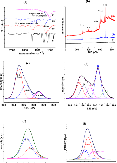
Surface hydrophilicity plays a key role in the efficiency and rate of scaffold–bone integration67,68 ; the schematic of this process is shown in Fig. 5a. During the cold plasma treatment of PVDF, multiple reactive species, such as ions, free radicals, and excited molecules, are produced61,69. When PVDF reacts with these species, hydroxyl (-OH)15 and carboxylic acid (COOH)15,70 are formed. The surface of PVDF newly created functional groups, is polar and very hydrophilic, meaning they have a strong attraction for water molecules. As a result, water spreads more uniformly across the PVDF surface, increasing wettability. Increased protein and cellular adhesion promote better bone development67. Cold atmospheric helium plasma jet was employed to treat PVDF and PVDF/10BTO scaffolds. Plasma treatment for 2 and 4 min significantly reduced the contact angles, indicating enhanced surface hydrophilicity (Table 3).
Figure 5(b and c) show an increase in surface roughness and a notable decrease in contact angle after cold plasma treatment. This indicates that the treated surface becomes both rougher and more hydrophilic. The increased roughness provides more surface area and topographical cues for cell attachment, while the reduced contact angle reflects enhanced wettability, both of which are beneficial for improving cell adhesion and overall biocompatibility of the scaffold71.

(a) schematic of the helium plasma jet, Images of contact angle and roughness map obtained by laser profilometer of PVDF/BTO nanofiber scaffolds (b) before surface treatment, and (c) cold plasma treatment.
The in vitro biocompatibility of PVDF and PVDF/BTO nanofibers containing 5%, 10%, and 15% BTO was evaluated using the MTT assay to assess cell viability. As shown in Fig. 6, the nanofiber scaffolds exhibited good cytocompatibility with mesenchymal stem cells. Consistent with the results mentioned above, all the PVDF/BTO nanofibers demonstrated greater cell viability than pure PVDF nanofibers. Among the PVDF/BTO nanofibers, cell viability was significantly enhanced on PVDF/15BTO compared to the other nanofibers, as shown in Fig. 6d. These results were associated with the content of the piezoelectric β-phase and high hydrophilicity in the PVDF/BTO nanofibers. The increased barium titanate content is responsible for the enhancement of the β phase, and electrospinning is an effective method for preparing polar surfaces with both positive and negative electrical charges. Therefore, the PVDF/15BTO nanofiber exhibited the highest cell viability, which was statistically significant compared to the control group (P < 0.01). Moreover, the addition of BTO nanoparticles to PVDF nanofibers also increased the formation of stronger O-H and F-C hydrogen bonds, which improved the hydrophilicity of the surface and promoted better protein and cellular adhesion. Thus, the increase of BTO as a filler in PVDF nanofibers can enhance their piezoelectric response and biocompatibility with living tissue68. Increased cell proliferation within scaffolds is a key driver of successful long-term in vivo performance. It promotes uniform tissue formation, accelerates healing, enhances vascularization, improves integration, and supports the functional replacement of the scaffold with native tissue72.

SEM images of mesenchymal stem cells cultured on (a) PVDF, (b) PVDF/ 5BTO, (c) PVDF/10BTO, and (d) PVDF/15BTO nanofibers after one day.

Proliferation behavior of MSC cells on PVDF/BTO nanofibers.
The behavior of cell proliferation on PVDF and PVDF/BTO nanofibers is shown in Fig. 7. After seven days of cell proliferation on control samples and treatment samples, PVDF/BTO treated nanofibers have more biocompatibility than control nanofibers.
The bioactivity of biomaterials in bone tissue engineering, particularly their capacity to form apatite structures on their surface, is essential for bonding with living bone tissue. This property can be evaluated by immersing the biomaterials in simulated body fluid (SBF) for extended periods. A schematic of this process is shown in Fig. 8a. Several studies and reviews have Previous studies have explored the use of natural polymers—such as collagen, alginate, silk, hyaluronic acid, and chitosan—in bone-related applications73,74; meanwhile, in this study, PVDF/BTO nanofibers were used .
Based on previous findings [12], PVDF is recognized as a hydrophobic polymer devoid of any bioactive characteristics. The bioactivity of a material is determined by its ability to prevent the formation of a fibrous capsule around it and to attract the deposition of body elements on its surface. Cold plasma treatment effectively enhanced scaffold bioactivity, as evidenced by increased mineral deposition on scaffold surfaces Fig. 8(b-d) and Inductively Coupled Plasma test (ICP) results Fig. 8e. Plasma treatment increased the surface energy and hydrophilicity of the nanofibers, thereby improving cell adhesion and facilitating calcium phosphate (CaP) deposition.
Electrospinning is an effective technique for producing PVDF nanofiber scaffolds with a high β-phase content without the need for additional high-voltage poling or mechanical stretching56,75. The use of polar β-PVDF and cold plasma treatment on nanofibers create distinct negative and positive charge centers that affect the behavior of SBF. These centers attract positively charged calcium ions (Ca2+) and negatively charged phosphate ions (PO43−), but these ions must be absorbed on the surface of the fibers to form apatite nuclei. SEM images showed that the PVDF/15BTO scaffold had more CaP structure deposition on the fiber surface after seven days of immersion than other scaffolds. Barium titanate not only promotes β-phase formation but also acts as a bioactive ceramic76; thereby increasing calcium deposition on the nanofiber surface, as shown in Fig. 8(c-e) and confirmed by ICP analysis Fig. 8b. BTO nanoparticles provide nucleation sites that accelerate hydroxyapatite (HA) formation. At 15 wt%, increased surface exposure of BTO facilitates rapid ion exchange, promoting the deposition of calcium phosphate precursors. This aligns with studies showing that kinetic factors, such as ion addition rates, critically influence nucleation pathways in calcium phosphate systems77. Therefore, BTO is not only a piezoelectric material but also possesses inherent bioactive and biocompatible properties.
The biodegradation results of the PVDF/BTO nanofiber scaffolds in PBS medium for 56 days are represented in Fig. 9. The results showed a reduction in sample weight across all four fibers after 56 days. The PVDF nanofiber had a higher degradation rate in the PBS solution and lost most of its initial weight after 56 days. In other words, PVDF/BTO nanofibers degrade more slowly than pure PVDF nanofibers. Because of this, BTO ceramic nanoparticles increase the β-phase and PVDF crystallite, decreasing the degradation rate of these fibers78. According to previous studies, the fibers were primarily used in the field of wound dressing79. However, results from biodegradation and antibacterial tests indicate that these nanofibers are also suitable for sustained drug release and bone scaffold applications. The PVDF/BTO nanofiber scaffolds were tested for their antibacterial properties against S. aureus and E. coli using the disc diffusion method. The result of the disk diffusion test is reported based on the diameter of the non-growing halo of bacteria around the disk containing the antimicrobial agent.

(a) schematic of soaking nanofibers in simulated body fluid. (b) ICP TEST results,
An apatite formation on the surface of (c) PVDF/ 5BTO, (d) PVDF/ 10BTO, (e) PVDF/ 15BTO with and without plasma treatment,

Biodegradation of nanofibers after 1, 4, 7, 14, 28, and 56 days.
The PVDF/BTO nanofiber scaffolds were tested for their antibacterial properties against S. aureus and E. coli using the disc diffusion method. The result of the disk diffusion test is reported based on the diameter of the non-growing halo of bacteria (inhibition zone) around the disk containing the antimicrobial agent.
The PVDF/BTO nanofiber scaffold without vancomycin (used as the control sample), had no antibacterial activity against E. coli and S. aureus. As shown in Fig. 10(b-1) and (a-1), no evidence exists of an inhibition zone around the samples (A, B and C). Previous studies report that vancomycin is effective against Gram-positive bacteria, including S. aureus, a common cause of bone infections80. Accordingly, the PVDF/BTO scaffold loaded with vancomycin (samples D, E, and F) produced visible inhibition zones against S. aureus, as shown in Fig. 10(b-2). However, no antibacterial effect was observed against E. coli, and no inhibition halo was seen, as presented in Fig. 10(a-2). Finally, Fig. 10(c) shows the inhibition zone diameters for PVDF/BTO nanofiber scaffolds loaded with vancomycin (samples D, E, and F) against S. aureus.

The disk diffusion antibacterial test results (a) against S. aureus and (b) E. coli was obtained for the PVDF/BTO nanofiber scaffold with/ no the drug, and (c) the diameters inhabitation zone for samples.
Conclusion
In this study, electrospun PVDF/BTO nanofiber scaffolds with multifunctional properties were successfully developed for bone tissue engineering applications. The scaffolds exhibited a highly porous and interconnected architecture with an average fiber diameter of approximately 180 nm, facilitating cellular infiltration and nutrient diffusion. The incorporation of barium titanate (BTO) nanoparticles enhanced the piezoelectric performance, with the highest voltage output (1.56 mV) achieved at 15 wt% BTO. However, increasing the BTO content beyond 10 wt% led to nanoparticle agglomeration, negatively impacting mechanical properties. The PVDF/5BTO scaffold demonstrated the best mechanical performance, with an ultimate tensile strength of 17.2 MPa and a Young’s modulus of 34.5 MPa. Surface treatment via cold plasma significantly improved scaffold hydrophilicity, decreasing the contact angle from 73° to 39°, which enhanced cell adhesion and proliferation as confirmed by in vitro biocompatibility assays. In addition to their biocompatibility, the scaffolds exhibited bioactivity, suggesting their ability to support bone tissue regeneration. Moreover, vancomycin-loaded PVDF/BTO scaffolds displayed effective antibacterial activity against Staphylococcus aureus, indicating their potential for preventing infections during bone healing. While higher BTO content promoted greater piezoelectric response and bioactivity, it slightly compromised mechanical strength and influenced biodegradation behavior. Overall, these results highlight the potential of PVDF/BTO nanofiber scaffolds as promising candidates for bone regeneration, particularly in the context of infected bone defects. Importantly, this study is among the first to successfully integrate piezoelectric stimulation, cold plasma tratment, and controlled antibiotic release into a single nanofibrous scaffold. This multifunctional design offers a unified strategy to address the critical limitations of previous bone scaffolds that tackled osteoinduction and infection control separately. By combining structural support, electrical cues for osteogenesis, enhanced bioactivity, and localized antibacterial action, our approach provides a more comprehensive and effective platform for the regeneration of infected bone defects.
Data availability
The data used to support the findings of this study are available from the corresponding author upon reasonable request.
References
-
Polo-Corrales, L., Latorre-Esteves, M. & Ramirez-Vick, J. E. Scaffold design for bone regeneration. J. Nanosci. Nanotechnol. 14(1), 15–56 (2014).
-
Curry, A. S., Pensa, N. W., Barlow, A. M. & Bellis, S. L. Taking cues from the extracellular matrix to design bone-mimetic regenerative scaffolds. Matrix Biol. 52, 397–412 (2016).
-
Filippi, M., Born, G., Chaaban, M. & Scherberich, A. Natural polymeric scaffolds in bone regeneration. Front. Bioeng. Biotechnol. 8, 474 (2020).
-
Baino, F. et al. Recent trends in design, manufacturing and challenges of bone-like bioceramic scaffolds. Ceram. Int. (2025).
-
Yamada, K. M., Doyle, A. D. & Lu, J. Cell–3D matrix interactions: recent advances and opportunities. Trends Cell Biol. 32(10), 883–895 (2022).
-
Choudhury, S., Das, D., Roy, S. & Chowdhury, A. R. Piezoelectric biomaterials for use in bone tissue engineering—A narrative review. J. Biomedical Mater. Res. Part. B: Appl. Biomaterials. 113(4), e35564 (2025).
-
Mariano, L. C., Fernandes, M. H. R. & Gomes, P. S. Antimicrobial biomaterials for the healing of infected bone tissue: A systematic review of microtomographic data on experimental animal models. J. Funct. Biomaterials. 13(4), 193 (2022).
-
Lee, S. S., Du, X., Kim, I. & Ferguson, S. J. Scaffolds for bone-tissue engineering. Matter 5(9), 2722–2759 (2022).
-
Covaci, C. & Gontean, A. Piezoelectric energy harvesting solutions: A review. Sensors 20(12), 3512 (2020).
-
Tandon, B., Blaker, J. J. & Cartmell, S. H. Piezoelectric materials as stimulatory biomedical materials and scaffolds for bone repair. Acta Biomater. 73, 1–20 (2018).
-
Jacob, J., More, N., Kalia, K. & Kapusetti, G. Piezoelectric smart biomaterials for bone and cartilage tissue engineering. Inflamm. Regeneration. 38(1), 2 (2018).
-
Swain, S., Lenka, R. & Rautray, T. Synthetic strategy for the production of electrically polarized polyvinylidene fluoride-trifluoroethylene—co‐polymer osseo‐functionalized with hydroxyapatite scaffold. J. Biomedical Mater. Res. Part. A. 112(10), 1675–1687 (2024).
-
Donate, R. et al. An overview of polymeric composite scaffolds with piezoelectric properties for improved bone regeneration. Mater. Des. 112085. (2023).
-
Liu, R. et al. Progress of fabrication and applications of electrospun hierarchically porous nanofibers. Adv. Fiber Mater. 4(4), 604–630 (2022).
-
Li, Y., Liao, C. & Tjong, S. C. Electrospun polyvinylidene fluoride-based fibrous scaffolds with piezoelectric characteristics for bone and neural tissue engineering. Nanomaterials 9(7), 952 (2019).
-
Szewczyk, P. K. et al. Surface-potential-controlled cell proliferation and collagen mineralization on electrospun polyvinylidene fluoride (PVDF) fiber scaffolds for bone regeneration. ACS Biomaterials Sci. Eng. 5(2), 582–593 (2018).
-
Ruan, L. et al. Properties and applications of the β phase poly (vinylidene fluoride). Polymers 10(3), 228 (2018).
-
Swain, S., Padhy, R. N. & Rautray, T. R. Electrically stimulated hydroxyapatite–barium titanate composites demonstrate immunocompatibility in vitro. J. Korean Ceram. Soc. 57(5), 495–502 (2020).
-
Sharma, R., Singh, R. & Batish, A. Study on barium titanate and graphene reinforced PVDF matrix for 4D applications. J. Thermoplast. Compos. Mater. 34(9), 1234–1253 (2021).
-
Liu, M., Liu, Y. & Zhou, L. Novel flexible PVDF-TrFE and PVDF-TrFE/ZnO pressure sensor: fabrication, characterization and investigation. Micromachines 12(6), 602 (2021).
-
Ahbab, N. & Xu, T-B. (eds) Multifunctional polyvinylidene fluoride (PVDF)-based polymer, copolymer, and compo-sites for aerospace applications: a comprehensive technical review. In ASME Aerospace Structures, Structural Dynamics, and Materials Conference ( American Society of Mechanical Engineers, 2024).
-
Yue, T. et al. Core-sheath PVDF Hollow porous fibers via coaxial wet spinning for energy harvesting. Compos. Commun. 102019 (2024).
-
Almeida, S. D., Silva, J. C., Borges, J. P. & Lança, M. C. Characterization of a biocomposite of electrospun PVDF membranes with embedded BaTiO3 Micro-and nanoparticles. Macromol 2(4), 531–542 (2022).
-
Ardeshiri, F. et al. PVDF membrane assisted by modified hydrophobic ZnO nanoparticle for membrane distillation. Asia-Pac. J. Chem. Eng. 13(3), e2196 (2018).
-
Duca, M. D., Plosceanu, C. L. & Pop, T. Surface modifications of polyvinylidene fluoride (PVDF) under Rf ar plasma. Polym. Degrad. Stab. 61(1), 65–72 (1998).
-
Kitsara, M. et al. Cyto-and bio-compatibility assessment of plasma-treated polyvinylidene fluoride scaffolds for cardiac tissue engineering. Front. Bioeng. Biotechnol. 10, 1008436 (2022).
-
Dos Santos, G. G. et al. 4 Th generation biomaterials based on PVDF-Hydroxyapatite composites produced by electrospinning: processing and characterization. Polymers 14(19), 4190 (2022).
-
Rimondini, L., Fini, M. & Giardino, R. The microbial infection of biomaterials: A challenge for clinicians and researchers. A short review. J. Appl. Biomaterials Biomech. 3(1), 1–10 (2005).
-
Ahmed, W., Zhai, Z. & Gao, C. Adaptive antibacterial biomaterial surfaces and their applications. Mater. Today Bio. 2, 100017 (2019).
-
Swain, S., Padhy, R. N. & Rautray, T. R. Polarized piezoelectric bioceramic composites exhibit antibacterial activity. Mater. Chem. Phys. 239, 122002 (2020).
-
Yao, C., Li, X., Neoh, K., Shi, Z. & Kang, E. Antibacterial activities of surface modified electrospun poly (vinylidene fluoride-co-hexafluoropropylene)(PVDF-HFP) fibrous membranes. Appl. Surf. Sci. 255(6), 3854–3858 (2009).
-
Zhao, C., Liu, W., Zhu, M., Wu, C. & Zhu, Y. Bioceramic-based scaffolds with antibacterial function for bone tissue engineering: A review. Bioactive Mater. 18, 383–398 (2022).
-
Swain, S., Mangaraj, S. & Rautray, T. R. Assessment of polarized piezoelectric SrBi4Ti4O15 nanoparticles as alternative antibacterial agents. Inorg. Chem. Commun. 162, 111965 (2024).
-
Johnson, C. T. & García, A. J. Scaffold-based anti-infection strategies in bone repair. Ann. Biomed. Eng. 43, 515–528 (2015).
-
Chen, M. et al. The antibacterial effect, biocompatibility, and osteogenesis of Vancomycin-Nanodiamond composite scaffold for infected bone defects. Int. J. Nanomed. 1365–1380 (2023).
-
Dong, Q. et al. Design of functional vancomycin-embedded bio-derived extracellular matrix hydrogels for repairing infectious bone defects. Nanatechnol. Reviews. 12(1), 20220524 (2023).
-
Serrano-Aroca, Á. et al. Scaffolds in the microbial resistant era: fabrication, materials, properties and tissue engineering applications. Mater. Today Bio 100412 (2022).
-
Liyanage, A. T., Chen, A. J. & Puleo, D. A. Vancomycin-and poly (simvastatin)-loaded scaffolds with time-dependent development of porosity. ACS Appl. Bio Mater. 2(6), 2511–2519 (2019).
-
Fang, B. et al. Extracellular matrix scaffold crosslinked with vancomycin for multifunctional antibacterial bone infection therapy. Biomaterials 268, 120603 (2021).
-
Yi, H., Ur Rehman, F., Zhao, C., Liu, B. & He, N. Recent advances in nano scaffolds for bone repair. Bone Res. 4(1), 1–11 (2016).
-
Donate, R. et al. An overview of polymeric composite scaffolds with piezoelectric properties for improved bone regeneration. Mater. Design. 231, 112085 (2023).
-
Fathollahzadeh, V. & Khodaei, M. Effect of BaTiO3 nanoparticles contents on piezoelectric response of PVDF-BaTiO3 nanocomposite. J. Ultrafine Grained Nanostructured Mater. 56(2), 157–164 (2023).
-
Fathollahzadeh, V. & Khodaei, M. Enhanced piezoelectric response of PVDF by incorporating of BaTiO3 nanoparticles and surface treatment. J. Mater. Sci.: Mater. Electron. 35(2), 107 (2024).
-
Sorayani Bafqi, M. S., Sadeghi, A-H., Latifi, M. & Bagherzadeh, R. Design and fabrication of a piezoelectric out-put evaluation system for sensitivity measurements of fibrous sensors and actuators. J. Ind. Text. 50(10), 1643–1659 (2021).
-
Zhou, J. et al. Tunable degradation rate and favorable bioactivity of porous calcium sulfate scaffolds by introducing nano-hydroxyapatite. Appl. Sci. 6(12), 411 (2016).
-
Shirazi, P. et al. Size-dependent piezoelectric properties of electrospun BaTiO3 for enhanced energy harvesting. Adv. Sustainable Syst. 1(11), 1700091 (2017).
-
Güçlü, H., Kasım, H. & Yazici, M. Investigation of the optimum vibration energy harvesting performance of electrospun PVDF/BaTiO3 nanogenerator. J. Compos. Mater. 57(3), 409–424 (2023).
-
Fridrikh, S. V., Yu, J. H., Brenner, M. P. & Rutledge, G. C. Controlling the fiber diameter during electrospinning. Phys. Rev. Lett. 90(14), 144502 (2003).
-
Han, W-H. et al. Electrospun aligned nanofibers: A review. Arab. J. Chem. 15(11), 104193 (2022).
-
Kubin, M. et al. Effects of nano-sized BaTiO3 on microstructural, thermal, mechanical and piezoelectric behavior of electrospun PVDF/BaTiO3 nanocomposite Mats. Polym. Test. 126, 108158 (2023).
-
Oftadeh, R., Perez-Viloria, M., Villa-Camacho, J. C., Vaziri, A. & Nazarian, A. Biomechanics and mechanobiology of trabecular bone: a review. J. Biomech. Eng. 137(1), 010802 (2015).
-
Kurtz, T. et al. Method for evaluating cortical bone Young’s modulus: numerical twin reconstruction, Fe calculation and microstructure analysis. J. Biomech. Eng. 1–30 (2023).
-
Huo, Y. et al. A critical review on the design, manufacturing and assessment of the bone scaffold for large bone defects. Front. Bioeng. Biotechnol. 9, 753715 (2021).
-
Bose, S., Roy, M. & Bandyopadhyay, A. Recent advances in bone tissue engineering scaffolds. Trends Biotechnol. 30(10), 546–554 (2012).
-
Ouyang, T. & Hu, M. Thermal transport and thermoelectric properties of beta-graphyne nanostructures. Nanotechnology 25(24), 245401 (2014).
-
Bai, Y. et al. Processes of electrospun polyvinylidene fluoride-based nanofibers, their piezoelectric properties, and several fantastic applications. Polymers 14(20), 4311 (2022).
-
Morali, A., Mandal, A., Skorobogatiy, M. & Bodkhe, S. Unleashing the piezoelectric potential of PVDF: a study on phase transformation from gamma (γ) to beta (β) phase through thermal contact poling. RSC Adv. 13(44), 31234–31242 (2023).
-
Liu, L. et al. Boosting the piezoelectric response and interfacial compatibility in flexible piezoelectric composites via DET-doping BT nanoparticles. Polymers 16(6), 743 (2024).
-
Alvarez-Lorenzo, C. et al. Physical stimuli-emitting scaffolds: the role of piezoelectricity in tissue regeneration. Mater. Today Bio. 22, 100740 (2023).
-
Zaszczyńska, A., Zabielski, K., Gradys, A., Kowalczyk, T. & Sajkiewicz, P. Piezoelectric scaffolds as smart materials for bone tissue engineering. Polymers 16(19), 2797 (2024).
-
Lin, Y. et al. Studies on the electrostatic effects of stretched PVDF films and nanofibers. Nanoscale Res. Lett. 16(1), 79 (2021).
-
Walrafen, G. & Franks, F. Water: A Comprehensive Treatise. vol. 1, 151 (Prenum Press, 1972).
-
Dixon, D. T. & Gomillion, C. T. Conductive scaffolds for bone tissue engineering: current state and future outlook. J. Funct. Biomaterials. 13(1), 1 (2021).
-
Zhou, J. et al. Study on the influence of scaffold morphology and structure on osteogenic performance. Front. Bioeng. Biotechnol. 11, 1127162 (2023).
-
Ansari, M. Bone tissue regeneration: biology, strategies and interface studies. Prog. Biomater. 8(4), 223–237 (2019).
-
Xing, Z. et al. Altered surface hydrophilicity on copolymer scaffolds stimulate the osteogenic differentiation of human mesenchymal stem cells. Polymers 12(7), 1453 (2020).
-
Dufay, M., Jimenez, M. & Degoutin, S. Effect of cold plasma treatment on electrospun nanofibers properties: A review. ACS Appl. Bio Mater. 3(8), 4696–4716 (2020).
-
Sahrayi, H. et al. Cold atmospheric plasma modification and electrical conductivity induction in gelatin/polyvinylidene fluoride nanofibers for neural tissue engineering. Artif. Organs. 46(8), 1504–1521 (2022).
-
Sargent, M. & Koenig, J. Fourier transform infrared spectroscopic analysis of Poly (vinylidene fluoride)—poly (vinyl acetate) blends. Vib. Spectrosc. 2(1), 21–28 (1991).
-
Cai, X., Lei, T., Sun, D. & Lin, L. A critical analysis of the α, β and γ phases in poly (vinylidene fluoride) using FTIR. RSC Adv. 7(25), 15382–15389 (2017).
-
Majhy, B., Priyadarshini, P. & Sen, A. Effect of surface energy and roughness on cell adhesion and growth–facile surface modification for enhanced cell culture. RSC Adv. 11(25), 15467–15476 (2021).
-
Lovett, M., Lee, K., Edwards, A. & Kaplan, D. L. Vascularization strategies for tissue engineering. Tissue Eng. Part. B: Reviews. 15(3), 353–370 (2009).
-
Qu, H., Fu, H., Han, Z. & Sun, Y. Biomaterials for bone tissue engineering scaffolds: A review. RSC Adv. 9(45), 26252–26262 (2019).
-
Kokubo, T. & Takadama, H. How useful is SBF in predicting in vivo bone bioactivity? Biomaterials 27(15), 2907–2915 (2006).
-
Ribeiro, C. et al. Piezoelectric poly (vinylidene fluoride) microstructure and poling state in active tissue engineering. Eng. Life Sci. 15(4), 351–356 (2015).
-
Munakata, F. et al. α–β phase transition induced by self-assembly process of BaTiO3 aggregates in polyvinylidene fluoride composites. Mater. Lett. 312, 131567 (2022).
-
McDonogh, D. P., Kirupananthan, P. & Gebauer, D. Counterintuitive crystallization: rate effects in calcium phosphate nucleation at near-physiological pH. Cryst. Growth. Des. 23(10), 7037–7043 (2023).
-
Aghayari, S. PVDF composite nanofibers applications. Heliyon (2022).
-
He, T. et al. Electrospinning polyvinylidene fluoride fibrous membranes containing anti-bacterial drugs used as wound dressing. Colloids Surf., B. 130, 278–286 (2015).
-
Yang, Y. et al. Novel therapeutic strategy for bacteria-contaminated bone defects: reconstruction with multi‐biofunctional GO/Cu‐Incorporated 3D scaffolds. Adv. Ther. 5(7), 2200043 (2022).
Acknowledgements
The authors would like to express their gratitude to the Iran National Science Foundation (INSF) for their financial support of this work (INSF, Grant No. 4001120). We also extend our sincere thanks to Mohammad Mahdi Aghamolaei for generously providing his device and assisting with experimental procedures, which contributed significantly to the completion of this research.
Ethics declarations
Competing interests
The authors declare no competing interests.
Additional information
Publisher’s note
Springer Nature remains neutral with regard to jurisdictional claims in published maps and institutional affiliations.
Rights and permissions
Open Access This article is licensed under a Creative Commons Attribution-NonCommercial-NoDerivatives 4.0 International License, which permits any non-commercial use, sharing, distribution and reproduction in any medium or format, as long as you give appropriate credit to the original author(s) and the source, provide a link to the Creative Commons licence, and indicate if you modified the licensed material. You do not have permission under this licence to share adapted material derived from this article or parts of it. The images or other third party material in this article are included in the article’s Creative Commons licence, unless indicated otherwise in a credit line to the material. If material is not included in the article’s Creative Commons licence and your intended use is not permitted by statutory regulation or exceeds the permitted use, you will need to obtain permission directly from the copyright holder. To view a copy of this licence, visit http://creativecommons.org/licenses/by-nc-nd/4.0/.
About this article

Cite this article
Fathollahzadeh, V., Khodaei, M., Emadi, S. et al. Plasma activated PVDF-BaTiO3 composite nanofiber scaffolds loaded with vancomycin for enhancing biocompatibility and piezoelectric response. Sci Rep 15, 28515 (2025). https://doi.org/10.1038/s41598-025-14391-4
-
Received:
-
Accepted:
-
Published:
-
DOI: https://doi.org/10.1038/s41598-025-14391-4