Introduction
Root canal treatment (RCT) is an established procedure to address teeth with pulpal disease, either with or without apical tissue involvement1. Elimination of microorganisms from the infected canal space is paramount for a successful RCT outcome2. However, endodontically treated teeth (ETT) often have compromised coronal structure due to caries, previous restorations, access preparation, trauma, or instrumentation3making their restoration challenging and sometimes necessitating the use of a post to retain the final restoration.
The primary aim of the post is to offer retention and support for the final restoration4. According to Peroz et al.5when minimal dental structure remains, such as with only one or fewer intact walls, a post is recommended to retain the final restoration. Cast metal posts (CMP) have traditionally been used due to their strength, adaptability, and the strong integration between post and core6. However, CMPs require multiple clinical visits, may cause gingival shadowing, are prone to corrosion, and have a high modulus of elasticity (~ 200 GPa), which can lead to root fractures7.
To address these issues, fibre-reinforced composite (FRC) posts were introduced8. With an elastic modulus similar to dentine (15–20 GPa)9 FRC posts reduce stress concentrations and preserve more dentinal tissue during placement. They are also chemically inert, non-toxic, and offer good aesthetics due to their translucency3. However, they may be difficult to handle for inexperienced operators due to their sticky consistency and tendency for fibre separation7.
Zirconia was introduced as a computer-aided design and computer-aided manufacturing (CAD-CAM) material, enabling a fully digital or partially digital workflow for dental restorations10. One-piece zirconia post-and-core systems fabricated via CAD-CAM offer aesthetic advantages and comparable mean load-bearing capacity to cast posts, especially in the aesthetic zone11,12,13. However, due to their high elastic modulus (~ 200 GPa), zirconia posts may cause stress transmission to the surrounding dentine, leading to root fractures3.
Although zirconia post systems have been increasingly adopted, studies investigating their fracture resistance have yielded inconsistent findings. Some report superior performance14,15,16 while others suggest the opposite17. Furthermore, limited research has evaluated the adaptation of CAD-CAM fabricated zirconia posts within the root canal space. Given these uncertainties, this study aims to investigate the adaptation, fracture resistance, and fracture patterns of zirconia posts fabricated using CAD-CAM technology to provide evidence-based guidance for clinicians managing ETT with substantial structural loss. The null hypothesis states that there is no significant difference in adaptation, fracture resistance, and fracture patterns between zirconia posts fabricated using CAD-CAM technology and other commonly used post systems. The alternative hypothesis posits that significant differences do exist among these post systems in terms of the evaluated parameters.
Materials and methods
Sample size calculation
The sample size for this study was determined using PS software (version 3.1.6)18. For adaptation evaluation, assuming a standard deviation(σ) of 2.3 mg19 and to detect a mean difference of 2.9mg19 with 80% power and a significance level (α) of 0.05, 11 samples are needed for this study. With an anticipation of 10% of the samples that have problems during the procedure, the total samples were 12. Meanwhile, for fracture resistance and fracture pattern evaluations, the standard deviation was set at 131 N20. If the true difference in the experimental and control means is 229N20 6 experimental subjects and 6 control subjects were needed to be able to reject the null hypothesis that the population means of the experimental and control groups are equal with probability (power) 0.8. Thus, 6 samples per group were required.
Sample size standardisation
The selected teeth were uniform in size and had straight roots. Root dimensions of all teeth were measured using a digital calliper from the cemento-enamel junction (CEJ) both mesiodistally and buccolingually, as described by Newman et al.21 A metal ruler was used for root length measurement from the CEJ on the facial surface. The measured root lengths for all samples ranged from 14 to 16 mm, with mesiodistal widths between 4 and 6 mm, and buccolingual widths ranging from 6 to 9 mm.
Root canal preparation
An ultrasonic scaler was used to clean the extracted teeth, followed by soaking the teeth for 5 min in 5.25% aqueous sodium hypochlorite14. The teeth were randomly divided into 4 groups as in Table 1 using non-purposive random sampling, executed using Microsoft Excel.
In Groups 1–3, the coronal portion of the samples was reduced 2 mm above the CEJ to create a ferrule17utilising a hard tissue cutter (Exakt, Germany). In contrast, the coronal structure of the samples in Group 4 retained its intact coronal structure, with no post placement22. Samples from all four groups underwent root canal treatment using the step-back approach. As Group 4 has an intact coronal structure, an access cavity was carried out on the occlusal surface. The working length of each tooth was established by introducing the No. 10 file inside the canal till it reached the apex, then reducing the length by 0.5 mm23. The root canals were prepared up to the Master apical file (MAF) of ISO 40 (Maillefer, Dentsply, Switzerland) using 5.25% sodium hypochlorite as the irrigation solution. The canal was then obturated with the gutta-percha (Diadent, South Korea) along with AH26 sealer (Dentsply, Germany), utilising the lateral condensation technique17.
Post-space Preparation of groups 1–3
Post-space preparation was done using Gates Glidden drills size 2 and 3 (Diadent, Korea), leaving 5 mm of the remaining gutta-percha apically in the canal for apical seal23. The canal was then prepared using RelyX™ Fibre Post drills (3 M, USA) in sequence: blue (3), red (2), yellow (1), and white (0). The post space preparation was confirmed with a radiograph after completion of the procedure.
Coronal preparation for group 4
No post placement was carried out for Group 4. The occlusal access cavity made for the endodontic procedure was restored with a Class I restoration using Paracore (Coltene, USA) shade A2.
Fabrication of one-piece cast metal post (CMP) and core
An impression post from the ParaPostXP system (Coltene, USA) was used to support the impression during the impression-making procedure. The impression post was coated thinly with the polyvinyl siloxane (PVS) adhesive. The light body Examix™ NDS (GC, Japan) was injected inside the canal and coated on the surface of the impression post24. Then, the impression post was placed and gently pumped inside the canal. A custom tray filled with heavy-body Examix™ NDS (GC, Japan) was positioned over the sample, allowing the dental impression to set.
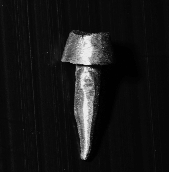
In the laboratory, the impression was poured using Type IV gypsum (Snowrock, Korea). Picosep die lubricant (Hilzingen, Germany) was applied before the construction of the wax pattern. Following that, a burnout post from the ParaPost system (Coltene, USA) was covered with sticky wax and inserted inside the canal space. The core was built up using the same modelling wax (Metrowax, UK). The core height was standardised at 3 mm15,23 while the margin was extended to 1 mm on the dentine rim. Then, wax sprue was added to the wax pattern, followed by casting using a lost wax technique at 850–900 Celsius. Type IV non-precious alloy (Nickel 58.4%, Chromium 26.9%) (System NH, Adentatec, Germany) was used for casting post and core (Fig. 1). The cast post and core surfaces were sandblasted with 50 μm aluminium oxide where the nozzle was positioned 10 mm away from the post surface and the pressure was set at 0.25 MPa.

Cast metal post and core (System NH, Adentatec, Germany).
Fabrication of one-piece zirconia post and core
Duralay, Reliance (Alsip, IL, USA) was used to construct the resin pattern for the zirconia post and core. The procedure includes the canal space being lubricated with a Picosep die lubricant (Hilzingen, Germany). Following that, a burnout post from the ParaPost system (Coltene, USA) was coated with Duralay, Reliance (Alsip, IL, USA) and inserted into the canal space. The height of the core was standardised as for CMP (System NH, Adentatec, Germany) (Fig. 2).

Example of duralay, reliance (Alsip, IL, USA) pattern.
The resin patterns were then digitised using the ultra-high-definition (UHD)-DOF (DOFLAB, USA) (Fig. 3), which provides scanning accuracy in the range of 5–7 μm, as specified by the manufacturer. The scanned data were exported in Standard Tessellation Language (STL) file format. These STL files were imported into DentalCAD software (version 3.1 Rejika)25where minor adjustments were made to ensure optimal fit and milling feasibility. Following the digital design process, the zirconia post and core were milled as a single unit using Explore Functional zirconia blocks (Upcera, China) via a computer-aided manufacturing (CAM) system (Fig. 4).

The scanned Duralay Reliance (Alsip, IL, USA) pattern for the milling process.

Example of one-piece zirconia Explore functional (Upcera, China) core and post.
Fabrication of everStick® post
The procedure for the everStick® post (GC, Japan) was based on Le Bell-Rönnlöf et al.26. The 1.5 mm fibre was fitted in the root canal, and each end was cut for the perfect fit while leaving 2 mm fibre above the coronal opening and then light-cured for 20 s. The post was then taken out and further cured outside the canal for an additional 40 s. The cured fibre post was repositioned back inside the canal. After that, fibres were added until the post fit perfectly in the canal and then cured according to the previously described steps. Once the post was ready, the Modelling Liquid (GC, Japan) was applied to coat the post surface and was allowed to dry. It was then light cured for 10 s.
Adaptation measurement
12 samples were used for adaptation analysis of all post systems. PVS was used to assess the adaptation between the post and the canal wall by measuring the space it occupied, which in clinical situations would typically be filled with cement. The protocol was based on Muttlib et al.27. The weight of the tooth was measured using AND GR200 digital scale (A&D Company Limited, Japan) as a reference to ensure that the PVS material was removed completely from the canal before the start of every weight measurement for different post systems. The weight of each tooth, along with its corresponding post, was then recorded. Next, light body Chromaclone™ (Ultradent, USA) was injected inside the canal, followed by post insertion. Once the PVS had been set, any excess impression material on the structure of the coronal tooth was carefully removed utilising a No. 15 blade. The total weight of the PVS and the post was then measured and documented. This measurement was taken 3 times to calculate an average value.
After completion of the adaptation test, the samples for each group (Group 1 and Group 2) with their respective post were used for the fracture resistance test. The sample for Group 3 was prepared to receive an everStick® (GC, Japan) post according to the procedure described for everStick® (GC, Japan) previously.
Cementation of post
A self-adhesive resin cement, Rely X™ U200 (3 M, USA), was utilised for post cementation in Groups 1–3 according to the protocol recommended by the manufacturer. The tack cure was done for 2 s, followed by the removal of excess cement. After that, each surface was cured for 20 s.
Core fabrication of samples in group 3
Core fabrications for teeth with everStick® (GC, Japan) posts were done using ParaCore (Coltene, USA) shade A2. The core height was standardised at 3 mm28 and the margin was extended to 1 mm on the dentine rim.
Tooth Preparation for crown construction
All samples were prepared for a metal crown. A 0.5 mm chamfer margin was prepared at the CEJ level, preserving a 2 mm height of tooth structure above CEJ and 1 mm thickness of axial dentine. The ferrule was standardised at 2 mm (Fig. 5a-d). Preparations were made freehand by 1 operator with a subjective convergence angle of 8 to 10 degrees for standardisation.

(a) Cemented one-piece zirconia post and core (b) Cemented cast metal post and core (c) Cemented everStick® (GC, Japan) with core build up (d) Sample in Group 4 (control) after crown preparation. Samples in all groups were prepared to receive a metal crown.
Crown fabrication and cementation
A metal crown was used as a final restoration. The impressions for metal crown fabrication were carried out using Chromoclone (Ultradent, USA) light and heavy body. Polishing and finishing of the crown were carried out with diamond burs under water cooling. The final crowns were cemented using similar luting cement as for posts.
Simulation of fatigue phenomenon for groups 1–4
All specimens were subjected to thermocycling (Zecttron, Malaysia) for 6000 cycles in 5˚C − 50˚C water baths, with a 20-second dwell period and a transfer time of 2 seconds29.
Simulating supporting tissues for groups 1–4
The protocol to stimulate supporting tissues was based on Palepwad and Kulkarni15. The root surfaces were dipped in molten wax and removed to create a thin layer of wax 2 mm beneath the CEJ, imitating the periodontal ligament. Auto polymerising polymethyl methacrylate (PMMA) was poured into a square mould measuring 18 mm X 18 mm. Teeth were mounted at 2 mm below the CEJ and were removed from PMMA once the initial polymerisation effect took place. Acrylic resin blocks were ready after the completion of resin polymerisation. The residual wax was cleaned from the surfaces of the root as well as the acrylic resin block. A light-body Chromaclone™ (Ultradent, USA) was used to coat the root surface, followed by reinsertion of the teeth into the individual resin blocks. Excess silicone was cut and cleaned with a surgical blade to ensure a flat surface 2 mm beneath the facial CEJ of each sample.
Evaluation of fracture resistance and fracture pattern
Loads were then introduced to the samples via a universal testing machine (Shimadzu, Japan) equipped with a 20 kN strain gauge load cell. The chisel wedge was positioned on the central fossa of the tooth samples at a crosshead speed of 0.5 mm/min, angled at a 45-degree angle to the long axis of the roots17.
The fracture resistance assessment was initiated by selecting the ‘start test’ icon on the computer. The process continued until the machine sensed an abrupt drop in the force, triggering the machine to stop loading. To ensure accuracy and prevent errors in test values, the machine was calibrated before each test.
After the fracture load analysis, the fracture pattern of the specimens was analysed for either favourable (Fig. 6a) or unfavourable (Fig. 6b) fracture patterns. Fractures were considered unfavourable if they were oblique or vertical and extended into or below the surrounding acrylic resin block. In contrast, fractures occurring above the resin block were classified as favourable22.

(a) Favourable fracture (b) Unfavourable fracture.
Statistical analysis
The data for all tests were tabulated and analysed using IBM SPSS version 26 (SPSS Inc, Chicago, IL, USA). For the objectives 1 and 2, the data were analysed to obtain the mean and standard deviation. The categorical data for objective 3 was then analysed to obtain frequency along with percentage.
Results
Adaptation
ANOVA and the post-hoc Bonferroni test were utilised for data evaluation since the normality test confirmed a normal distribution. Group 2 (CMP) had the lowest weight of PVS material [Mean (SD) = 0.006 (0.0042)], followed by Group 3 (everStick®) [Mean (SD) = 0.009 (0.0034)], and Group 1 (Zirconia) [Mean (SD) = 0.015 (0.0054)], which recorded the heaviest weight of PVS light body. The F-statistics test [F stats = 13.10, p < 0.001] showed there is a significant difference in the mean weight between groups (Table 2). Therefore, the null hypothesis regarding the adaptation of the post systems employed in this study was rejected.
Fracture resistance
The data followed a normal distribution. Therefore, ANOVA together with the post-hoc Bonferroni test were utilised for data analysis, as reported in Table 3. Group 4 (control) has the highest fracture resistance [Mean (SD) = (955.837 N (119.380 N)], followed by Group 2 (CMP) [Mean (SD) = (772.429 N (210.910 N)], then Group 1 (Zirconia) [Mean (SD) = (545.653 N (117.769 N)]. Group 3 (everStick®) has the lowest fracture resistance [Mean (SD) = (303.681 N (117.310 N)] in this study. The F-statistic test [F-stats = 22.159, p = 0.001] showed there is a significant difference in the mean weight between groups. Therefore, the null hypothesis was rejected since there was a significant difference in the fracture resistance between the different post systems.
Fracture patterns
Categorical data for fracture patterns were first analysed via the chi-square test. However, Fisher’s exact test was used since more than 20 per cent of the cells had expected frequencies below 5. There was statistically no significant difference (p = 0.76) in fracture patterns in the study groups, as shown in Table 4. Therefore, the null hypothesis regarding the fracture pattern was accepted.
Discussion
Adaptation
The weight of the PVS material reflects the amount of space occupied within the canal, which would be filled by cement material during clinical cementation. In this procedure, the PVS was used to replicate the cement layer, thereby simulating actual post cementation. It reflects the fit of the posts to the dentinal wall within the canal. A higher PVS weight indicates a thicker cement layer, signifying a poorer adaptation of the post to the canal. CMP recorded the lowest PVS weight, which reflects a better adaptation of CMP to the canal wall compared to the other posts. Although weight does not directly equate to volume, this weight-based method provides a reproducible, indirect assessment of internal post adaptation, particularly when using PVS with consistent handling and setting properties.
This finding is similar to the research by Muttlib et al.27which reported no difference in the adaptation of CMP when compared with the everStick® posts (GC, Japan). The everStick® (GC, Japan) post can be moulded to follow the canal shape and is able to allow light cure to penetrate, thus allowing polymerisation to occur within the root canal, ensuring precise adaptation26. Additionally, it allows the incorporation of extra fibres into the main fibre structure, which further enhances its adaptation to the canal30. Similarly, CMP is designed to conform to the canal shape during fabrication, leading to improved adaptation.
Perucelli et al.31 investigated the adaptability between CMP and one-piece CAD-CAM composite resin posts and cores fabricated using a part-digital workflow. Their findings indicated superior adaptability of CMP when compared with CAD-CAM composite resin post and core, although the CAD-CAM method still achieved clinically acceptable adaptation. The inferior adaptability of the posts, which were fabricated using milling technology, may be due to fabrication inaccuracies, particularly those resulting from the limitations of milling burs, which are unable to accurately reproduce sharp angles due to their round-ended design31. This was evident in the study by Bittner et al.13where they conducted a comparative analysis of the marginal fit of one-piece zirconia posts and resin patterns, identifying a significant discrepancy between the two.
Good adaptation of a post and core in the root canal enhanced bond strength and produced micromechanical interlocking through the hybrid layer31. Inadequate adaptation of the post results in the formation of marginal gaps, which, when coupled with insufficient cementation, can result in microleakage32which caused the debonding of the post from the canal wall, resulting in the failure33.
Fracture resistance
In this study, metal crowns were used as the final restorations to ensure that any recorded failures could be attributed to the post system rather than the crown material. This decision was based on a previous study17 reporting that restorations using ceramic crowns often failed at the crown level, thereby limiting the ability to accurately assess the performance of the underlying post system.
The control group (Group 4) demonstrated the highest fracture resistance in this study, with a mean value of 955.837 N ± 119.380 N. According to Sirimai et al.23the structural integrity of endodontically treated teeth is closely correlated with the quantity of residual dentine. Minimal tooth structure loss in the control group, as only occlusal access cavity was performed during the preparation is likely to contribute to its superior fracture resistance. This finding aligns with prior research, which has reported similar trends in control groups34,35.
The group restored with CMP (System NH, Adentatec, Germany) exhibited higher fracture resistance (772.429 N ± 210.910 N) than those restored with other post types. Several studies support this observation, particularly in comparisons involving CMPs, zirconia posts, and FRCs15,28,36.
Several research have studied the fracture resistance of various post materials. Alkhatri et al.36 found no significant difference in fracture resistance for CAD-CAM fabricated zirconia and metal posts, attributing this to zirconia’s high strength and toughness. Similarly, Palepwad and Kulkarni15 reported that teeth restored with prefabricated FRCs had significantly lower fracture resistance compared to zirconia posts and CMPs, with no significant difference between the latter two. Saritha et al.28 have concluded that prefabricated zirconia posts have superior fracture resistance, followed by fibre-reinforced posts and carbon posts. These findings collectively suggest that zirconia posts and CMPs offer superior mechanical performance in terms of fracture resistance, likely due to their high structural integrity and material properties.
Fibre-reinforced composite (FRC) posts have an elastic modulus similar to dentine, which helps reduce stress on the root. However, in this study, FRC showed the lowest fracture resistance. This is likely because the core was built separately using composite resin, unlike the cast metal and zirconia groups, where the post and core were one piece. Having a separate core can create a weak spot where the post and core meet, making it more likely to fail under pressure. Also, the composite core material is not as strong as metal or zirconia. This finding is consistence with a study by Vano, M. et al.37 where they also found that samples with a separate core have a higher risk of failure.
Fracture pattern
The fracture patterns of all groups in this study were found to have no significant difference statistically (p > 0.005). Unfavourable fractures occurred in 66.7% of samples in both the CMP (System NH, Adentatec, Germany) and one-piece zirconia post groups, while 50% of samples in the everStick® (GC, Japan) group exhibited unfavourable fractures. Notably, all samples in the control group sustained unfavourable fractures.
The control group (Group 4) showed a significantly higher rate of unfavourable fractures (100%) compared to the post-restored groups, which had more favourable fracture patterns. This can be partly explained by the fracture load applied to normal human bite force. According to Ustrell-Barral et al.38the maximum bite force in healthy adults typically ranges from 670 N in females and 807 N in males. In this study, the control group withstood a mean load of 955.84 N, which is above the normal physiological range. This suggests that under normal chewing forces, these teeth would likely remain intact. However, in conditions involving excessive forces, such as bruxism, teeth without a post and core lack internal reinforcement and stress distribution. This makes them more prone to catastrophic failures, as seen in this study, where fractures extended deep into the root, resulting in unfavourable patterns in all control specimens.
In contrast, the experimental groups had more favourable fracture outcomes despite low fracture resistance. The post-core systems likely helped absorb and spread out the forces, reducing the chance of severe root fractures. This reflects a key clinical principle, in which while posts may not significantly increase fracture strength, they can positively influence the type of fracture, making it more manageable and restorable.
The findings from this study also suggest that the design of the final restoration plays a critical role in the prognosis of endodontically treated teeth39,40. Crown placement over the post and core facilitates an even distribution of the masticatory forces across the root, along with the post-core complex41. However, variations in load distribution introduced by the crown can influence fracture patterns42potentially increasing the incidence of unfavourable root fractures43. The results from this study support this concept, as at least 50% or more of the samples in each group experienced unfavourable fractures.
Anweigi et al.14 found that milled zirconia posts had more favourable fracture patterns than CMPs and glass-fibre posts. Similarly, Palepwad and Kulkarni15 reported that zirconia and fibre-reinforced posts showed better fracture resistance than CMPs. However, a key methodological difference in both studies is the absence of crown placement as the final restoration. In their protocols, dual-cure resin core materials were used for zirconia and fibre-reinforced posts, while CMPs were restored as one-piece cast designs without coronal coverage.
In contrast, the present study incorporated standardised metal crowns for all groups, which more closely simulate clinical conditions and affect how occlusal forces are transferred. Crown placement offers coronal reinforcement, altering stress distribution through the ferrule effect, particularly when engaging sound dentine42. The omission of crowns in earlier studies may have led to stress being concentrated directly at the post-core interface, potentially resulting in different fracture patterns35,44. These differences underscore the importance of including full restorations in in vitro testing to ensure clinically relevant outcomes.
However, it is important to acknowledge that the use of metal crowns in this study, while effective for standardising testing conditions and isolating post-related failures, limits its applicability to aesthetic zones where ceramic crowns are typically preferred. Metal and ceramic crowns differ in mechanical behaviour, particularly in terms of brittleness and stress transmission, which could affect the failure modes of underlying restorations. Therefore, future research should incorporate ceramic crowns to more accurately simulate clinical situations in aesthetic regions and further explore how different crown materials interact with various post systems under functional loading.
Metal post, which is known to be rigid, is believed to prevent the bending action of the post during lateral forces, but this can transfer stress to the surrounding dentin, enhancing the chance of root fractures. Metal posts are highly rigid and often cause deep oblique or vertical fractures42. In contrast, fibre posts are thought to flex under load, distributing stress more evenly between the post and dentin, which may help prevent fractures43. Ideally, a post must have an elastic modulus equal to dentin to allow even stress distribution and reduce fracture risk45.
Zhou and Wang46 found that metal posts had an increased incidence of catastrophic root fractures than fibre posts, most likely due to differences in the modulus of elasticity and the stress-absorbing effect of the resin cement layer used with fibre posts. However, clinical studies were unable to consistently demonstrate significant differences in root fracture rates between metal and glass-fibre posts47,48. One recent systematic review, along with a meta-analysis, found that failure patterns were similar across cast metal, prefabricated metal, and fibre posts49.
Mastrogianni et al.22 investigated the failure of a few types of posts under axial and oblique loading. Their findings revealed no statistically significant differences in fracture modes. However, root fractures were the predominant failure mode under oblique loading. Oblique forces generate greater stress due to bending moments, transmitting tensile stress from the cervical margin of the crown to the root, the post-dentine interface, and the cement layer50. Mastrogianni et al.22 suggested that these stresses could lead to early cement loss, resulting in uncontrolled forces that increase the risk of root fracture. In the present study, oblique loading conditions were applied, which may explain the high prevalence of unfavourable fractures.
These findings underscore the complex interplay between post material properties, restoration design, and loading conditions in determining fracture behaviour in endodontically treated teeth.
Clinically, the findings of this study suggest that a one-piece zirconia post and core system produced using CAD-CAM technology is a viable alternative to conventional metal posts in cases requiring a custom-made solution. The use of CAD-CAM offers potential advantages in terms of precision and material properties. However, the authors acknowledge a current lack of in vivo studies on CAD-CAM fabricated custom posts, indicating that further clinical research is necessary to fully establish their effectiveness and long-term outcomes in patient care.
This study has a few limitations which need to be addressed. Thermocycling was performed in this study to simulate the thermal stresses experienced intraorally due to temperature fluctuations. While it provides a degree of ageing simulation, it does not replicate the cyclic mechanical forces experienced during mastication. The omission was based on the study’s primary focus on evaluating static fracture resistance under controlled conditions. Therefore, the absence of mechanical fatigue testing is acknowledged as a limitation. Future studies incorporating both thermal and mechanical cyclic loading would provide a more comprehensive assessment of long-term clinical performance and durability of post systems under functional conditions.
Another limitation of the study was the use of two different laboratories for fabricating the indirect posts, which was necessary due to availability and resource constraints. As a result, different methods of pattern fabrication were employed, where an indirect technique was employed for the custom metal posts and a direct technique for the one-piece zirconia posts. This was done to facilitate effective communication between the research team and the respective laboratories.
While this introduces a methodological variation, a previous study by Rayyan et al.6 has reported that the choice between direct and indirect pattern fabrication does not significantly influence the fit accuracy of post and core systems. Nevertheless, the difference in fabrication techniques may still act as a confounding factor, potentially influencing the adaptation results and limiting the validity of direct comparisons between the two post types. This limitation is acknowledged, and future research is recommended to either standardise the fabrication technique across all groups or evaluate the impact of each method separately to better isolate their effects on post adaptation.
Furthermore, although the oblique static loading protocol applied at a 45° angle and 0.5 mm/min crosshead speed is a standard method for evaluating fracture resistance in vitro, it does not accurately mimic the complex, dynamic occlusal forces encountered in the oral cavity. Thus, extrapolation of these results to clinical performance must be made with caution. Incorporating dynamic fatigue loading and varied loading conditions in future research would yield a more representative evaluation of failure behaviour and mechanical longevity under real clinical conditions.
Conclusion
Numerous studies have reported on the fracture resistance and properties of metal, zirconia, and fibre-reinforced composite posts, but findings have been inconsistent. Both adaptation and fracture behaviour are crucial in determining the overall performance of post systems. This study found no difference in adaptation in custom-made posts (CMPs) and fibre-reinforced composite posts, whereas milled zirconia posts exhibited significantly different adaptation compared to both CMPs and fibre-reinforced composite posts.
In terms of fracture resistance, no differences were observed among CMP and zirconia posts or between zirconia and fibre-reinforced composite posts. However, fibre-reinforced composite posts demonstrated significantly lower fracture resistance than CMPs. Additionally, teeth with intact cavity walls (control) exhibited the highest fracture resistance, emphasizing the importance of preserving dentin during endodontic and restorative procedures. Lastly, no differences in fracture patterns were found between the tested post systems.
Data availability
The data related to this study will be made available upon request from the corresponding author.
Abbreviations
- RCT:
-
Root canal treatment
- CMP:
-
Cast metal post
- ETT:
-
Endodontically treated teeth
- FRC:
-
Fibre-reinforced composite
- CAD-CAM:
-
Computer-aided design and computer-aided manufacturing
- CEJ:
-
Cemento-enamel junction
- MAF:
-
Master apical file
- PVS:
-
Polyvinyl siloxane
- UHD:
-
Ultra high definition
- STL:
-
Standard Tessellation Language
- PMMA:
-
Polymethyl methacrylate
References
-
Ho, C. & Argáez, C. Endodontic Therapy Interventions for Root Canal Failure in Permanent Dentition: A Review of Clinical Effectiveness, Cost-Effectiveness, and Guidelines. Ottawa: CADTH 1–9 (2017).
-
Gulabivala, K. & Ng, Y. L. Factors that affect the outcomes of root Canal treatment and retreatment—A reframing of the principles. Int. Endod J. 56, 82–115 (2023).
-
Bonchev, A., Radeva, E. & Tsvetanova, N. Fiber reinforced composite Posts-A review of literature. Int. J. Sci. Res. 6, 1887–1893 (2017).
-
Martino, N. et al. Retrospective analysis of survival rates of post-and-cores in a dental school setting. J Prosthet. Dent 123 (3), 434–441 (2019).
-
Peroz, I., Blankenstein, F., Lange, K. P. & Naumann, M. Restoring endodontically treated teeth with posts and cores-A review. Quintessence Int. 36, 737–746 (2005).
-
Rayyan, M. R., Aldossari, R. A., Alsadun, S. F. & Hijazy, F. R. Accuracy of cast posts fabricated by the direct and the indirect techniques. J. Prosthet. Dent. 116, 411–415 (2016).
-
Amižić, I. P. & Baraba, A. Estetski intrakanalni Kolčići. Acta Stomatol. Croat. 50, 143–150 (2016).
-
Duret, B., Reynaud, M. & Duret, F. {A new concept of corona-radicular reconstruction, the composipost (2)}. Chir. Dent. Fr. 60, 69–77 (1990).
-
V, L., Reddy, S., Garapati, V., Sudhamashetty, S. & Yadla, P. Fracture fragment reattachment using projectors and anatomic Everstick post™: an ultraconservative approach. J. Int. Soc. Prev. Community Dent. 7, 52 (2017).
-
Ban, S. Chemical durability of high translucent dental zirconia. Dent Mater. J 39 (1),12–23 (2019).
-
Awad, M. A. & Marghalani, T. Y. Fabrication of a custom-made ceramic post and core using CAD-CAM technology. J. Prosthet. Dent. 98, 161–162 (2007).
-
Streacker, A. B. & Geissberger, M. The milled ceramic post and core: A functional and esthetic alternative. J. Prosthet. Dent. 98, 486–487 (2007).
-
Bittner, N., Hill, T. & Randi, A. Evaluation of a one-piece milled zirconia post and core with different post-and-core systems: an in vitro study. J. Prosthet. Dent. 103, 369–379 (2010).
-
Anweigi, L., Noah, R., Alessa, L., Al-Madi, E. & Aldegheishem, A. Structural integrity of extracted teeth restored using three different post-and-core systems: an in vitro comparative study. Saudi Dent. J. 33, 63–68 (2021).
-
Palepwad, A. & Kulkarni, R. In vitro fracture resistance of zirconia, glass-fiber, and cast metal posts with different lengths. J. Indian Prosthodontic Soc. 20, 202 (2020).
-
Abduljabbar, T. et al. Fracture resistance of three post and core systems in endodontically treated teeth restored with All-ceramic crowns. King Saud Univ. J. Dent. Sciences 3, 33–38 (2012).
-
Habibzadeh, S., Rajati, H. R., Hajmiragha, H., Esmailzadeh, S. & Kharazifard, M. Fracture resistances of zirconia, cast Ni-Cr, and fiber-glass composite posts under all-ceramic crowns in endodontically treated premolars. J. Adv. Prosthodont. 9, 170–175 (2017).
-
Dupont, W. D., Plummer, W. D., Power & Size, S. and (2024). https://cqsclinical.app.vumc.org/ps/
-
Pitigoi-Aron, G., Streacker, A., Schulze, K. & Geissberger, M. Accuracy of cast posts and cores using a new investigative method. Gen. Dent. 60, e153–e157 (2012).
-
Özcan, M. & Valandro, L. F. Fracture strength of endodontically-treated teeth restored with post and cores and composite cores only. Oper. Dent. 34, 429–436 (2009).
-
Newman, M., Yaman, P., Dennison, J., Rafter, M. & Billy, E. Fracture resistance of endodontically treated teeth restored with composite posts. J. Prosthet. Dent. 89, 360–367 (2003).
-
Mastrogianni, A. et al. Fracture strength of endodontically treated premolars restored with different post systems and metal-ceramic or monolithic zirconia crowns. Dent Mater. J 40 (3), 606–614 (2021).
-
Sirimai, S., Riis, D. N. & Morgano, S. M. An in vitro study of the fracture resistance and the incidence of vertical root fracture of pulpless teeth restored with six post-and-core systems. J. Prosthet. Dent. 81, 262–269 (1999).
-
Trebilcock, C. & Evans, D. A two-stage impression technique for the indirect fabrication of multiple cast dowel and cores. J. Prosthet. Dent. 66, 422–425 (1991).
-
DentalCAD. (2022). https://exocad.com/our-products/dentalcad-rijeka
-
Le Bell-Rönnlöf, A. M., Lassila, L., Kangasniemi, I. & Vallittu, P. Load-bearing capacity of human incisor restored with various fiber-reinforced composite posts. Dent. Mater. 27, e107–e115 (2011).
-
Muttlib, N. A., Azman, A. N., Seng, Y. T., Alawi, R. & Ariffin, Z. Intracanal adaptation of a fiber reinforced post system as compared to a cast post-and-Core. Acta Stomatol. Croat 50 (4), 329–336 (2016).
-
Saritha, M. et al. Comparative evaluation of fracture resistance of different post systems. J. Int. Soc. Prev. Community Dent. 7, 356 (2017).
-
Ozcan, N. & Sahin, E. In vitro evaluation of the fracture strength of all-ceramic core materials on zirconium posts. Eur. J. Dent. 7, 455–460 (2013).
-
Salim, N. A., Muttlib, A., Alawi, N. A., Abd Rahman, R., Arrifin, Z. & N. & Evaluation of microleakage between different post and core systems under gradual loading: an In-Vitro study. Acta Stomatol. Croat. 52, 218–226 (2018).
-
Perucelli, F., Costa, R., Souza, E. & Rached, R. Effect of half-digital workflows on the adaptation of customized CAD-CAM composite post-and-cores. J Prosthet. Dent 126 (6), 756–762 (2020).
-
Ayyildiz, S. T. & Bagci, E. Ç. Evaluation of coronal microleakage in two different Post-Core systems. Int. J. Prosthodont. Restor. Dentistry. 1, 163–168 (2011).
-
Coleman, A. J., Moses, M. S. & Rickerby, H. H. D. Macromolecular leakage beneath full cast crowns: A two-year in vitro investigation. J. Prosthet. Dent. 85, 20–25 (2001).
-
Heydecke, G. & Peters, M. The restoration of endodontically treated, single-rooted teeth with cast or direct posts and cores: A systematic review. J. Prosthet. Dent. 87, 380–386 (2002).
-
Libman, W. & Nicholls, J. Load fatigue of teeth restored cast posts and cores and complete crowns. Int. J. Prosthodont. 8, 155–161 (1995).
-
Alkhatri, R., Saleh & Kheder, W. Evaluating fracture resistance and failure modes of root filled teeth restored with CAD/CAM-Fabricated post and core. Clin. Cosmet. Investig Dent. 11, 349–355 (2019).
-
Vano, M. et al. The adhesion between fibre posts and composite resin core: the evaluation of microtensile bond strength following various surface chemical treatments to posts. Int. Endod J. 39, 31–39 (2006).
-
Ustrell-Barral, M., Olave, Z., Khoury-Ribas, C., Rovira, L. & Martínez-Gomis, J. B. Reliability, reference values and factors related to maximum bite force measured by the innobyte system in healthy adults with natural dentitions. Clin Oral Investig 28 (11), 620 (2024).
-
Fernandes, A. & Dessai, G. Factors affecting the fracture resistance of Post-Core reconstructed teeth: A review. Int. J. Prosthodont. 14, 355–363 (2001).
-
Wahab, F. et al. Restoration of root filled teeth; current opinions and techniques. Open Dent. J 15, 71–83 (2021).
-
Deutsch, A. S., Musikant, B. L., Cavallari, J. & Lepley, J. B. Prefabricated dowels: A literature review. J. Prosthet. Dent. 49, 498–503 (1983).
-
Butz, F., Lennon, A., Heydecke, G. & Strub, J. S. Rate and fracture strength of endodontically treated maxillary incisors with moderate defects restored with different Post-and-Core systems: an in vitro study. Int. J. Prosthodont. 14, 58–64 (2001).
-
Bateman, G., Ricketts, D. & Saunders, W. Fibre-based post systems: A review. Br. Dent. J. 195, 43–48 (2003). discussion 37.
-
Sorensen, J. A. & Engelman, M. J. Ferrule design and fracture resistance of endodontically treated teeth. J. Prosthet. Dent. 63, 529–536 (1990).
-
Akkayan, B. & Gulmez, T. Resistance to fracture of endodontically treated teeth restored with different post systems. J. Prosthet. Dent. 87, 431–437 (2002).
-
Zhou, L. & Wang, Q. Comparison of fracture resistance between cast posts and fiber posts: A Meta-analysis of literature. J. Endod. 39, 11–15 (2013).
-
Parcina Amizic, I., Baraba, A. & Amižić & Esthetic intracanal posts. Acta Stomatol. Croat. 50, 143–150 (2016).
-
de Figueiredo, F., Martins-Filho, P. R. & Faria-e-Silva, A. Do metal Post–retained restorations result in more root fractures than fiber Post–retained restorations? A systematic review and Meta-analysis. J Endod 41 (3), 309–16 (2014).
-
Martins, M. et al. Is a fiber post better than a metal post for the restoration of endodontically treated teeth? A systematic review and meta-analysis. J. Dent. 112, 103750 (2021).
-
Schmitter, M. et al. Teeth restored using fiber-reinforced posts: in vitro fracture tests and finite element analysis. Acta Biomater. 6, 3747–3754 (2010).
Funding
This project is funded by the Research University Grant, Universiti Sains Malaysia (1001/PPSG/8012367).
Ethics declarations
Competing interests
The authors declare no competing interests.
Ethics, consent to participate, and consent to publish
The study was initiated after getting ethical approval from the Human Research Ethics Committee of Universiti Sains Malaysia (HREC-USM) (USM/JEPeM/20030157). The study was carried out following the Declaration of Helsinki, along with all participants giving written informed consent before participating in the study.
Consent to participate
Prior written informed consent was obtained for the collection of teeth used in this study.
Additional information
Publisher’s note
Springer Nature remains neutral with regard to jurisdictional claims in published maps and institutional affiliations.
Rights and permissions
Open Access This article is licensed under a Creative Commons Attribution-NonCommercial-NoDerivatives 4.0 International License, which permits any non-commercial use, sharing, distribution and reproduction in any medium or format, as long as you give appropriate credit to the original author(s) and the source, provide a link to the Creative Commons licence, and indicate if you modified the licensed material. You do not have permission under this licence to share adapted material derived from this article or parts of it. The images or other third party material in this article are included in the article’s Creative Commons licence, unless indicated otherwise in a credit line to the material. If material is not included in the article’s Creative Commons licence and your intended use is not permitted by statutory regulation or exceeds the permitted use, you will need to obtain permission directly from the copyright holder. To view a copy of this licence, visit http://creativecommons.org/licenses/by-nc-nd/4.0/.
About this article

Cite this article
Rahim, N.F.A., Alawi, R., Ariffin, A. et al. Comparative evaluation of adaptation and fracture resistance of CAD-CAM fabricated zirconia posts in endodontically treated teeth. Sci Rep 15, 29792 (2025). https://doi.org/10.1038/s41598-025-15348-3
-
Received:
-
Accepted:
-
Published:
-
DOI: https://doi.org/10.1038/s41598-025-15348-3