
Introduction
Marine and freshwater filter-feeding animals with modular organization, such as sponges, cnidarians, bryozoans, and tunicates, have emerged as valuable sources of bioactive compounds1,2,3. Among these, the phylum Bryozoa, comprised of microscopic, almost exclusively colonial invertebrates, holds a substantial untapped potential for producing secondary metabolites. Despite the existence of approximately 6700 described extant species4 bryozoans remain a relatively understudied group in this respect5. Bryozoans are typically sessile animals consisting of repetitive morpho-functional modules referred to as zooids6,7. Like other epibiotic organisms, bryozoans face constant threats from grazing and overgrowth pressures, compelling them to employ both mechanical and chemical defense mechanisms8,9,10. Notably, the symbiotic bacteria associated with some bryozoan species were shown to biosynthesize specialized metabolites, which have ecological functions such as defense against predators, UV protection, antifouling, and protection against infections and parasites11,12.
While our understanding of the associations between bryozoan hosts and their symbiotic microbiota remains limited, prior studies have delved into the biodiversity of cultivable bacteria associated with the phylum Bryozoa and assessed their potential to produce bioactive compounds13,14,15. A noteworthy example is the complex polyketide bryostatin-1, produced by the Gammaproteobacterium ‘Candidatus Endobugula sertula’ symbiotic of the marine bryozoan Bugula neritina16,17. This compound has anticancer properties, and shows significant therapeutic effects in pre-clinical studies of Alzheimer’s disease and other neurological disorders, and promising effects in anti-HIV therapy18,19,20. Earlier research on the dynamic concentration levels of bryostatin within both ‘B. neritina’ colonies and larvae revealed significantly higher levels of these polyketides in the larvae, likely playing a role in chemical defense, protecting vulnerable larvae from predators11,21,22. Additionally, sexual reproduction in ‘B. neritina’ is influenced by its defensive symbiont. Absence of the symbiont suppresses female reproductive processes in the host colony, presumably due to a lack of bryostatins23,24.
Apart from polyketides, several secondary metabolites of other compound classes with cytotoxic, antibacterial, antiviral, and antifungal as well as some other medically relevant biological activities were identified in bryozoans5,11,25,26. For example, tambjamines, a group of 4-methoxypyrrolic alkaloids found in the bryozoans Bugula longissimi, Sessibugula translucens, and Virididentula dentata, are assumed to originate from symbiotic bacteria22,27,28. Metabolomic analyses revealed production of chemically diverse tambjamines (tambjamine A, C, and D) by V. dentata that inhibit its nudibranch predators, suggesting that these compounds can act as a chemical defense in these marine invertebrates29. Evidence for the symbiotic origin of the analog tambjamine YP1 comes from the production of these alkaloids by Serratia marcescens and Pseudoalteromonas tunicata, bacteria associated with tunicates and bryozoans, in response to grazing and predation30. Members of the tambjamines compound class possess a diverse range of biological activities, including antifouling, antimicrobial, antitumoral, and immunosuppressive15,26,29. While these insights derive largely from marine bryozoans, the untapped potential of freshwater species, particularly their microbiomes, remain unexplored.
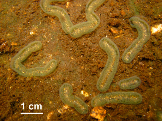
All bryozoans that were so far examined for the presence of secondary metabolites were marine, and members of the class Gymnolaemata. Here, we aimed to unlock the secondary metabolite biosynthesis potential of bacteria associated with the freshwater bryozoan Cristatella mucedo Cuvier, 1798 from the class Phylactolaemata (Fig. 1). C. mucedo is the only known species in the family Cristatellidae, common in lakes and rivers of the Northern Hemisphere. To accomplish our goal, we adopted a multiphasic approach that integrates both culture-dependent and -independent methods alongside comprehensive genome analysis and metabolomics.

Free-living colonies of Cristatella mucedo bryozoan on silty bottom. (photo by A. Ostrovsky).
Results and discussion
Bacterial and archaeal communities of C. mucedo and surrounding environmental samples
Cristatella mucedo bryozoan host colonies and samples of water and sediments from the same location were collected at the Laxenburg ponds in Austria. The bacterial and archaeal community associated with the bryozoan was investigated by recovering a diverse collection of isolates and genome analysis of representative strains. The cultivation-based approach was complemented by 16 S rRNA gene amplicon sequencing of C. mucedo-associated bacterial communities and surrounding water and sediment samples.
The distribution of 16 S rRNA-gene amplicon sequence variants (ASVs) across bryozoan, sediment, and water matrices showed that the majority of ASVs detected in the bryozoan samples also were detectable in the two environmental matrices, while only 25 ASVs (3.4%) were unique to the bryozoan samples (Fig. 2A). However, relative taxon abundances differed between the samples. Bryozoan samples were dominated by ASVs affiliated to the bacterial classes Cyanobacteria, Bacteroidia and Gammaproteobacteria (Fig. 2B). The sediments harbored more diverse microbial communities, showing higher relative abundances of taxa affiliated with Gammaproteobacteria and Anaerolineae compared to bryozoan and water samples, and a lower relative abundances of Cyanobacteria compared to bryozoan samples. The water community was primarily composed of Alphaproteobacteria and Actinomycetes. These findings highlight the distinct microbial composition within each community while emphasizing shared microbial taxa that may play pivotal roles in ecosystem interactions. List of all AVSs with taxonomic assignments and relative abundances per sample type are given in Table S1 (Supplementary material).

(A): Shared and unique ASVs across bryozoan, sediment, and water samples (data from two biological replicates per sample type). Average values across biological replicates per sample type were used to estimate the number and proportion of shared and unique ASVs. (B) Top 8 most abundant microbial classes across the different sample types across the whole dataset. Microbial classes not included in the top 8 were assigned to “others”.
Isolation and genome-based taxonomic identification of C. mucedo-associated bacteria
A homogenized sample of C. mucedo bryozoan host colonies was used to isolate bacteria by plating serial dilutions of the homogenate on various agar media. The plates were incubated over a period of up to 6 weeks in the dark at room temperature or at 28 °C. After 16 S rRNA gene screening, a total of 29 isolates representing 26 bacterial genera were selected for further studies (see Table S2, Supplementary material). The genomes of all 29 were sequenced and were used to construct genome-based phylogenetic trees (Fig. 3). The names of the isolates, beside genera, were designated according to the guidelines suggested by Ishaqu et al. (2024)31. The sizes of the genomes of bacterial isolates ranged from 2.5 to 9.4 Mb and the GC content from 35 to 73%, and the detailed information on the genmomes is given in the supplementary Excel file “Genome statistics”. Among the 29 isolates, 19 were classified as members of the Gram-negative classes Alphaproteobacteria, Gammaproteobacteria, and Bacteroidia. According to a comparison against TYGS genome database, most of the newly isolated strains may represent new species, in particular Roseomonas sp. CML04 (Cristatella mucedo isolate 4), Bosea sp. CML06, Aureimonas sp. CML07, Paracoccus sp. CML02, Gemmobacter sp. CML18, Rhizobium sp. CML03, Serratia sp. CML12, Kinneretia sp. CML19 and Deefgea sp. CML13 (Fig. 3A). An isolate CML21 representing a potentially new genus within the family Enterobacteriaceae was also identified. The other ten isolates were classified as members of the Gram-positive classes Actinomycetes and Bacilli. Within this group, Bacillus spp. CML08, CML11, Metabacillus sp. CML26, Streptomyces sp. CML16, Rhodococcus sp. CML10 and Micromonospora sp. CML27 also potentially represented new species (Fig. 3B). The prevalence of members from the Pseudomonadota phylum, especially Alphaproteobacteria and Gammaproteobacteria, within the cultivated bacteria associated with C. mucedo is consistent with ecological patterns observed across various terrestrial and aquatic invertebrate taxa31. Previous research on species-specific endosymbionts from the bryozoan Flustra foliacea has shown a predominance of Gammaproteobacteria, such as Pseudomonas32 while Alphaproteobacteria were identified as the most abundant group in other bryozoan genera33,34. For instance, in Watersipora arcuata, bacteria such as Candidatus Endowatersipora, Rhizobium, and Agrobacterium were prevalent, while in Bugula plumosa bryozoan, species like Paracoccus were dominant35. Enterobacteriaceae species (Gammaproteobacteria), also identified in the isolates from C. mucedo, have been recognized for their significant role in enhancing host nutrition and fitness through processes such as carbohydrate fermentation, degradation of organic matter, and nitrogen-fixation. This taxonomic group has repeatedly occurred in the gut of various invertebrates36,37. Among the Gram-positive bacteria, we identified distinct members of the Actinomycetes and Bacilli classes, which are well-known producers of secondary metabolites. Previous studies have documented the isolation of these bacterial groups from marine invertebrates. For instance, several strains of Brevibacterium spp. were isolated from the sponge Dendrilla nigra38 and Streptomyces spp. from the freshwater sponge Spongilla lacustris39. Members of the Bacilli, such as Bacillus, have been frequently associated with sponges and coral specimens40,41,42. These findings are consistent with the dominant groups observed in our study.


Genome-based phylogenetic trees and Biosynthetic Gene Clusters (BGCs) detected in corresponding genomes. (A): Gram-negative isolates; (B): Gram-positive isolates.
In addition to the isolation of bacteria after plating serial dilutions of the C. mucedo homogenate, mixed enrichment cultures were obtained after direct inoculation of the bryozoan sample homogenate in various liquid media (CMR2). 16 S rRNA gene amplicon sequence analyses of the enrichment cultures revealed additional genera not identified previously amongst the isolates, including Aeromonas, Klebsiella, Pseudomonas, Salmonella, and Acinetobacter (Fig. 4). Mixed enrichment cultures were serially diluted to isolate single strains, which were subsequently identified by 16 S rRNA gene amplicon sequencing and compared to the overall 16 S rRNA gene diversity of the enrichment cultures. However, not all of the newly detected genera were successfully isolated on plates, while this isolation attempt yielded more representatives of genera (Enterobacter, Pseudomonas, Elizabethkingia, Chryseobacterium, and Bacillus) which were already obtained after homogenate dilution and plating. This suggests that our inability to cultivate representatives of all detected genera may be due to putative obligatory associations with other bacteria from the same community43,44,45.

Relative abundance of the top 10 most abundant ASVs across the different enrichment media.
Secondary metabolite biosynthesis potential of C. mucedo bacterial isolates
To reveal their biosynthetic potential, the genomes of bacterial isolates obtained by serial dilutions of homogenized C. mucedo host colonies were analyzed for the presence of secondary metabolite biosynthesis gene clusters (BGCs) using the antiSMASH 7.0 online bioinformatics tool46. This analysis facilitated the selection of isolates with the highest number of potentially unique BGCs, thereby enhancing the chance of identifying novel secondary metabolites in our study. A total of 256 BGCs of various classes was identified across all genomes (Fig. 3). The number of detected BGCs per genome ranged from 2 to 26. The Micromonospora sp. CML27 genome contained the highest BGC number among the Gram-positive bacteria, with 26 BGCs, followed by Rhodococcus sp. CML10, with 20 BGCs. Among the Gram-negative isolates, the Enterobacteriaceae strain CML21 potential representing a new genus, along with Paracoccus sp. CML02 and Paucibacter sp. CML20 had the highest number of BGCs (11). The predominant BGC types were ribosomally synthesized and posttranslationally modified peptides (RiPPs), terpenes and nonribosomal peptide synthetases (NRPSs). Identified BGCs were compared against the MIBiG repository to annotate those involved in the biosynthesis of known secondary metabolites. While antiSMASH is a valuable tool for identifying previously described BGCs, it may occasionally fail to accurately delimit gene cluster borders, leading to lower similarity scores with their corresponding entries in the MIBiG database. To uncover BGCs that may specify potentially novel compounds, a stringent similarity threshold of < 60% identity to the already characterized BGCs was used to select such clusters. Under these criteria, only 43 of the 256 detected BGCs were identified as known clusters. In contrast, 213 BGCs showed no significant match to any known clusters when compared with the online databases (Table S3–S4, Supplementary material).
We observed distinct differences in the BGC patterns between isolates that belong to Gram-negative and Gram-positive taxa. Specifically, within the C. mucedo cultured bacterial community, Gammaproteobacteria and Alphaproteobacteria were the only groups containing previously uncharacterized homoserine lactone (HSL) BGCs. HSLs are compounds serving as signaling molecules in bacterial quorum sensing, facilitating intercellular communication among Gram-negative bacteria47. RiPPs were a predominant type of BGC identified among Gram-negative isolates, particularly in Paucibacter sp. CML20 and Paracoccus sp. CML02. Notably, these RiPP BGCs did not match any entries in MiBIG database. Higher counts of NRPS BGCs were detected in Gram-positive bacteria, especially in Rhodococcus sp. CML10 and Micromonospora sp. CML27. The only BGC detected in Rhodococcus sp. with a similarity above 65% was an NRPS gene cluster closely associated with corynecin III. Corynecins are a group of N-acyl derivatives of D-threo-2-amino-1-(p-nitrophenyl)-1,3-propanediol compounds, which share structural similarities with the antibiotic chloramphenicol48,49.
Selection of isolates for secondary metabolites profiling
Upon closer examination of genomic profiles of all strains, we identified Bacillus sp. CML08 Bacillus sp. CML11, Micromonospora sp. CML15 the Enterobacteriaceae strain CML21 and Streptomyces sp. CML16 as particularly interesting candidates by having exhibited the highest diversity, uniqueness, and number of BGCs. To validate these computational predictions and assess their potential to biosynthesize secondary metabolites, the selected isolates were grown in different conditions, and crude methanolic extracts of these cultures were tested for antimicrobial activity against standard test bacteria and yeast (Table S5, Supplementary material), as well as other bacteria isolated from C. mucedo. Notably, the extract from Bacillus sp. CML11 grown in liquid ISP2 medium showed bioactivity against Rhodococcus sp. CML08. The extract from the Micromonospora sp. CML15 culture grown in 5334 medium exhibited considerable activity against B. subtilis DSMZ 10 and partial inhibition of E. coli and S. carnosus DSMZ 20,501. No antimicrobial activity was shown for the extracts from Streptomyces sp. CML16 and the unclassified Enterobacteriaceae CML21 cultures in the conditions tested (Table S6, Figures S1–S4, Supplementary Material).
To investigate their secondary metabolomes in more detail, the abovementioned crude methanolic extracts (Table S6) were subjected to non-targeted LC-MS analysis followed by a previously established dereplication workflow50. The results of these analyses are shown in Tables S7–S8 (Supplementary material), whereby only one or a few main congeners are listed for each group of biosynthetically closely related natural products.
Bacillus sp. CML11 produced polyhydroxybutyrates (PHBs), previously reported from Bacillus wiedmannii51. PHBs are biotechnologically relevant for the production of biodegradable plastics. Through BGC identification, we found Bacillus sp. CML11 produced bacillibactin, a catechol-type siderophore commonly produced by Bacillus spp.52 and a non-ribosomal peptide putatively representing an unknown congener of bacitracin A. The MS/MS data for the latter suggest an identical linear N-terminal sequence53 but amino acid substitution(s) in the cyclized peptide part (Figure S7, Supplementary Material), which fits the antiSMASH predictions for the corresponding BGC. Furthermore, dihexosyldiacylglycerols were detected, presumably gentiobiosyldiacylglycerol described as cell wall lipids of several Bacillus spp54.
The other Bacillus isolate, CML08, produced schizokinen as the main hydroxamate-type siderophore55 and an array of lipodepsipeptides identified as pumilacidin congeners56. The pumilacidins are apparently biosynthesized by the NRPS modules 1–7 encoded by the genes ctg3_2017 to ctg3_2015 BGC 3.7 of the Bacillus sp. CML08 genome. The function of the NRPS modules 8–11 encoded by ctg3_2014 to ctg3_2013 apparently being the part of BGC3.7, is not yet described57. In the Bacillus sp. CML08 culture extract, a small linear depsipeptide was identified, which production co-occurred with that of pumilacidins under all tested growth conditions. This potentially new natural product was assigned the sum formula C22H40N2O7, consisting of a chain containing the amino acids Ile or Leu and Val and two hydroxy acyl groups with five and six carbons (Figure S8, Supplementary Material). The organization of NRPS modules 8–11 encoded in the BGC3.7 is similar to those involved in the biosynthesis of the cereulide58. This, along with the antiSMASH prediction for module 8 as being specific for 2-oxo-isovaleric and the sum formula of the depsipeptide led us to hypothesize that this new natural product has a structure very similar to (D-Hiv)-(D-Val)-(L-Hic)-(L-Ile/Leu), where Hiv is an α-hydroxyisovalyl and Hic an α-hydroxyisocapryl group. In addition, dihexosyldiacylglycerols and lysylphosphatidyldiacylglycerols, lipids typical for Bacillus spp59. were detected.
No known secondary metabolites could be identified in the extracts from the Rhodococcus sp. CML10 cultures. The Streptomyces sp. CML16 was found to produce common hydroxamate-type siderophores, in particular desferrioxamine B. Furthermore, the volatile compound 2-isopentyl-3,6-dimethylpyrazine60 and ribocitrin, a tririboside of (S)-homocitric acid whose biosynthesis pathways has yet to be established61 were tentatively identified.
Hydroxamate-type siderophores, mainly desferrioxamine E and G, were also present in the extracts of the Micromonospora sp. CML15. Furthermore, several potentially new natural products were found, including a 4811.67 Da peptide, which could not be further classified since no sequence information could be obtained by collision-induced dissociation. A much smaller, sulfur-containing compound with the sum formula C20H22N4O4S, presumably representing a novel non-ribosomal peptide, was identified. The MS/MS data indicate a tryptamine moiety (Figure S9, Supplementary Material), suggesting structural similarity to the antialgal bacillamides62. Bacillamides are mainly reported from members of the order Bacillales, but also from a Microbispora aerata (Streptosporangiales) strain63. However, we could not identify a BGC for the biosynthesis of this compound. Finally, a blue-colored compound was discovered in the Micromonospora sp. CML15 extract and assigned the sum formula C27H18ClNO8. The MS/MS data indicated a very stable polycyclic structure (Figure S10, Supplementary material), most likely biosynthesized by the PKS type II BGC 1.7 containing a halogenase required for installing the chlorine atom. Based on the partial similarity of this BGC and MS-derived structural information, we hypothesize that this compound is related to the xantholipins, hexacyclic xanthones, which usually exhibit potent cytotoxic and antibacterial activity64.
LC-MS analysis of the extracts from the Enterobacteriaceae strain CML21 did not identify any secondary metabolites. However, we tentatively identified N-acetyl-1,6-anhydro-β-muramic acid, a metabolite produced upon cell-wall recycling by various Enterobacteriaceae and other Gram-negative bacteria65 as well as several ornithine lipids59. This result suggests that none of the 13 BGCs identified in the genome of this isolate is expressed during the incubation conditions used, or that the detection methods applied were not suitable for the produced compounds.
Secondary metabolites identified in co-culturing experiments
Co-cultivation of bacteria may stimulate production of secondary metabolites by the co-cultured partners66. Thus, three bacterial isolates, Bacillus spp. CML08 and CML11, and Rhodococcus sp. CML10, which showed comparable growth curves in several liquid media, were selected for experiments where these isolates were co-cultured in pairs. The methanolic extracts from these co-cultures were assessed for bioactivity against the same test organisms as for monocultures. While none of the monoculture extracts showed bioactivity against the same test organisms, the co-culture extract from Bacillus spp. CML11 plus CML08 (ISP2 medium) showed significant bioactivity against Bacillus sp. CML11 grown in pure culture (Figure S5, Supplementary Material). Additionally, bioactivity was observed with the co-culture extract of Bacillus sp. CML11 plus Rhodococcus sp. CML10 (ISP2 medium) against Rhodococcus sp. CML10 (Figure S6, Supplementary Material).
The comparative LC-MS analyses of these co-culture extracts with the corresponding monocultures did not indicate any additional secondary metabolites that were unique or differentially produced (Table S8, Supplementary Material). However, upon co-culturing the Rhodococcus sp. CML10 with either Bacillus spp. CML11 or CML08, two peaks representing large peptides produced by CML10 in monoculture were almost absent, while peaks of smaller peptide fragments thereof appeared (Figures S11 and S12, Supplementary Material). This finding strongly indicated that the two large peptides with monoisotopic neutral masses of 3405.78 and 4610.20 Da are exported by the Rhodococcus sp. into the medium, where proteases from the two Bacillus spp. degrade them. Partial de novo sequencing of the cleavage products and intact peptides allowed us to identify the corresponding ORFs in the CML10 genome, namely ctg1_2935 and ctg2_140, respectively. The MS/MS data fitted perfectly to the translated sequences fMSAILYDYLLPLMGHDAATYWATLLVIKPV and fMSWWNVIIAALNAQLAPAPAPAPAPAPAPAPNPNPCWGDTICYF with an N-formyl group as a modification on both and a disulfide bond in the latter. The two peptides were chemically synthesized, and their identities confirmed through comparative LC-MS analysis (Figures S13–S16, Supplementary Material). The synthetic peptides were tested for antibiotic activity against a standard test organism panel as well as several C. mucedo isolates, but no significant activity under the tested conditions was revealed.
Finally, we also analyzed the extracts of the mixed enrichment culture CMR2 grown in ISP2 and R2 media. In both media, we found two known siderophores in high abundance, namely aerobactin and bisucaberin, and two in traces only, namely desferrioxamine E and desferrioxamine G (Table S8 and Figures S17–S23, Supplementary Material). This fits the data shown in Fig. 4, since aerobactin is known to be a main siderophore of many members of the Enterobacteriaceae family, including Klebsiella spp67,68. which are dominant in CMR2 cultures, while the desferrioxamines are characteristic for Streptomyces spp. and Micromonospora spp69. which were isolated from the C. mucedo homogenate but seem to be present in very low amounts only in CMR2. Desferrioxamine E and G were also found as main siderophores in the monocultures of Micromonospora sp. CML15. The occurrence of bisucaberin is less well studied but was reported from several Gammaproteobacteria such as Shewanella spp., Aliivibrio spp. and Alteromonas holoplanktis and might thus also be produced by Klebsiella spp70. We also detected several ornithine lipids, phosphorus-free aminolipids that are common in bacteria but absent in archaea or eukaryotes. They were only very recently confirmed to be produced by Klebsiella spp71. More interestingly, we found abundant aminolipids that were so far only described as synthetic compounds72. The MS/MS-data strongly suggest that they are N-alkyl (C16) and N-alkenyl (C18:1) derivatives of putrescin, whereby no information on branching or localization of the double bond is obtained (Figures S18–S19, Supplementary Material). Furthermore, two other prominent compounds with the sum formulas C14H27N5O5 and C18H31NO5 could not be identified as any known bacterial metabolites and thus represent potentially new natural products (Figures S20–S23, Supplementary Material).
Conclusion
In this study, the bacterial community of the freshwater bryozoan Cristatella mucedo was investigated for the first time using a combination of culture-dependent and -independent methods. We have demonstrated that, although bryozoan bacterial community composition overlaps to a large extent with those of the surrounding water and sediment, there appear to be bacterial taxa specific for C. mucedo. Our culture-dependent approach yielded representatives of at least 26 bacterial genera, with one candidate presumably representing a new one. Genome sequencing of representative isolates revealed a significant potential of C. mucedo microbiome to synthesize various classes of secondary metabolites, for which both structures and biosynthesis routes remain unknown. Metabolomics of extracts derived from selected isolates, their co-cultures as well as enriched bryozoan-derived cultures identified both known and potentially novel secondary metabolites, some of which may be of interest for drug discovery. These new insights into the composition and biosynthetic capabilities of C. mucedo microbiome are also relevant from the point of view of environmental microbiology, and may help to understand complex interactions between the members of bacterial community of this bryozoan and potential host-microbe interactions.
Materials and methods
Collection and treatment of bryozoan and environmental samples
The collection of Cristatella mucedo bryozoan host colonies and environmental samples was conducted manually at the Laxenburg ponds in Austria (6th of August 2020, at specific coordinates 48° 3′ 57.33″ N, 16° 22′ 9.019″ E) at the depth of 1 meter. A portion of the filtered water, bryozoan host colonies, and surface sediment samples was immediately transferred to sterile 50 mL Falcon tubes and cryopreserved in liquid nitrogen for subsequent omics studies. For 16S rRNA gene amplicon based microbial community analysis, the water samples were initially passed through a 0.45 µm filter membrane to remove large debris and algae, followed by filtration through a 0.2 µm membrane. The filtered water was discarded, and the filter papers were folded into a 50 mL sterile Falcon tube and stored at -80 °C. The remaining environmental samples and bryozoan host colonies were preserved in a 20% sterile glycerol solution and stored at -80 °C for bacterial isolation.
DNA extraction and 16 S rRNA gene amplicon sequencing of bryozoan, water and sediment samples
For all three sample types, DNA was extracted from two biological replicates per sample type (total n = 6) using the PowerSoil Pro DNA extraction kit (Qiagen), following the manufacturer’s protocol. The V4 region of bacterial and archaeal 16 S rRNA genes was amplified using primers 515F73 and 806R74 applying a 2-step amplicon barcoding approach75 and sequenced on an Illumina MiSeq (V3 chemistry, 2 × 300 bp). The FASTQ workflow (default settings, BaseSpace; Illumina) was used to extract amplicon pools, and BBDuk (B. Bushnell, https://sourceforge.net/projects/bbmap) was used to remove phiX contamination. Demultiplexing, allowing for one mismatch to barcodes and two missmatches to primer/linker sequences, was performed with the python package demultiplex (J. F. J. Laros, https://github.com/jfjlaros/demultiplex). The DADA2 R package v. 1.20.0 (R 4.1.1)76,77 was used to infer amplicon sequence variants (ASVs) applying the recommended workflow (https://f1000research.com/articles/5-1492). ASVs were subsequently classified against the SILVA database 138.1 using the classifier implemented78 in DADA2 (https://zenodo.org/records/4587955). Prior to downstream analysis, ASVs without classification, or classified as eukaryotes, mitochondria, or chloroplasts were removed. DNA extraction, amplicon generation, sequencing and sequence analysis was performed at the Joint Microbiome Facility of the Medical University of Vienna and the University of Vienna (JMF) under project IDs JMF-2008-6 and JMF-2203-11. Downstream analyses were performed using R v4.3.2 and Bioconductor v3.16 packages SummarizedExperiment v1.32, SingleCellExperiment v1.24, TreeSummarizedExperiment v2.8 (https://doi.org/10.12688/f1000research.26669.2), mia v1.8 (https://github.com/microbiome/mia), vegan v2.6-4 (https://CRAN.R-project.org/package=vegan), phyloseq v1.44 (https://doi.org/10.1371/journal.pone.0061217) and microViz v0.10.8 (https://doi.org/10.21105/joss.03201). Stacked bar plots depicting the top 8 most abundant classes across the dataset were plotted using function comp_barplot() from R package microViz, The remaining taxa were assigned to ‘others’. Heatmaps were plotted using the amp_heatmap() function or R package ampvis2.
Bacterial isolation and growth conditions
The colonies of C. mucedo were ground, homogenized in 20% sterile glycerol for 40s at 4500 rpm (Precellys 24 lysis and homogenization, Bertin Technologies) and plated via serial dilutions in sterile H2O (1:10− 1 to 1:10− 5) on six different nutrient agar media. Initially, BTT agar, tryptic soy agar (TSA), actinomycete isolation agar (AIA; Sigma-Aldrich), Czapek´s agar (Sigma-Aldrich), international Streptomyces project-2 agar (ISP2; Difco) and international Streptomyces project-4 agar (ISP4; Difco) media were selected to isolate a broad spectrum of bacterial genera. To prevent fungal growth, the agar plates were supplemented with 50 µg/mL of both nystatin and cycloheximide and, in some cases, with 30 µg/mL nalidixic acid to inhibit the growth of Gram-negative bacteria in mixed cultures (Table S1, Supplementary Material). The plates were incubated in the dark at room temperature and were monitored for up to two months. In addition to the use of various media, morphologically distinct colonies and phenotypic features such as spore formation and pigment production were criteria for isolating a diverse array of organisms. Colonies were meticulously selected from the initial dilution plates using a sterile toothpick and streaked onto their corresponding original growth media. Subsequent sub-culturing, often conducted multiple times, ensured the attainment of pure cultures. The isolates were designated according to the guidelines by Ishaq et al. (2024)31 as CML01 = Cristatella mucedo Laxenburg isolate 1. Pure cultures were harvested by scraping biomass from the plates, followed by resuspension in 2 ml cryotubes containing 20% sterile glycerol. These cell suspensions were promptly stored at -80 °C for long-term preservation. Actinomycetes exhibiting tridimensional morphology and spore formation were subcultured on soy flour mannitol agar (SFM) or CP-6 medium and preserved as spore suspension in 20% glycerol at -80 °C. To recover sufficient biomass of isolates for genomic DNA extraction, 10–15 mL of the selected medium (TSB, ISP2, liquid R2A, or 2xYT) was inoculated with 150 mL of cell or spore suspensions and incubated at 28 °C with agitation at 200 rpm.
DNA extraction and genome sequencing
Genomic DNA was extracted from bacterial pellets using the PowerSoil Pro Kit (Qiagen, Germany) following the manufacturer’s instructions. DNA extraction and sequencing was performed at the Joint Microbiome Facility of the Medical University of Vienna and the University of Vienna under project IDs JMF-2104-09, JMF-2109-09, and JMF-2307-07. Illumina short read libraries were prepared using the NEBNext Ultra II FS DNA Library Prep Kit for Illumina following the manufacturer’s instructions, pooled equimolarly, and sequenced on an Illumina MiSeq (Kit V3, 600 cycles, 2 × 300 bp). The raw Illumina reads were trimmed using cutadapt (v. 3.1)79 and used for polishing some of the long-read assemblies.
Nanopore long-read libraries were prepared in three batches. Briefly, the first two batches were prepared for sequencing using the rapid barcoding sequencing kit (SQK-RBK110-96, Oxford Nanopore Technologies) following the manufacturers protocol. The DNA was sequenced on a Promethion P24 (Oxford Nanopore Technologies) on a R9.4.1 flowcell (FLO-PRO002, Oxford Nanopore Technologies). Basecalling was done using Guppy (v. 5.0.17) using the super accurate basecalling model. Raw reads were assembled using flye with “–nano-hq” (v. 2.8.3-b1695)80 polished twice with minimap2 (v. 2.17)81 and racon (v. 1.4.3)82 followed by two rounds of polishing with medaka (v. 1.4.1, github.com/nanoporetech/medaka). The genomes were additionally polished with Illumina short-reads using minimap2 (v. 2.17) and racon (v. 1.4.3). The third batch of Nanopore long-read libraries was prepared using the rapid barcoding sequencing kit SQK-RBK114.96 (Oxford Nanopore Technologies) following the manufacturers protocol, loaded on a R10.4.1 flowcell (FLO-PRO114, Oxford Nanopore Technologies) and sequenced on a Promethion P24 (Oxford Nanopore Technologies). Basecalling was done using Guppy (v. 6.3.9) using the super accurate basecalling model. The nanopore reads were assembled using flye (v. 2.9.1)80 with “–nano-hq,” and polished once with medaka (v. 1.7.2). Polished assemblies were manually inspected and low coverage contaminants (< 10x coverage, if present) were removed.
Assemblies were quality checked using QUAST (v. 5.2.0) and CheckM (lineage workflow, v. 1.2.3)82 and classified using GTDBtk (v. 2.4.0)83.
Construction of genome-based phylogenetic tree
Taxonomic identification and preliminary dereplication of isolates via 16S rRNA gene sequencing were performed using the colony PCR. DNA templates were obtained directly from fresh bacterial colonies by picking them with a sterile toothpick and resuspending them in a PCR tube containing a small volume of sterile water. Approximately 1400 bp rRNA gene fragments were amplified using the standard forward 27F (5’-AGAGTTTGATCMTGGCTCAG-3’) and reverse 1492R (5’-TACGGY TACCTTGTTACGACTT-3’). The sizes of the amplified fragments were analyzed by gel electrophoresis. The final PCR products were purified using DNA Clean & Concentrator™-5 (Zymo Research, Irvine, CA, USA) and Zymoclean™ Gel DNA Recovery Kit (Zymo Research, Irvine, CA, USA). Purified PCR products were sent to Microsynth (Switzerland) for sequencing. Sequences were assembled using Clone Manager 10 software and queried against databases, including RDP classifier (Ribosomal Database Project)84 EzBioCloud database85 and NCBI´s nucleotide BLAST to enable taxonomic classification and dereplication at the genus level.
The taxonomic analyses based on whole-genome sequences obtained from the Joint Microbiome Facility were performed using the Type strain Genome Server (TYGS) web tool (Leibniz Institute, DSMZ-German Collection of Microorganisms and Cell Cultures GmbH, Braunschweig, Germany). The most closely related type strain genomes were identified via comparison between all available C. mucedo bacterial genomes and the complete set of type strain genomes accessible in the TYGS database using the MASH algorithm86. Subsequently, the ten type strains exhibiting the smallest MASH distances were selected for each C. mucedo bacterial genome. An additional set of ten closely related type strains was identified by analyzing the 16 S rRNA gene sequences. These sequences were extracted from C. mucedo bacterial genomes utilizing RNAmmer87 followed by BLAST analysis88,89 against the 16 S rRNA gene sequence of all 19,367 type strains available in the TYGS database as of April 2024. This approach served to identify the top 50 matching type strains for each genome. Genome similarities were calculated using the Genome BLAST Distance Phylogeny approach (GBDP)89. The closest related strain, together with each C. mucedo bacterial genome after dereplication, was uploaded to the TYGS server (https://tygs.dsmz.de/) to construct initial whole-genome based phylogenetic trees. Taxonomic analyses and examination of available genomes from closely related strains were simultaneously validated using the National Center for Biotechnology Information online database (NCBI). TYGS output provided a tentative suggestion on whether or not the analyzed genome may represent a new species90.
To construct the final whole-genome based phylogenetic trees, alignments of the genomes and closely related strains was generated using GTDB-TK v2.4.0 (https://gtdb.ecogenomic.org/). The alignments were used to generate phylogenies using IQ-Tree v2.3.6 with 1000 bootstraps, 1000 branch tests and a modeling test. The resulting phylogenies were visualized using iTOL (https://itol.embl.de/), where features such as bootstrap support values, scale bars, and taxon labels were annotated.
Mono- and co-culture growth conditions for secondary metabolites production
Monocultures were established in various media tailored to the requirements of each isolate for secondary metabolite production. Bacillus sp. CML08 and Bacillus sp. CML11 were cultured in TSB and ISP2 media for 24 h, while Rhodococcus sp. CML10 was cultured in the same media for 48 h. The unclassified Enterobacteriaceae CML21 was grown for 72 h in Landy, nutrient broth (NB), and ISP2 media. Micromonospora kangleipakensis CML15 was grown in 5334, 5304, and ISP2 media supplemented with three g/L CaCO₃ for 10 and 14 days. For Streptomyces sp. CML16, Gause liquid medium, MYM, and SM17 (Tables S9–S10, Supplementary material) were used as fermentation media. All cultures were maintained in 250 mL conical flasks with 25 mL medium, incubated at 28 °C with agitation at 200 rpm.
For the co-culture experiments, Bacillus sp. CML08, Bacillus sp. CML11, and Rhodococcus sp. CML10 were initially cultivated in monocultures for 24 h, except for Rhodococcus sp. CML10, which was grown for 48 h to reach a comparable growth stage. Then, 2 mL from the initial culture of each isolate was inoculated using the media and conditions mentioned above. Monocultures maintained under equal conditions served as controls. Growth was monitored by measuring the optical density (OD) at 600 nm until the cultures reached the stationary phase. The final OD values for the control monocultures were comparable to those of the mixed cultures.
To simulate a natural microbial community from C. mucedo (CMR2) under laboratory conditions, mixed cultures were prepared by inoculating 50 µl of homogenized bryozoa sample (obtained from glycerol stock containing 2–3 bryozoa host colonies) into 25 ml of MYM (0.1X), R2A (0.2X), and ISP2 (0.2X) media in 250 ml flasks. Mixed cultures were grown in duplicate at 30 °C with agitation at 120 rpm in an incubator shaker, and the growth of the microbial community was monitored by measuring optical density (OD) at 600 nm. Each culture was divided upon harvesting, and 10 ml was centrifuged at 5000 rpm for 10 min. One pellet was stored at -80 °C in 20% glycerol, while the other pellet was preserved for DNA isolation and subsequent 16 S rRNA gene amplicon sequencing. The glycerol stock (CMR2) was used to prepare serial dilutions ranging from 1:10 − 1 to 1:10 − 8. These dilutions were plated in triplicate on R2 agar containing nystatin and cycloheximide, and then incubated at 28 °C. Individual bacterial colonies were picked and subcultured to obtain monocultures, which were identified by 16 S rRNA gene sequencing.
To investigate the biosynthesis potential of this community, further cultivation was performed with ISP2 and R2 media. Initially, 150 µl from a previously prepared glycerol stock was seeded into 15 ml of R2 medium (n = 3). Optical density was measured daily until reaching at least an OD value > 1.5, achieving a final OD = 2.0 for all replicates. After 24 h, 5 ml from the initial cultures were transferred to 250 ml flasks containing 50 ml of ISP2 or R2 medium (n = 3). The cultures were incubated for five days at 200 rpm, 28 °C.
At the end of the fermentation period for both mono- and co-cultivations under study, the entire cultures (cells and supernatant) were flash-frozen in a dry ice/ethanol bath and lyophilized. Metabolites were extracted by adding methanol to the dried biomass at a 1:1 ratio, followed by agitation at 150 rpm for one hour at room temperature. Following filtration, the supernatants were transferred to 100 mL round-bottom flasks, and the solvent was removed under reduced pressure using a rotary evaporator. The dried residues were re-suspended in 2.5 mL of methanol and stored at -20 °C for further analyses.
Secondary metabolites identification and characterization
Genomes were analyzed using the antiSMASH 7.0 public server46 to identify Biosynthetic Gene Clusters (BGCs). Each genome file, in FASTA format, was uploaded to the antiSMASH server with relaxed detection parameters. Enhanced features were employed to improve prediction accuracy, including KnownClusterBlast, ClusterBlast, MiBiG cluster comparison, and ActiveSiteFinder, enabling comparative analyses with publicly available genomes and aiding in compounds identification.
LC-MS analyses to identify secondary metabolites in the bacterial extracts were conducted on two different systems. A Vanquish Horizon UHPLC system coupled to the electrospray ionization (ESI) source of an LTQ Orbitrap Velos mass spectrometer (both from Thermo Fisher Scientific) was used for initial experiments. Chromatographic and MS parameters were as described previously91. The majority of the LC-MS data were obtained on a Vanquish Horizon UHPLC system equipped with an Acquity Premier HSS T3 column, 2.1 × 150 mm, 1.8 μm (Waters) coupled to the ESI source of a timsTOF fleX mass spectrometer (Bruker Daltonics). Separation and high-resolution ESI-MS and data-dependent MS/MS spectra acquisition were performed as described previously50. The sum formulas of the detected ions were determined using either Thermo Xcalibur 4.1.31.9 Qual browser or Bruker Compass DataAnalysis 5.3, respectively, based on the mass accuracy (Δm/z ≤ 5 ppm) and isotopic pattern matching. Compounds represented in GNPS were identified by a MOLECULAR-LIBRARYSEARCH-V2 (release_28) workflow92. Identification of additional known compounds was accomplished with the aid of The Natural Products Atlas93 and CAS SciFinder (American Chemical Society) based on the predicted sum formula and plausible match of MS/MS and DAD data. For ribosomal peptides, sufficiently long sequence tags were obtained by de novo sequencing which were then matched to the corresponding ORFs in the genome followed by validation of the consistency of the complete peptides sequence with the MS/MS data.
Peptides synthesis
Peptides were synthesized on a CEM Liberty Prime microwave peptide synthesizer (CEM GmbH, Kamp-Lintfort, Germany) at 0.1 mmol scale using preloaded Wang resins with substitutions of 0.28 mmol/g (P1) and 0.31 mmol/g (scP1 and P2). Coupling reactions were performed with a 5-fold excess of a 0.5 M solution of Fmoc-amino acid in DMF also containing 2 M DIC and 0.25 M Oxyma Pure at 105 °C for 90 s (P1 and P2) and at 90 °C for 120 s (scP1). Fmoc removal was performed using 20% piperidine in DMF at 110 °C for 40 s (P1 and P2) and at 90 °C for 60 s (scP1).
N-terminal formylation was performed by adding three eq p-nitrophenylformate (25 mg in 1.2 mL DMF) to the resin for three h.
Peptides were cleaved from resin with reagent K (TFA/TIS/H2O/EDT/phenol (87.5/5/5/2.5) for four h followed by ether precipitation, re-solubilization in a 1:1 mixture of water and solvent D (solvent D: acetonitrile/isopropanol/H2O – 6:3:1 + 20% trifluoroethanol and 0.08% TFA) and lyophilization.
RP-HPLC purification of the crude peptides was performed at a flow rate of 5 mL/min on a C4-column (250 × 10 mm) with H2O + 0.1% TFA as solvent A and solvent D. A gradient of 5–40% D in A in 10 min and 40–70% D in A over 30 min was used. Following purification, the purity of the final product was assessed by ESI-MS (on a Waters 3100 Mass Detector) and analytical RP-HPLC (on a Dionex Ultimate 3000 system) using a C4 analytical column (4.6 × 50 mm) with solvent A (H2O + 0.1% TFA) and solvent D.
Bioactivity testing: disk diffusion/drop assays
The antimicrobial susceptibility testing of bacterial extracts against a diverse array of test organisms, including Escherichia coli DH5α, Pseudomonas putida KT2440, Bacillus subtilis DSMZ 10, Micrococcus luteus DSMZ 1790, Staphylococcus carnosus DSMZ 20,501 and Saccharomyces cerevisiae BY4742, was conducted using the agar disc diffusion method. In some cases, strains from the Cristatella mucedo collection, such as Bacillus sp. CML08, Bacillus sp. CML11, Bacillus sp. CML24, Rhodococcus sp. CML10, Agrobacterium tumefaciens CML01, Chryseobacterium gambrini CML17 and Micrococcus luteus CML25 were also employed as test organisms, providing a comprehensive assessment of the extracts’ antimicrobial activity across different microbial groups.
To conduct the assays, Lennox agar (LA), tryptic soy agar medium (TSA), and extract peptone dextrose (YPD) agar plates were inoculated with 150 µL of the respective test organism from a thawed 20% glycerol cryo-culture or a fresh culture in LB or TSB broth medium. Subsequently, 9 mm sterile filter paper discs (Whatman GE Healthcare Life Sciences, USA) impregnated with 50 µL of crude methanolic extract were placed onto the agar surface after drying the disc under sterile conditions and ensuring complete solvent evaporation. The plates were placed in an incubator at 28–37 °C, depending on the test organism (Table S6, Supplementary Material). After a set incubation period, the diameters of inhibition growth zones were measured to estimate the antimicrobial activity of the compound extract.
To evaluate the bioactivity of the synthesized peptides, a drop assay method was employed. Small droplets of 5–7 µL were dispensed onto the surface of agar plates that had been inoculated with the target microorganism. Metabolite extracts were dissolved in dimethyl sulfoxide (DMSO) to ensure their complete solution. Negative controls included the same volume of pure DMSO and a scrambled synthesized peptide P1SCR with the amino acid sequence fMNPVPPPGNAAWAPFNIYACPPAWDALIPIACPNQPWASTLAMA. The droplets were spaced adequately to prevent interference with diffusion and to facilitate accurate measurement of inhibition zones.
Data availability
The 16 S rRNA gene amplicon sequencing data, as well as the bacterial genomes and associated raw reads have been deposited at the Sequence Read Archive under the BioProject accession PRJNA1223330.
References
-
Haygood, M. G. & Davidson, S. K. Bacterial symbionts of the Bryostatin-Producing bryozoan Bugula neritina. in New Developments in Marine Biotechnology 281–284 (Springer US, 1998).
-
Carroll, A. R., Copp, B. R., Davis, R. A., Keyzers, R. A. & Prinsep, M. R. Marine natural products. Nat. Prod. Rep. 37, 175–223 (2020).
-
Tan, L. T. Impact of marine chemical ecology research on the discovery and development of new pharmaceuticals. Mar. Drugs 21, 174 (2023).
-
Denisenko, N. V. Species richness and diversity patterns of bryozoans of the Arctic region: Spatial variation of bryozoan diversity. in Bryozoan Studies 2022 9–16 (CRC Press, 2023).
-
Ciavatta, M. L. et al. The phylum bryozoa: From biology to biomedical potential. Mar. Drugs 18, 200 (2020).
-
Schwaha, T. Phylum Bryozoa (De Gruyter, 2021).
-
Ryland, J. S. Bryozoa: An Introductory Overview. www.biologiezentrum.at.
-
Lidgard, S. Predation on marine bryozoan colonies: Taxa, traits and trophic groups. Mar. Ecol. Prog Ser. 359, 117–131 (2008).
-
Lidgard, S., Carter, M. C., Dick, M. H., Gordon, D. P. & Ostrovsky, A. N. Division of labor and recurrent evolution of polymorphisms in a group of colonial animals. Evol. Ecol. 26, 233–257 (2011).
-
Bogdanov, E. A., Vishnyakov, A. E. & Ostrovsky, A. N. From prokaryota to eumetazoa: Symbiotic associations in fossil and recent bryozoans. Paleontol. J. 56, 836–851 (2022).
-
Sharp, J. H., Winson, M. K. & Porter, J. S. Bryozoan metabolites: An ecological perspective. Nat. Prod. Rep. 24, 659 (2007).
-
Flórez, L. V., Biedermann, P. H. W., Engl, T. & Kaltenpoth, M. Defensive symbioses of animals with prokaryotic and eukaryotic microorganisms. Nat. Prod. Rep. 32, 904–936 (2015).
-
Kittelmann, S. & Harder, T. Species- and site-specific bacterial communities associated with four encrusting bryozoans from the North sea, Germany. J. Exp. Mar. Biol. Ecol. 327, 201–209 (2005).
-
Paul, V. J., Arthur, K. E., Ritson-Williams, R., Ross, C. & Sharp, K. Chemical defenses: From compounds to communities. Biol. Bull. 213, 226–251 (2007).
-
Figuerola, B. & Avila, C. The phylum bryozoa as a promising source of anticancer drugs. Mar. Drugs. 17, 477 (2019).
-
Pettit, G. R. et al. Isolation and structure of Bryostatin 1. J. Am. Chem. Soc. 104, 6846–6848 (1982).
-
Davidson, S. K., Allen, S. W., Lim, G. E., Anderson, C. M. & Haygood, M. G. Evidence for the biosynthesis of bryostatins by the bacterial symbiont candidatus Endobugula sertula of the bryozoan Bugula neritina. Appl. Environ. Microbiol. 67, 4531–4537 (2001).
-
Gutiérrez, C. et al. Bryostatin-1 for latent virus reactivation in HIV-infected patients on antiretroviral therapy. AIDS 30, 1385–1392 (2016).
-
Alkon, D. L., Sun, M. K., Tuchman, A. J. & Thompson, R. E. Advanced alzheimer’s disease patients show safe, significant, and persistent benefit in 6-Month Bryostatin trial. J. Alzh Dis. 96, 759–766 (2023).
-
Tian, Z., Lu, X. T., Jiang, X. & Tian, J. Bryostatin-1: A promising compound for neurological disorders. Front Pharmacol 14, (2023).
-
Lopanik, N., Gustafson, K. R. & Lindquist, N. Structure of Bryostatin 20: A Symbiont-Produced chemical defense for larvae of the host bryozoan, Bugula neritina. J. Nat. Prod. 67, 1412–1414 (2004).
-
Lopanik, N. B. Chemical defensive symbioses in the marine environment. Funct. Ecol. 28, 328–340 (2013).
-
Mathew, M. et al. Influence of symbiont-produced bioactive natural products on holobiont fitness in the marine bryozoan, Bugula neritina via protein kinase C (PKC). Mar. Biol. 163, (2016).
-
Mathew, M., Schwaha, T., Ostrovsky, A. N. & Lopanik, N. B. Symbiont-dependent sexual reproduction in marine colonial invertebrate: Morphological and molecular evidence. Mar. Biol. 165, (2017).
-
Maltseva, A. L. et al. Novel brominated metabolites from bryozoa: A functional analysis. Nat. Prod. Res. 31, 1840–1848 (2016).
-
Tian, X. R. et al. Review of bioactive secondary metabolites from marine bryozoans in the progress of new drugs discovery. Future Med. Chem. 10, 1497–1514 (2018).
-
Blackman, A. J. & Li, C. P. New Tambjamine alkaloids from the marine bryozoan Bugula dentata. Aust. J. Chem. 47, 1625 (1994).
-
Takaki, M. et al. Metabolomics reveals minor tambjamines in a marine invertebrate food chain. J. Nat. Prod. 84, 790–796 (2020).
-
Sakai-Kawada, F. E., Ip, C. G., Hagiwara, K. A. & Awaya, J. D. Biosynthesis and bioactivity of prodiginine analogs in marine bacteria, Pseudoalteromonas: A mini review. Front. Microbiol. 10, (2019).
-
Franks, A. et al. Isolation and structure Elucidation of a novel yellow pigment from the marine bacterium Pseudoalteromonas tunicata. Molecules 10, 1286–1291 (2005).
-
Ishaq, S. E. et al. Cultivation of diverse novel marine bacteria from deep ocean sediment using spent culture supernatant of ca. Bathyarchaeia enrichment. J. Microbiol. 62, 611–625 (2024).
-
Engel, P. & Moran, N. A. The gut microbiota of insects – diversity in structure and function. FEMS Microbiol. Rev. 37, 699–735 (2013).
-
Peters, L. et al. Secondary metabolites of Flustra foliace a and their influence on bacteria. Appl. Environ. Microbiol. 69, 3469–3475 (2003).
-
Heindl, H., Wiese, J., Thiel, V. & Imhoff, J. F. Phylogenetic diversity and antimicrobial activities of bryozoan-associated bacteria isolated from mediterranean and Baltic sea habitats. Syst. Appl. Microbiol. 33, 94–104 (2010).
-
Anderson, C. M. & Haygood, M. G. α-Proteobacterial symbionts of marine bryozoans in the genus Watersipora. Appl. Environ. Microbiol. 73, 303–311 (2007).
-
Pukall, R. et al. Paracoccus seriniphilus sp. nov., an l-serine-dehydratase-producing Coccus isolated from the marine bryozoan Bugula Plumosa. Int. J. Syst. Evol. Microbiol. 53, 443–447 (2003).
-
Su, Q. et al. The endosymbiont Hamiltonella increases the growth rate of its host Bemisia tabaci during periods of nutritional stress. PLoS One 9, e89002 (2014).
-
Heidari Latibari, M., Arias-Penna, C., Ghafouri Moghaddam, D., Butcher, B. A. & M. & Bacterial symbiont as game changers for Aphis craccivora koch’s fitness and survival across distinct climate types. Sci. Rep. 15, 14208 (2025).
-
Kiran, G. S., Sabarathnam, B. & Selvin, J. Biofilm disruption potential of a glycolipid biosurfactant from marine Brevibacterium casei. FEMS Immunol. Med. Microbiol. 59, 432–438 (2010).
-
Graffius, S. et al. Secondary metabolite production potential in a Microbiome of the freshwater sponge Spongilla lacustris. Microbiol. Spectr. 11, (2023).
-
Rizzo, C. et al. The demosponge Halichondria (Halichondria) Panicea (Pallas, 1766) as a novel source of biosurfactant-producing bacteria. J. Basic. Microbiol. 58, 532–542 (2018).
-
Padmavathi, A. R. & Pandian, S. K. Antibiofilm activity of biosurfactant producing coral associated bacteria isolated from Gulf of Mannar. Indian J. Microbiol. 54, 376–382 (2014).
-
Mabrouk, M. E. M., Youssif, E. M. & Sabry, S. A. Biosurfactant production by a newly isolated soft coral-associated marine Bacillus Sp.E34: Statistical optimization and characterization. Life Sci. J. 11, 756–768. http://www.lifesciencesite.comhttp://www.lifesciencesite.com.123 (2014).
-
Stewart, E. J. Growing unculturable bacteria. J. Bacteriol. 194, 4151–4160 (2012).
-
Rappé, M. S. & Giovannoni, S. J. The uncultured microbial majority. Annu. Rev. Microbiol. 57, 369–394 (2003).
-
Achtman, M. & Wagner, M. Microbial diversity and the genetic nature of microbial species. Nat. Rev. Microbiol. 6, 431–440 (2008).
-
Blin, K. et al. AntiSMASH 7.0: New and improved predictions for detection, regulation, chemical structures and visualisation. Nucleic Acids Res. 51, W46–W50 (2023).
-
Xiao, Y. et al. Impact of quorum sensing signaling molecules in gram-negative bacteria on host cells: Current Understanding and future perspectives. Gut Microbes 14, (2022).
-
Nakano, H., Tomita, F., Yamaguchi, K., Nagashima, M. & Suzuki, T. Corynecin (chloramphenicol analogs) fermentation studies: Selective production of Corynecin I by Corynebacterium Hydrocarboclastus grown on acetate. Biotechnol. Bioeng. 19, 1009–1018 (1977).
-
Undabarrena, A. et al. Rhodococcus comparative genomics reveals a phylogenomic-dependent non-ribosomal peptide synthetase distribution: Insights into biosynthetic gene cluster connection to an orphan metabolite. Microb. Genom. 7, 000621 (2021).
-
Malfent, F., Zehl, M., Kirkegaard, R. H., Oberhofer, M. & Zotchev, S. B. Genomes and secondary metabolomes of Streptomyces spp. Isolated from Leontopodium Nivale ssp. Alpinum. Front.Microbiol. 15, (2024).
-
Danial, A. W. et al. Bioplastic production by Bacillus wiedmannii AS-02 OK576278 using different agricultural wastes. Microorganisms 9, 2395 (2021).
-
May, J. J., Wendrich, T. M. & Marahiel, M. A. The Dhb Operon of Bacillus subtilis encodes the biosynthetic template for the catecholic siderophore 2,3-Dihydroxybenzoate-Glycine-Threonine trimeric ester bacillibactin. J. Biol. Chem. 276, 7209–7217 (2001).
-
Pittenauer, E. et al. Comparison of CID spectra of singly charged polypeptide antibiotic precursor ions obtained by positive-ion vacuum MALDI IT/RTOF and TOF/RTOF, AP‐MALDI‐IT and ESI‐IT mass spectrometry. J. Mass. Spectr. 4, 421–447 (2006).
-
Wu, C. H. et al. Bacillus subtilis YngB contributes to wall teichoic acid glucosylation and glycolipid formation during anaerobic growth. J. Biol. Chem. 296, 100384 (2021).
-
Mullis, K. B., Pollack, J. R. & Neilands, J. B. Structure of schizokinen, an iron-transport compound from Bacillus megaterium. Biochemistry 10, 4894–4898 (1971).
-
Saggese, A. et al. A marine isolate of Bacillus pumilus secretes a Pumilacidin active against Staphylococcus aureus. Mar. Drugs 16, 180 (2018).
-
Domingos, D. F. et al. Genomic and chemical insights into biosurfactant production by the mangrove-derived strain Bacillus safensis CCMA-560. Appl. Microbiol. Biotechnol. 99, 3155–3167 (2015).
-
Magarvey, N. A., Ehling-Schulz, M. & Walsh, C. T. Characterization of the cereulide NRPS α-Hydroxy acid specifying modules: Activation of α-Keto acids and chiral reduction on the assembly line. J. Am. Chem. Soc. 128, 10698–10699 (2006).
-
Vences-Guzmán, M. Á., Geiger, O. & Sohlenkamp, C. Ornithine lipids and their structural modifications: From A to E and beyond. FEMS Microbiol. Lett. 335, 1–10 (2012).
-
Citron, C. A., Rabe, P. & Dickschat, J. S. The scent of bacteria: Headspace analysis for the discovery of natural products. J. Nat. Prod. 75, 1765–1776 (2012).
-
Takashio, M., Ohnuki, T. & Okami, Y. Studies on ribocitrin, a new inhibitor of Streptococcus mutans dextransucrase. Part III. The structure of ribocitrin and its structure-activity relationship in the Inhibition of dextransucrase of Streptococcus mutans E49. Agric. Biol. Chem. 46, 2449–2456 (1982).
-
Socha, A. M., Long, R. A. & Rowley, D. C. Bacillamides from a hypersaline microbial mat bacterium. J. Nat. Prod. 70, 1793–1795 (2007).
-
Ivanova, V. et al. Microbiaeratin, a new natural Indole alkaloid from a Microbispora aerata strain, isolated from Livingston island, Antarctica. Prep Biochem. Biotechnol. 37, 161–168 (2007).
-
Zhang, W. et al. Unveiling the Post-PKS redox tailoring steps in biosynthesis of the type II polyketide antitumor antibiotic Xantholipin. Chem. Biol. 19, 422–432 (2012).
-
Zhang, W. et al. Reactions of the three AmpD enzymes of Pseudomonas aeruginosa. J. Am. Chem. Soc. 135, 4950–4953 (2013).
-
Arora, D. et al. Expanding the chemical diversity through microorganisms co-culture: Current status and outlook. Biotechnol. Adv. 40, 107521 (2020).
-
Martinez, J. L., Cercenado, E., Baquero, F. & Pérez-Diaz, J. C. Delgado-Iribarren, A. Incidence of aerobactin production in Gram-negative hospital isolates. FEMS Microbiol. Lett. 43, 351–353 (1987).
-
Russo, T. A., Olson, R., MacDonald, U., Beanan, J. & Davidson, B. A. Aerobactin, but not yersiniabactin, salmochelin, or enterobactin, enables the growth/survival of hypervirulent (Hypermucoviscous) Klebsiella pneumoniae ex vivo and in vivo. Infect. Immun. 83, 3325–3333 (2015).
-
Arulprakasam, K. R. & Dharumadurai, D. Genome mining of biosynthetic gene clusters intended for secondary metabolites conservation in actinomycetes. Microb. Pathog. 161, 105252 (2021).
-
Soe, C. Z., Telfer, T. J., Levina, A., Lay, P. A. & Codd, R. Simultaneous biosynthesis of putrebactin, avaroferrin and Bisucaberin by Shewanella putrefaciens and characterisation of complexes with iron(III), molybdenum(VI) or chromium(V). J. Inorg. Biochem. 162, 207–215 (2016).
-
Olea-Ozuna, R. J. et al. Alternative lipid synthesis in response to phosphate limitation promotes antibiotic tolerance in Gram-negative ESKAPE pathogens. PLoS Pathog. 21, e1012933 (2025).
-
Murata, Y., Ueda, M. & Miyamoto, E. Antimicrobial activity of 7V-Alkylpolymethylenediamines against some dental plaque bacteria. Caries Res. 24, 33–35 (1990).
-
Parada, A. E., Needham, D. M. & Fuhrman, J. A. Every base matters: Assessing small subunit rRNA primers for marine microbiomes with mock communities, time series and global field samples. Environ. Microbiol. 18, 1403–1414 (2015).
-
Apprill, A., McNally, S., Parsons, R. & Weber, L. Minor revision to V4 region SSU rRNA 806R gene primer greatly increases detection of SAR11 bacterioplankton. Aquat. Microb. Ecol. 75, 129–137 (2015).
-
Pjevac, P. et al. An economical and flexible dual barcoding, Two-Step PCR approach for highly multiplexed amplicon sequencing. Front. Microbiol. 12, (2021).
-
Callahan, B. J., Sankaran, K., Fukuyama, J. A., McMurdie, P. J. & Holmes, S. P. Bioconductor workflow for microbiome data analysis: From raw reads to community analyses. F1000Res 5, 1492 (2016).
-
Callahan, B. J. et al. DADA2: High-resolution sample inference from illumina amplicon data. Nat. Methods 13, 581–583 (2016).
-
Pruesse, E., Peplies, J. & O. Glöckner, F. SINA: Accurate high-throughput multiple sequence alignment of ribosomal RNA genes. Bioinformatics 28, 1823–1829 (2012).
-
Martin, M. Cutadapt removes adapter sequences from high-throughput sequencing reads. EMBnet J. 17, 10 (2011).
-
Kolmogorov, M., Yuan, J., Lin, Y. & Pevzner, P. A. Assembly of long, error-prone reads using repeat graphs. Nat. Biotechnol. 37, 540–546 (2019).
-
Li, H. Minimap2: Pairwise alignment for nucleotide sequences. Bioinformatics 34, 3094–3100 (2018).
-
Vaser, R., Sović, I., Nagarajan, N. & Šikić, M. Fast and accurate de Novo genome assembly from long uncorrected reads. Genome Res. 27, 737–746 (2017).
-
Parks, D. H., Imelfort, M., Skennerton, C. T., Hugenholtz, P. & Tyson, G. W. CheckM: Assessing the quality of microbial genomes recovered from isolates, single cells, and metagenomes. Genome Res. 25, 1043–1055 (2015).
-
Chaumeil, P. A., Mussig, A. J., Hugenholtz, P. & Parks, D. H. GTDB-Tk: A toolkit to classify genomes with the genome taxonomy database. Bioinformatics 36, 1925–1927 (2019).
-
Maidak, B. L. et al. The RDP (Ribosomal database Project). Nucleic Acids Res. 25, 109–110 (1997).
-
Yoon, S. H. et al. Introducing ezbiocloud: A taxonomically united database of 16S rRNA gene sequences and whole-genome assemblies. Int. J. Syst. Evol. Microbiol. 67, 1613–1617 (2017).
-
Ondov, B. D. et al. Mash: Fast genome and metagenome distance Estimation using MinHash. Genome Biol. 17 (2016).
-
Lagesen, K. et al. RNAmmer: Consistent and rapid annotation of ribosomal RNA genes. Nucleic Acids Res. 35, 3100–3108 (2007).
-
Meier-Kolthoff, J. P., Auch, A. F., Klenk, H. P. & Göker, M. Genome sequence-based species delimitation with confidence intervals and improved distance functions. BMC Bioinform. 14 (2013).
-
Camacho, C. et al. BLAST+: Architecture and applications. BMC Bioinform. 10 (2009).
-
Oberhofer, M. et al. Endophytic Akanthomyces sp. LN303 from Edelweiss produces emestrin and two new 2-Hydroxy-4 pyridone alkaloids. ACS Omega 6, 2184–2191 (2021).
-
Wang, M. et al. Sharing and community curation of mass spectrometry data with global natural products social molecular networking. Nat. Biotechnol. 34, 828–837 (2016).
Acknowledgements
We would like to thank Julia Ramesmayer, Gudrun Kohl and Jasmin Schwarz for technical assistance with sample processing at the JMF and Anna Fabisikova from the Mass Spectrometry Centre of the Faculty of Chemistry, University of Vienna, for her assistance with LC-MS data acquisition. We are thankful to Jay Osvatic for help with building genome-based phylogenetic trees. The computational results of this work have been achieved using the Life Science Compute Cluster (LiSC) of the University of Vienna. This work was supported by the University of Vienna via the Research Platform Secondary Metabolomes of Bacterial Communities (MetaBac).
Funding
Open access funding provided by University of Vienna.
Ethics declarations
Competing interests
The authors declare no competing interests.
Additional information
Publisher’s note
Springer Nature remains neutral with regard to jurisdictional claims in published maps and institutional affiliations.
Supplementary Information
Rights and permissions
Open Access This article is licensed under a Creative Commons Attribution-NonCommercial-NoDerivatives 4.0 International License, which permits any non-commercial use, sharing, distribution and reproduction in any medium or format, as long as you give appropriate credit to the original author(s) and the source, provide a link to the Creative Commons licence, and indicate if you modified the licensed material. You do not have permission under this licence to share adapted material derived from this article or parts of it. The images or other third party material in this article are included in the article’s Creative Commons licence, unless indicated otherwise in a credit line to the material. If material is not included in the article’s Creative Commons licence and your intended use is not permitted by statutory regulation or exceeds the permitted use, you will need to obtain permission directly from the copyright holder. To view a copy of this licence, visit http://creativecommons.org/licenses/by-nc-nd/4.0/.
About this article

Cite this article
Tocino-Márquez, I., Zehl, M., Séneca, J. et al. The bacterial community of the freshwater bryozoan Cristatella Mucedo and its secondary metabolites production potential. Sci Rep 15, 31456 (2025). https://doi.org/10.1038/s41598-025-17084-0
-
Received:
-
Accepted:
-
Published:
-
DOI: https://doi.org/10.1038/s41598-025-17084-0