Introduction
One of the most important modern sciences that are applied to many different technologies is nanotechnology, whose influence has been more noticeable recently. This field has garnered more interest in recent years from a variety of scientific disciplines within the research community1,2. Metals and metal oxides are among the materials that can produce nanoparticles (NPs). Due to their particular properties, metal and metal oxide NPs bear substantial relevance and show potential for applications spanning from basic research to industrial development3,4. Numerous industries, including food processing, agriculture, nanomedicine, and more, have used nanomaterials5,6,7,8. An inventive nanotechnological approach that has drawn more attention is the incorporation of living cells and bioresources in the creation of NPs. Numerous methods, as chemical, physical method, and biological ones, are used to create metal nanoparticles9,10,11. However, NPs mediated by plant extracts have been found to be safer for medicinal purposes, less poisonous, and more biocompatible than those made by physicochemical means12,13. The biological approach, also referred to as “green synthesis,” gained popularity because to its many benefits, including low toxicity of the resultant NPs, cost-effectiveness, safety considerations, removal of hazardous chemicals, and decrease of byproduct waste14. Therefore, a viable substitute for chemical and physical method is the biological creation of NPs using plant-cell extracts, which act as both capping and reducing agents15. The physical characteristics of nanomaterials as shape and particle diameter affected by the external reaction conditions as pH and temperature16,17. However, the synthesis of nanomaterials for certain applications mostly faces numerous limitations, as pronounced instability, potential toxicity, and insufficient understanding of the reaction mechanisms and behavior of the synthesized nanomaterials. While plant-mediated synthesis offers a straightforward approach, microbial synthesis requires additional steps, which can hinder NP production efficiency18.
Using NPs to increase the bioavailability of active herbal components is a practical approach. Alkaloids, flavonoids, phenols, tannins, ascorbic acids, and other secondary metabolites are among the phytochemical components of plant leaves that are essential for the creation of metal and metal oxide-based nanomaterials19. These phytocompounds help produce metal nanoparticles (MNPs) by stabilizing and reducing metal salts, which ultimately produces NPs with improved stability and biocompatibility. Additionally, these NPs have antibacterial and antioxidant properties20,21. Natural products and metal nanoparticles have been shown to have therapeutic benefits in the treatment of a range of infectious and metabolic conditions, such as those associated with inflammation, bacterial infection, and oxidative stress22. Numerous research have demonstrated the antibacterial capabilities of CuO-NPs, demonstrating that their efficacy varies according to synthesis techniques, surface charge, and size23,24. Bacterial and fungal cell death can result from interactions between CuO-NPs and microbial cell membranes that cause structural damage. Additionally, their compact size enhances their antibacterial effect by allowing for higher cellular uptake. When CuO-NPs are mixed with other natural substances, increased activity is frequently seen, indicating a synergistic effect that may be used therapeutically25. Traditional cancer treatments frequently come with negative side effects, pharmacokinetic difficulties, and diagnostic constraints. Nanotechnology offers a promising answer to these challenges. Nanoparticles, characterized by their high surface charge, dimensions, and shapes, have transformed cancer diagnosis and therapeutic approaches26,27. Significantly, metal nanoparticles like CuO-NPs have attracted interest because of their advantageous safety profile and lower toxicity28. The use of nanoparticles has led to remarkable progress in drug delivery, resulting in extremely precise systems that can target and deliver therapeutic agents to specific cancer cells.
This study aimed to synthesize CuO-NPs quickly and simply via the bioreduction process using Opuntia ficus indica leaves extract. Furthermore, the characteristics of these CuO-NPs were examined, along with their potential in medical applications with docking study related to antimicrobial, antibiofilm, antioxidant, antiviral, anti-inflammatory and antidiabetic activities.
Materials and methods
Chemicals
Copper acetate (Cu(CH3COO)2), Dimethyl sulfoxide (DMSO), DPPH (2,2-diphenyl-1-picrylhydrazyl), Muller Hinton agar and broth medium, Potatoes Dextrose broth medium, MTT (3-(4,5-Dimethylthiazol-2-yl)-2,5-Diphenyltetrazolium Bromide), Phosphate-buffered saline (PBS), Dulbecco’s Modified Eagle Medium (DMEM), 3,5-dinitrosalicylic acid (DNSA), pNPG )chromogenic substrate)and sodium-carbonate (Na2CO3) were procured from Sigma Aldrich (St. Louis, MO, USA).
Preparation of Opuntia ficus indica leaf extract
As the previous study with some alteration29, the plant leaves were collected from the garden of the Faculty of Science at Al-Azhar university, followed by established protocol, and permission was obtained. Further, the plant material was identified at Department of Botany and Microbiology, Faculty of Science, Al-Azhar University, Egypt. It was deposited in the department’s herbarium. The plant leaves were washed three times by D.H2O then dried in air. The dried leaves of Opuntia ficus indica had been crushed with a mortar and pestle. After more crushed fluids were moved, they underwent a 10 min. centrifugation operation at 10,000 rpm to eliminate debris. Boiling the mixture for 45 min at 60 °C, this solution was collected and put in a 250 mL beaker along with 100 mL of purified water. After filtration via filter paper several times, the resultant solution was kept for twenty-four h at room temperature.
Green synthesis of CuO-NPs
Next, whilst stirring at 60 °C, we mixed 45 mL of 0.1 M copper acetate to 100 mL of Opuntia ficus-indica produced water extract. We developed green manufactured CuO-NPs, which were centrifuged for 10 min. at 5000 rpm to eliminate the impurities. The obtained pellet had been heated at 200 °C for 3 h30. This sample was stored at room temperature for additional studies.
Characterization of CuO-NPs
CuO-NPs were characterised at a spatial resolution of 1 nm and also in the 200–800 nm wavelength spectrum employing the Tokyo, Japan, JASCOـV 560 UV–vis spectroـphotometer. Prior to estimating them, the samples had been diluted ten times with deionized water to investigate particle size. TEM microscopy using the JEOL JEM-100 CX model (Peabody, MA, USA) was utilized to assess the size and shape of the produced CuO-NPs. Drop coating the CuO-NPs on carbon-coated TEM layers allowed for TEM imaging to be performed. SEM coupled to a JEOL JSM ـ6510 LV energy dispersive spectroscopy (EDS) instrument was used to examine the surface morphology as well as elemental compositions of CuO-NPs. The size of the particles of CuO-NPs distributed in water was measured using dynamic-light scattering (DLS). FTIR employing the potassium bromide (KBr) technique was used to identify the functional groups present in the biomass of bacterium filtrate and how they are used in the production of CuO-NPs. A sample of CuO-NPs powder was combined with KBr for this study, and the mixture was thoroughly compressed to create a disk. The disk was then scanned within 400–4,000 cm−1. The rectified sample was centrifuged, and the precipitate was removed for XRD examination after being vacuum-dried. XRD-6000 series was used to produce X-ray diffraction patterns, which were then superimposed to analyze materials and perform stress-analysis, residual austenite quantification, NPs crystallite size, and crystallinity computation. The Shimadzu apparatus, manufactured by Shimadzu-Scientific Instruments (SSI) in Kyoto, Japan, uses a nickel-filter and a Cu-Ka target. The Debye ـScherrer equation, D= kλ/β Cos θ, was additionally employed to calculate the median crystalline size of the CuO-NPs. In this instance, λ is the X-ray wavelength, β is the full width at half maximum, D is the mean crystalline size (nm), k is the Scherrer constant (0.9–1), and θ is the Bragg diffraction angle (degrees). These estimates comprised materials evaluation using superimposed X-ray diffraction models, stress research, residual austenite quantitation, crystallite capacity, and crystallization consideration5,28.
Antimicrobial efficacy
Agar well diffusion technique
CuO-NPs were evaluated versus nine different microbial strains (Staphylococcus aureus (MRSA &MSSA), Proteus vulgaris, Bacillus subtilis, Streptococcus pneumoniae, E. faecalis, E. coli, K. pneumoniae and C. albicans) CuO-NPs were evaluated for their antimicrobial activity utilizing the agar well diffusion technique31. The bacterium suspension of 1.5 × 10⁸ cfu/mL was achieved by adjusting the microbial-cultures to a standard concentration of 0.5 McFarland. Employing the spread plate approach, Petri plates received inoculation with the chosen microbes to supply every trial after being filled approximately 25 mL of Muller ـHinton agar medium. Following a 30 min, the 6 mm-diameter wells were properly cut into the agar plates and subsequently loaded with 100 µL of dissolved CuO-NPs in DMSO. All the plates were then incubated for 24 and 72 h at 37 °C and 28 °C. Three duplicates of each experiment were conducted to guarantee accuracy. To reduce mistakes and provide trustworthy findings on CuO-NPs’ antimicrobial activity versus the investigated microbial strains, this technique was used. Using the micro-well diluted test, the MICs of CuO-NPs were ascertained towards all microbial strains. After the turbidity of the Muller Hinton broth and Potatoes Dextrose broth (OXOID Ltd., Basingstoke, UK) equaled 0.5 McFarland, the microbial specimens were added. Ten microliters of the suspensions of microbes and ninety microliters of broth media were added to each well of 96-well sterile microplates to conduct the test. Next, 100 µL of CuO-NPs, with concentrations varying (0.8, 0.7, 0.6, 0.5, 0.4, 0.3, 0.2, 0.1 and 0.05 mg/mL), were introduced into the wells. The microbial plates were incubated at 37 °C and 28 °C for a 24 and 72 h. Using an ELISA microtiter plate reader from Thermo FisherـScientific, Inc. in the Chinese city of Shanghai, the optical density (O.D.) has been determined at 595 nm. The MIC was found to be the lowest concentration of CuO-NPs that prevented microbes from growing visibly. Every experiment was run three times32.
Bioflm inhibition assay
Based to the previous work, the MTP technique was utilized to assess CuO-NPs’ capacity to prevent or lessen the development of bacterial biofilms versus clinical bacterial biofilm producer isolates S. aureus and P. aeruginosa33,34, with a few changes. In summary, CuO-NPs were disseminated at varying quantities into a flat- MTP that contained trypticـsoy broth medium (TSB) enriched by 1% glucose. Test strains diluted 1:100 in TSB with an inoculum size of 1.5 × 10⁶ CFU/ml were cultured overnight and then placed onto MTP, where they were incubated for 48 h at 37 °C. Planktonic cells were transferred over the dishes following the incubation tim. After that, the well materials were moved without affecting the biofilms that had grown, and the MTP wells subsequently repeatedly cleaned with Phosphate-buffered saline (PBS), which has a pH of 7.4, to get rid of any remaining floating unbounded cell debris. For 10 min, 200 µl of 95% methanol was administered to fix the layers of biofilm that had grown in each well. Using a multichannel micropipette (CAPP), 0.3% w/v of crystal violet was introduced to every well. The plates were then allowed to sit at room temperature for 15 min. Furthermore, the plates underwent a gentle washing with sterile distilledـwater after the excess crystal violet stain was eliminated. At this stage, the biofilm-bound crystal violet was inspected and captured on camera using an Olympus Ck40 × 150 inverted microscope. In order to identify quantitative biofilm development, 30% acetic acid was applied to each well, and an automated microplate reader (Tecan Elx800) was used to quantify the absorbance color at 540 nm. The wells that received treatment and those that did not were contrasted.
Molecular docking simulation
The molecular docking simulation process for CuO-NPs was performed using Molecular Operating Environment (MOE) version MOE 2019.0901. The 3D structure of target receptors beta-1,3-glucanase (PDB: 2CYG) was downloaded from the protein data bank. The active site of receptors was generated using only one chain (chain A). Beta-1,3-glucanase (PDB: 2CYG) was constructed with an active site generated by selecting (chain A) and generating the active site according to usual methodology. Triangle matcher placement and London dG and GBVI/WSAdG as Rescoring 1 and 2, respectively, were used to finish the docking process. Forcefield was also utilized for post-placement refining. With a negative value, the docking stance had the most binding energy.
Antioxidant assay of CuO-NPs
The 1,1-diphenyl-2-picryl hydrazyl (DPPH) method was used to measure the free radical-fighting capacity of different concentrations of CuO-NPs, hence evaluating their efficacy as antioxidants. A 0.1 mM DPPH in ethanol was produced. A mL of the resultant mixture was combined with 3 mL of varying concentrations of CuO-NPs, ranging from 500 to 7.8 µg/mL, in Ethanol prepared by the dilutionmethod. Following thorough agitation of the chemical-reaction mixture including DPPH and CuO-NPs, allow it to rest at 20 °C for 30 min. The absorbance value at 517 nm was measured. This series of experiments used ascorbic acid as the principal standard reagent. The CuO-NP dosage required to inhibit 50% of the free radical produced by DPPH is referred to as the IC50 value, which has been calculated using a logarithmic dose-inhibition curve.
Antiviral activity
Distribute 103 cells using 200 mL of water each well in a 96-well plate. To facilitate controls, all of the wells must stay unoccupied. Incubate nightly at 37 °C with 5% CO2 to facilitate cell adhesion in the plate wells. Incubate the material under study for an h with an innocuous dosage of power source mixed in a 1: 1 V/v ratio with the virus. Introduce 100 µl then let the viral mixture to incubate for a period. Position it on an unstable and rotate at a velocity of 150 rpm for a duration of 5 min. To initiate the virus’s activity, incubate it for 24 h at 37 °C with 5% CO2. Prepare a minimum of 2 ml of the mixture using 5 mg/ml of MTT in PBS for each 96-well plate. Dispense 20 µL of the solution for MTT into every well. To thoroughly incorporate the MTT into the medium, put it on an unstable surface and spin at 150 rpm for five min. Place the MTT reagent at 37 °C in a 5% CO2 atmosphere for one to five h to facilitate metabolic activity. If required, eliminate any residues by use tissue paper to absorb the medium from the dry plate. Formazan, an MTT metabolic product, may be reconstituted in 200 µL of DMSO. Combine the formazan and solvent in a shaker, then agitate at 150 rpm for 5 min to achieve complete homogenization. Assess optical density at roughly 560 nm while eliminating interference at 620 nm. A definite link should exist between cell amount and optical density35.
Anti-diabetic assay
α-amylase test
Using the 3,5-dinitrosalicylic acid (DNSA) method, the study was conducted. After initially dissolving in 10% dimethyl sulfoxide (DMSO), the CuO-NPs were incorporated to buffer phospate at pH 7.0 to more thoroughly dissolve it, giving levels (1.9 to 1000 µg/mL). After mixing 200 µL of CuO-NPs to a 2 units/mL α-amylase liquid form, the mixture was let to sit at 32 °C for a duration of 9.0 min. Subsequently, 200 µL of the 1% liquefied starch (w/v) solution was added to tubes, and they remained vacant for three min. The reaction that was performed had been heated in a boiling water bath about 10 min at 95 °C afterwards it was stopped with 200 µL of DNSA. After allowing the liquid cool down to 20 °C, 5.0 mL of distilled water was added for diluting it. After that, a UV-Visible MiltonـRoy310 (Tokyo; Japan) spectro-photometer was used to find the wavelength approximately 540 nm36.
α-glucosidase test
The specimens undergoing study were to have their α-glucosidase activity tested. As stated throughout the α-amylase experiment, 10 µL of the sample comprising a variety of dosages were placed in the 0.1 M phosphate buffer (pH 6.8) and the α-glucosidase enzyme solution (1.0 U/mL) and allowed to sit for 20 min at 35 °C. The chemical process was initiated by injecting 20 µL of 1 M pNPG )chromogenic substrate) after 20 min. The mixture’s ingredients were all left for thirty-five min. The method needed the addition of 50 µL of 0.1 N sodium-carbonate (Na2CO3), and the MiltonـRoy (Tokyo; Japan) 310 plus spectrophotometer was used to determine the wavelength at 405 nm37.
Results and discussion
Charcterization
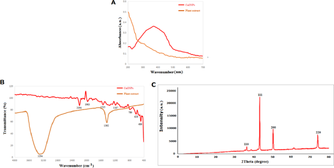
The CuO-NPs’ surface light absorption was examined using UV-Vis. This nanoparticle’s UV range was 200–700 nm, with a maximum absorption around 370 nm (Fig. 1A). It is comparable to CuO-NPs’ UV range. It makes the existence of CuO-NPs known38,39. The extract from Opuntia ficus indica may function as a capping, reducer, and stabilizer, converting the copper salt into copper oxide nanoparticles. Nonetheless, an absorption peak at around 272 nm was displayed by the biosynthesized nanoparticles made with E. alata extract, indicating the successful production and pure phase of CuO-NPs40,41. The Opuntia ficus indica extract and prepared CuO-NPs’ FTIR spectra revealed a sharp band at 3294, 2194, 1963, 1535, 1392, 1187, 941, 786, 620, and 486 cm−1. These bands, respectively, correspond to -O-H bending,-C-O stretching, Amid bonds and -C = C- stretching, Metal-OH (Fig. 1B). The CuO-NPs that were generated had functional groups in them. According to this IR spectrum, flavonoids, proteins, nucleic acids,, phenols, and alkaloids reduce and stabilize those nanoparticles by acting as a capping-agent30. The structure of the nanoparticles is preserved under alkaline circumstances by those metabolites as well as chemicals42. Green biosynthesised CuO-NPs’ XRD spectra showed a variety of difraction-patterns at 36.35°,43.33°, 50.45°, and 74.1° corresponding to the (110), (111), (200), and (220), respectively (JCPDS, Card code. 65-9743)43 (Fig. 1C). The mean particle size of the formed CuO-NPs was determined employing the Scherrer equation, yielding an average crystallite-size of 42.75 nm. The results of the diffraction analysis suggests that the peaks of CuO-NPs exhibit significant good peaks, signifying that the products that are generated are extremely crystalline43. The planes found in XRD indicate the creation of a pure monoclinic-structure of CuO-NPs, with no additional peaks detected in the plots44. Therefore, it may be proposed which the plant extract significantly influences the size within the bio-reduced CuO-NPs.

UV–Visible (A), FTIR (B), and XRD (C) spectra of biosynthesized CuO-NPs.
The morphology and dimensions of the CuO-NPs have been investigated through SEM, while EDX confirmed the presence of elements inside the CuO-NPs. The leaf extracts of Opuntia ficus indica facilitated the formation of CuO-NPs, which exhibit coarse agglomerates and irregular spherical morphologies (Fig. 2A). EDX analysis confirmed that the highest amounts of elemental Cu and O peaks were seen in CuO-NPs (Fig. 2B). These findings validated previous study data about CuO-NPs derived from leaves extract of Psid. guajava45. The dimension of nanoparticles made of CuO was throughout the acceptable range of previously synthesized dispersion nanoparticles46. Furthermore, the nanoparticles were aggregated, maybe attributable their diminutive size, enhanced area of coverage, and diverse molecules47.

SEM (A) And EDX analysis (A) of biofabricated CuO-NPs.
The typical particle dimension and surface electrical charge of nanoparticles has been determined through DLS analysis. Figure 3A indicates that the mean particle size variation among green generated nanoparticles composed of copper oxide was 123.3 nm, which is equivalent to the 50 nm size of CuO-NPs produced with Sida acuta extract48. TEM pictures of produced CuO-NPs are in Fig. 3B. A TEM investigation of phytosynthesize CuO-NPs sized particles as well as shape on the surface showed polydisperse, spherical CuO-NPs with avarage size 65 nm. Padmavathi et al. found that manufactured CuO-NPs are surface components and can reduce CuO to NPs49. The solution of sodium hydroxide serves as a catalytic agent, preventing the aggregation of CuO-NPs. The TEM findings of CuO-NPs were entirely congruent with the XRD pattern of the acquired CuO-NPs. This discovery was supported by the findings of Fardood et al., who observed the FCC structure of CuO-NPs by TEM and SAED patterns derived from CuO-NPs generated using Morinda citrifolia extract of leaves50.

DLS (A) and TEM (B) of biofabricated CuO-NPs.
Antimicrobial activity
The antimicrobial activity of CuO-NPs towards several pathogenic microbes was assessed, and the width of the inhibition region within each well is shown in Fig. 4. Specifically, CuO-NPs obtained from Opuntia ficus indica leaf extract exhibited a greater zone of inhibition against P. vulgaris, C. albicans and B. subtilis, while demonstrating the least zone of inhibition for MRSA and E. faecalis. The results suggest that CuO-NPs obtained from Opuntia ficus indica leaf extract are a more promising source of antibacterial chemicals. The findings illustrate the MIC values of phyto-synthesized CuO-NPs. The findings indicate that CuO-NPs synthesized from Opuntia ficus indica leaf extract demonstrated concentrations of 0.05, 0.3, 0.4, 0.3, 0.1, 0.8, 0.4, 0.2 and 0.05 mg/mL against P. vulgaris, E. coli, K. pneumoniae, E. faecalis, B. subtilis, MRSA, MSSA, S. pneumoniae and C. albicans respectively. CuO-NPs synthesized using Mangifera indica fluid extract have shown significant antibacterial efficacy against both E. coli and S. aureus strains51. Furthermore, copper oxide nanoparticles produced from Curcuma longa shown significant-antibacterial efficacy against P. aeruginosa and P. vulgaris52. CuO-NPs synthesized via Allahabad Safeda extraction shown enhanced antibacterial activity attributable to their superior chemical reactivity and chemical composition on the surface, facilitating effective contact with microbes53. Their little size augments their capacity to engage with bacteria and bolster overall antimicrobial capabilities. CuO-NPs affect the external membrane, resulting in structural alterations, disintegration, and cell lysis54. They additionally impede the DNA reproduction of bacteria, leading to detrimental consequences55. Furthermore, the charge they carry facilitates electrostatic forces with the conductive membranes of cells of microbes, resulting in their demise. Furthermore, CuO-NPs obtained from herbal extracts generate reactive oxygen species (ROS) that may harm the DNA and cellular membranes of microorganisms, hence impeding their proliferation54. Moreover, powerful metabolites derived from CuO-NPs may interfere with bacterial quorum-sensing systems, hence inhibiting their ability to induce outbreaks. CuO-NPs produce copper cations that by electrostatic force adhere to bacterial cell envelopes and permeate the bacteria’s membrane, therefore enhancing their bactericidal efficacy56. In overall, CuO-NPs generated from extracts from plants exhibit many antimicrobial techniques, involving chemical interactions with cell membranes, suppression of DNA replication, production of reactive oxygen species, and disruption to microbial quorum sensing. These features render CuO-NPs effective in combating harmful bacteria57.

Antimicrobial activity of biofabricated CuO-NPs via agar well.
Antibiofilm
The antibiofilm impact of copper oxide nanoparticles was assessed by cultivating biofilms with various amounts of nanoparticles, thereafter identifying the adherent cells with crystal violet34,58. Figure 5 illustrates a dose-dependent reduction in the biofilm-forming ability of the target bacterium, with an increase in nanoparticle concentration considerably affecting MRSA more than P. aeruginosa. CuO-NPs had a slight inhibitory effect versus P. aeruginosa, whereas demonstrating the most significant impact on biofilm formation of MRSA, inhibiting up to 59.3% at 200 µg/mL and decreasing to 8.3% at 3.12 µg/mL for P. aeruginosa, but for MRSA, the inhibition reached 89.4% and decreased to 25.4% at 3.12 µg/mL. The creation of biofilms is a primary factor contributing to the establishment of drug resistance in pathogenic bacteria. To address infections triggered by biofilm-forming bacteria, it is essential to interfere with or eradicate the biofilm, given that these microorganisms are unresponsive to traditional antibiotics, necessitating the pursuit of new biofilm objectives59. Consequently, this work examined not only the antibacterial efficacy of CuO-NPs but also their antibiofilm activity against multidrug-resistant target microorganisms. The findings indicated that CuO-NPs markedly suppressed biofilm development by the target bacteria in a dose-dependent manner. Numerous research have shown the efficacy of CuO-NPs as biofilm inhibitors60,61. The outcomes they obtained corroborated the findings of Shehabeldine et al., who similarly demonstrated the antibiofilm efficacy of mycogenic CuO-NPs, seeing 59% and 49% suppression of biofilms versus K. oxytoca and E. coli, respectively, at subinhibitory doses of nanoparticles60. Chari et al. also observed over 60% inhibition of the creation of biofilm in all examined marine pathogens by CuO-NPs manufactured utilizing a one-pot approach62.

Antibiofilm activity of biofabricated CuO-NPs versus MRSA and P. auroginosa.
Docking simulation
Molecular docking simulation is a crucial method when using computer models to predict the three-dimensional structure of protein-ligand complexes and understand their interactions63,64. Furthermore, the nanomaterials docking simulation might shed light on possible action paths and response mechanisms. The CuO-NPs in the beta-1,3-glucanase (PDB: 2CYG) active sites were subjected to docking simulation in this study (Fig. 6A). To find potential targets for antibacterial action, the study set out to ascertain how CuO-NPs interacted with specific enzymes. Initially, the co-crystallized ligand and beta-1,3-glucanase (PDB: 2CYG) root mean square deviation (RMSD) values were acquired. The corresponding values were 0.9412 and 1.17 Å. In the active site of the beta-1,3-glucanase enzyme (PDB: 2CYG), the CuO-NPs demonstrated hydrophobic interactions with numerous amino acid residues, including Pro41,Val46, Ser48, Arg57, and Lys332 (Fig. 6B and C), and a binding energy S = -2.8688 kcal/mol. In the end, we may conclude that CuO-NPs antibacterial action is due to their suppression of beta-1,3-glucanase. However, the binding energy indicates that beta-1,3-glucanase is more preferred by CuO-NPs, and that hydrophobic interactions in the active site pocket, which is located less than 10.8 Å away, indicate their activity.

(A) 3D crystallography of beta-1,3-glucanase, (B) 3D, and (C) 2D Structure of the CuO-NPs within the active site of beta-1,3-glucanase (MOE version MOE 2019.0901).
Antioxidant activity
The contemporary examination of the antioxidant properties of nanoparticles for integration with biological systems is a crucial issue. Free radicals are unstable molecules that are generated in several biological processes as a result of the relationship among molecular oxygen as well as biomolecules65. The aforementioned radicals possess a number of unpaired electrons that are marked by high instability, resulting in destruction of biological molecules by extracting electron within them to achieve stability55. Different processes, including peroxide breakdown, chain initiation blocking, molecular oxygen abstraction prevention, free radical scavenging, and reductive capacity, have contributed to the antioxidant activity of both synthetic and natural substances66. The DPPH scavenge test is the predominant technique for assessing the antioxidant properties of novel active substances. The present work examined the antioxidant capacity of green produced CuO-NPs employing the DPPH scavenging technique. The antioxidant capability of CuO-NPs appeared directly correlated to the quantity of CuO-NPs, as seen in Fig. 7. This finding aligns with the existing literature about the antioxidant properties of green produced CuO-NPs 55,65. At an amount of 500 µg mL−1, the naturally produced CuO-NPs exhibited a scavenging effectiveness of 72.1 ± 1.4%, whereas ascorbic acid demonstrated a scavenging efficiency of 95.3 ± 1.3% (Fig. 7). The proportions were reduced at minimal concentrations. The scavenge ratios were 58.7 ± 1.6% for CuO-NPs and 88.2 ± 1.3% with ascorbic-acid at a dosage level of 125 µg mL−1. The CuO-NPs synthesized using the water-soluble extract of Suaeda maritima the heartwood exhibited antioxidant property, as measured by DPPH scavenging techniques, having a value of 83.9% at an amount of 40 µg mL−1, in comparison to ascorbic acid, which exhibited 95.2% at the exact same dosage56. According the findings of the authors, at the dosage of 5 µg mL−1, the scavenging capacity of ascorbic acid along with CuO-NPs dropped according to measured readings of 6.5% and 3.5%, correspondingly. Additionally, at an amount of 300 µg mL−1, CuO-NPs made from plant extracts of Prunus africana as well as Camellia sinensis demonstrated antioxidant effects with proportions of 28.8% along with 28.5%, consequently, in contrast to ascorbic acid’s value of 70.8% at the identical dosage67. It was determined the effective amount of CuO-NPs required to scavenge 50% for the free radicals (IC50). As demonstrated, the IC50 amount for ascorbic acid remained 4.21 µg mL−1, whereas that of CuO-NPs was 57.3 µg mL−1. Comparable to ascorbic acid, which had an IC50 of 23.7 µg mL−1, produced CuO-NPs had an IC50 of 28.1 µg mL−156.

Antioxiant activity of biofabricated CuO-NPs via DPPH.
Anti-diabetic activity of CuO-NPs
The degradation of α-amylase as well as the gastric enzymes α-glucosidase leads to the formation of di-saccharides and oligo-saccharides68. Type-2 diabetes is a medical condition that involves hyperglycemia and polyuria. It is often induced by insufficient insulin levels. It refers to a chronic condition marked by elevated blood glucose and insulin levels. It results from diminished insulin efficacy and the increased hepatic glucose generation69,70. Due to insulin ineffectiveness, blood glucose levels in diabetic persons stay high. Consequently, inhibiting α-amylase and α-glucosidase enzymes is crucial for regulating glucose concentrations. Presently, several medications are available to block the α-amylase and α glucosidase enzymes, such as miglitol, acarbose, and voglibose, although with some adverse side effects. This work examines the synthesis of CuO-NPs as an alternative. The inhibition (%) of the enzymes α amylase Fig. 8A and α glucosidase Fig. 8B by CuO-NPs. The reduction in activity proportion for α-amylase varied from 91.7% at 1 mg/ml to 34.1% at 0.0019 mg/ml, while for α-glucosidase, it varied from 81.8% at 1 mg/ml to 32.3% at 0.0019 mg/ml. Prior studies demonstrated CuO-NPs’ capacity to inhibit α-amylase, and the inhibition manner goes along with our results68,71,72,73.

The effect of biofabricated CuO-NPs α-amylase (A) & α-glucosidase (B) enzymes.
Antiviral activity
This research examined the efficacy of CuO-NPs in reducing the growth of HAV and CoxB4 viruses. To determine the MNTC, CuOـNPs were assayed for cytotoxicity versus cell lines at 125 µg/mL. CuO-NPs were reported to exhibit promising antiviral properties against both HAV and CoxB4. At the same dose, COX B4 was more active than HAV. CuO-NPs showed 41.1% antiviral activity against COXB4 and 25.2% effectiveness against HAV at 125 µg/mL (Figs. 9 and 10 ). CuO-NPs shown remarkable antiviral efficacy versus both HAV and COXـB4, showing their potential for usage in biological areas. The antiviral efficacy of nanoparticles (NPs) has been elucidated through various mechanisms, which includes the inhibition of viral and cellular reactions that obstruct infection, connections between nano and specific cell-surfaces or receptors that impede viral entry, suppression of viral propagation, avoidance of viral dissemination, augmentation of oxidative stress through ROS generation, induction of cellular-apoptosis, and improvement of the host cell’s defenses against infection74,75,76. The decrease in virus frequency after administration with CuO-NPs underscores the pivotal function of copper, suggesting that copper nanoparticles provide significant potential for the development of potent antiviral therapies77,78. Copper is effective versus immuno-deficiency virus (HIV-1), as well as SARS-CoV-2. Direct 1 h interaction with SARS-CoV-2 onto surfaces coated in Cu NPs resulted in a considerable reduction of the virus’s titer (antiviral agents). The antiviral action of Cu included the destruction of virions by oxidation of virus capsid proteins, impairment with respect to their envelope, and denatured state of viral nucleic acids79. CuO-NPs might be useful in inactivating virus H1N1-influenza. Bronchial and renal cells infection with H1N1 and stimulated with 100 nm spherical CuO-NPs exhibited increased resistance to the cytopathic consequences of the virus. In the course of viral production, a markedly reduced amount of influenza virus nucleoproteins was observed after a 30 min exposure to Cu NPs. This validates the relevance of Cu NPs in the protection of virus-associated infectious illnesses25,80.

The effect of biofabricated CuO-NPs on HAV.

The effect of biofabricated CuO-NPs on CoXB4 virus.
Conclusion
The unique technology characterisation of CuO-NPs in this study demonstrates its remarkable potential as multifunctional bioactive materials with a variety of eco-friendly properties. CuO-NPs biosynthesis from Opuntia ficus-indica extract produces encouraging application outcomes, demonstrating exceptional effectiveness across a range of tests. Characterize of CuO-NPs was done via Uv, FTIR, XRD, SEM-EDX, DL and TEM. The antimicrobial effectiveness of CuO-NPs was performed against bacterial and fungal strain and MIC between 62.5 and 500 µg/mL. CuO-NPs shown an antioxidant activity via DPPH method with an IC50 of 165.5 µg/ml. Moreover, CuO-NPs demonstrated antibiofilm advantage versus MRSA and P. aeruginosa. The inhibitory effect of CuO-NPs towards bacterial strains may be caused by beta-1,3-glucanase, which interacts hydrophobically with amino acid residues in the active site, according to a molecular docking modelling. Furthermore, CuO-NPs displayed a noteworthy antiviral property against COXB4 and HAV at a dosage of 125 µg/mL, with antiviral efficacy of 40.9% and 28.6%, respectively. Additionally, 91.5% suppression of α-amylase and 82.3% inhibition of α-glucosidase were demonstrated by the CuO-NPs at 1000 µg/mL, confirming their antidiabetic qualities. CuO-NPs therefore hold great potential as an anti-inflammatory medication. The development of CuO-NPs in clinical and commercial applications is based on these discoveries, which validate its potential in antibacterial, antibiofilm, antiviral, anticancer, and antioxidant treatments.
Data availability
The data used to support the findings of this study are available from the corresponding author upon request.
References
-
Schiavo, L. et al. An overview of the advanced nanomaterials science. Inorg. Chim. Acta 559, 121802 (2024).
-
Salem, S. S. A mini review on green nanotechnology and its development in biological effects. Arch. Microbiol. 205(4) (2023).
-
Salem, S. S. & Fouda, A. Green synthesis of metallic nanoparticles and their prospective biotechnological applications: An overview. Biol. Trace Elem. Res. 199(1), 344–370 (2021).
-
Elfadel, R. G. et al. Preparation of new surface coating based on modified oil-based polymers blended with ZnO and CuZnO NPs for steel protection. Sci. Rep. 13(1), 7268 (2023).
-
Badawy, A. A., Abdelfattah, N. A. H., Salem, S. S., Awad, M. F. & Fouda, A. Efficacy assessment of biosynthesized copper oxide nanoparticles (CuO-NPs) on stored grain insects and their impacts on morphological and physiological traits of wheat (Triticum aestivum l.) plant. Biology 10(3) (2021).
-
Salem, S. S. Bio-fabrication of selenium nanoparticles using baker’s yeast extract and its antimicrobial efficacy on food borne pathogens. Appl. Biochem. Biotechnol. 194(5), 1898–1910 (2022).
-
Dezfuli, A. A. Z., Abu-Elghait, M. & Salem, S. S. Recent insights into nanotechnology in colorectal Cancer. Appl. Biochem. Biotechnol. 196(7), 4457–4471 (2024).
-
Saqib, S. et al. Organometallic assembling of chitosan-Iron oxide nanoparticles with their antifungal evaluation against rhizopus oryzae. Appl. Organomet. Chem. 33(11), e5190 (2019).
-
Salem, S. S., Hammad, E. N., Mohamed, A. A. & El-Dougdoug, W. A comprehensive review of nanomaterials: Types, synthesis, characterization, and applications. Biointerface Res. Appl. Chem. 13(1) (2023).
-
Saqib, S. et al. Postharvest disease Inhibition in fruit by synthesis and characterization of Chitosan iron oxide nanoparticles. Biocatal. Agric. Biotechnol. 28, 101729 (2020).
-
Hamed, A., El-Gebaly, A., Refaey, A., Sofy, A. & Youssef, A. Green synthesized silver and zinc oxide nanoparticles using Ficus carica leaf extract and their activity on human viruses. Egypt. J. Chem. 67(13), 847–857 (2024).
-
Said, A., Abu-Elghait, M., Atta, H. M. & Salem, S. S. Antibacterial activity of green synthesized silver nanoparticles using Lawsonia inermis against common pathogens from urinary tract infection. Appl. Biochem. Biotechnol. 196(1), 85–98 (2024).
-
Soliman, M. K. Y., Salem, S. S. Uncovering the potential of biofabricated Ananas comosus peel selenium nanoparticles for antibacterial, antibiofilm, suppression of virulence genes (can and LuxS), anticancer, and antioxidant properties. BMC Biotechnol. 25, 51 (2025). https://doi.org/10.1186/s12896-025-00999-x
-
Puri, A. et al. From nature to nanotechnology: the interplay of traditional medicine, green chemistry, and biogenic metallic phytonanoparticles in modern healthcare innovation and sustainability. Biomed. Pharmacother. 170, 116083 (2024).
-
Soni, V. et al. Sustainable and green trends in using plant extracts for the synthesis of biogenic metal nanoparticles toward environmental and pharmaceutical advances: A review. Environ. Res. 202, 111622 (2021).
-
Al-Rajhi, A. M. H., Salem, S. S., Alharbi, A. A. & Abdelghany, T. M. Ecofriendly synthesis of silver nanoparticles using Kei-apple (Dovyalis caffra) fruit and their efficacy against cancer cells and clinical pathogenic microorganisms. Arab. J. Chem. 15(7) (2022).
-
Abdelghany, T. M. et al. Phytofabrication of zinc oxide nanoparticles with advanced characterization and its antioxidant, anticancer, and antimicrobial activity against pathogenic microorganisms. Biomass Convers. Biorefinery 13(1), 417–430 (2023).
-
Alsaiari, N. S. et al. Plant and microbial approaches as green methods for the synthesis of nanomaterials: Synthesis, applications, and future perspectives. Molecules 28(1), 463 (2023).
-
Antonio-Pérez, A., Durán-Armenta, L. F., Pérez-Loredo, M. G. & Torres-Huerta, A. L. Biosynthesis of copper nanoparticles with medicinal plants extracts: From extraction methods to applications. Micromachines 14(10), 1882 (2023).
-
Bulut Kocabas, B., Attar, A., Peksel, A. & Altikatoglu Yapaoz, M. Phytosynthesis of cuonps via Laurus nobilis: Determination of antioxidant content, antibacterial activity, and dye decolorization potential. Biotechnol. Appl. Chem. 68(4), 889–895 (2021).
-
Soliman, M. K. Y., Amin, M. A. A., Nowwar, A. I., Hendy, M. H. & Salem, S. S. Green synthesis of selenium nanoparticles from Cassia javanica flowers extract and their medical and agricultural applications. Sci. Rep. 14(1) (2024).
-
Elnosary, M. E. et al. Uncovering and evaluating coconut oil-loaded silica nanoemulsion as anti-viral, bacterial, and fungal: synthesis, fabrication, characterization, and biosafety profiles. Beni Suef Univ. J. Basic. Appl. Sci. 13(1), 56 (2024).
-
Tyagi, S., Kumar, A., Tyagi, P. K. & Hatami, M. Development and characterization of biogenic copper oxide nanoparticles, with an exploration of their antibacterial and antioxidant potential. 3 Biotech 14(1), 20 (2023).
-
Alizadeh, S. R. & Ebrahimzadeh, M. A. Characterization and anticancer activities of green synthesized CuO nanoparticles, a review. Anti Cancer Agents Med. Chem. Former. Curr. Med. Chem. Anti Cancer Agents 21(12), 1529–1543 (2021).
-
Woźniak-Budych, M. J., Staszak, K. & Staszak, M. Copper and copper-based nanoparticles in medicine—perspectives and challenges. Molecules 28(18), 6687 (2023).
-
Chen, G., Roy, I., Yang, C. & Prasad, P. N. Nanochemistry and nanomedicine for nanoparticle-based diagnostics and therapy. Chem. Rev. 116(5), 2826–2885 (2016).
-
Hussein, A. S., Hashem, A. H. & Salem, S. S. Mitigation of the hyperglycemic effect of streptozotocin-induced diabetes albino rats using biosynthesized copper oxide nanoparticles. Biomol. Concepts 14(1), 20220037 (2023).
-
Shaheen, T. I., Fouda, A. & Salem, S. S. Integration of cotton fabrics with biosynthesized CuO nanoparticles for bactericidal activity in the terms of their cytotoxicity assessment. Ind. Eng. Chem. Res. 60(4), 1553–1563 (2021).
-
Neiva, J., Benzarti, Z., Carvalho, S. & Devesa, S. Green synthesis of CuO Nanoparticles—Structural, morphological, and dielectric characterization. Materials 17(23), 5709 (2024).
-
Rajamma, R., Gopalakrishnan Nair, S., Abdul Khadar, F. & Baskaran, B. Antibacterial and anticancer activity of biosynthesised CuO nanoparticles. IET Nanobiotechnol. 14(9), 833–838 (2020).
-
Guleria, S., Simsek, H., Chawla, P., Relhan, A. & Bhasin, A. Evaluation of Cladophora and Chlamydomonas microalgae for environmental sustainability: A comparative study of antimicrobial and photocatalytic dye degradation. Environ. Pollut. 340, 122806 (2024).
-
Mezher, M., El Hajj, R. & Khalil, M. Investigating the antimicrobial activity of essential oils against pathogens isolated from sewage sludge of Southern Lebanese villages. Germs 12(4), 488 (2022).
-
Mohamed, A. A., Abu-Elghait, M., Ahmed, N. E. & Salem, S. S. Eco-friendly mycogenic synthesis of ZnO and CuO nanoparticles for in vitro antibacterial, antibiofilm, and antifungal applications. Biol. Trace Elem. Res. 199(7), 2788–2799 (2021).
-
Soliman, M. K., Hashem, A. H., Al-Askar, A. A., AbdElgayed, G. & Salem, S. S. Green synthesis of silver nanoparticles from Bauhinia variegata and their biological applications. Green Process. Synth. 13(1), 20240099 (2024).
-
Sethi, P. Activity of Turbinaria ornata (turner) J. Agade against blue tongue virus (Btv). IOSR J. Pharm. 6(7), 93–95 (2016).
-
Ameena, S. et al. Antioxidant, antibacterial, and anti-diabetic activity of green synthesized copper nanoparticles of Cocculus hirsutus (Menispermaceae). Appl. Biochem. Biotechnol. 194(10), 4424–4438 (2022).
-
Visvanathan, R., Jayathilake, C., Liyanage, R. & Sivakanesan, R. Applicability and reliability of the glucose oxidase method in assessing α-amylase activity. Food Chem. 275, 265–272 (2019).
-
Faisal, S. et al. In vivo analgesic, anti-inflammatory, and anti-diabetic screening of Bacopa monnieri-synthesized copper oxide nanoparticles. ACS Omega 7(5), 4071–4082 (2022).
-
Saligedo, T. S., Muleta, G. G., Tsega, T. W. & Tadele, K. T. Green synthesis of copper oxide nanoparticles using Eichhornia crassipes leaf extract, its antibacterial and photocatalytic activities. Curr. Nanomater. 8(1), 58–68 (2023).
-
Weldegebrieal, G. K. Photocatalytic and antibacterial activityof CuO nanoparticles biosynthesized using Verbascum thapsus leaves extract. Optik 204, 164230 (2020).
-
Banu, S. N. Comparative study of photocatalytic degradation of methylene blue and Methyl orange in presence of solar light using green synthesized Cuo nanocatalyst. Int. J. Eng. Appl. Sci. Technol. 4(8), 365–370 (2019).
-
Verma, N. & Kumar, N. Synthesis and biomedical applications of copper oxide nanoparticles: An expanding horizon. ACS Biomater. Sci. Eng. 5(3), 1170–1188 (2019).
-
Cengiz, M. et al. Biogenic synthesized bare and Boron-Doped copper oxide nanoparticles from Thymbra spicat ssp. Spicata: In Silico and in vitro studies. J. Cluster Sci. 35(1), 265–284 (2024).
-
Navada, K. M., K N, G., D’Souza, J. N. & Kouser, S. Synthesis, characterization of phyto-functionalized CuO nano photocatalysts for mitigation of textile dyes in waste water purification, antioxidant, anti-inflammatory and anticancer evaluation. Appl. Nanosci. 11, 1313–1338 (2021).
-
Singh, J., Kumar, V., Kim, K-H. & Rawat, M. Biogenic synthesis of copper oxide nanoparticles using plant extract and its prodigious potential for photocatalytic degradation of dyes. Environ. Res. 177, 108569 (2019).
-
Noor, S. et al. A fungal based synthesis method for copper nanoparticles with the determination of anticancer, antidiabetic and antibacterial activities. J. Microbiol. Methods 174, 105966 (2020).
-
Ashar, A. et al. Synthesis, characterization and photocatalytic activity of ZnO flower and pseudo-sphere: Nonylphenol ethoxylate degradation under UV and solar irradiation. J. Alloys Compd. 678, 126–136 (2016).
-
Sathiyavimal, S. et al. Biogenesis of copper oxide nanoparticles (CuONPs) using Sida acuta and their incorporation over cotton fabrics to prevent the pathogenicity of gram negative and gram positive bacteria. J. Photochem. Photobiol. B 188, 126–134 (2018).
-
Padmavathi, A. R. et al. Copper oxide nanoparticles as an effective anti-biofilm agent against a copper tolerant marine bacterium, Staphylococcus lentus. Biofouling 35(9), 1007–1025 (2019).
-
Taghavi Fardood, S., Ramazani, A., Asiabi, P. & Joo, S. A novel green synthesis of copper oxide nanoparticles using a Henna extract powder. J. Struct. Chem. 59, 1737–1743 (2018).
-
Chinnathambi, A., Awad Alahmadi, T. & Ali Alharbi, S. Biogenesis of copper nanoparticles (Cu-NPs) using leaf extract of Allium noeanum, antioxidant and in-vitro cytotoxicity. Artif. Cells Nanomed. Biotechnol. 49(1), 500–510 (2021).
-
Faisal, S. et al. Curcuma longa mediated synthesis of copper oxide, nickel oxide and Cu-Ni bimetallic hybrid nanoparticles: Characterization and evaluation for antimicrobial, Anti-Parasitic and cytotoxic potentials. Coatings 11(7), 849 (2021).
-
Relhan, A., Guleria, S., Bhasin, A., Mirza, A. & Zhou, J. L. Biosynthesized copper oxide nanoparticles by Psidium guajava plants with antibacterial, antidiabetic, antioxidant, and photocatalytic capacity. Biomass Convers. Biorefinery (2024).
-
Kaningini, A. G., Motlhalamme, T., More, G. K., Mohale, K. C. & Maaza, M. Antimicrobial, antioxidant, and cytotoxic properties of biosynthesized copper oxide nanoparticles (CuO-NPs) using athrixia phylicoides DC. Heliyon 9(4) (2023).
-
Rehana, D., Mahendiran, D., Kumar, R. S. & Rahiman, A. K. Evaluation of antioxidant and anticancer activity of copper oxide nanoparticles synthesized using medicinally important plant extracts. Biomed. Pharmacother. 89, 1067–1077 (2017).
-
Peddi, P., Ptsrk, P. R., Rani, N. U. & Tulasi, S. L. Green synthesis, characterization, antioxidant, antibacterial, and photocatalytic activity of Suaeda maritima (L.) Dumort aqueous extract-mediated copper oxide nanoparticles. J. Genet. Eng. Biotechnol. 19(1), 131 (2021).
-
Dabhane, H. et al. A novel approach toward the bio-inspired synthesis of CuO nanoparticles for phenol degradation and antimicrobial applications. Biomass Convers. Biorefinery 14(15), 17235–17250 (2024).
-
Soliman, M. K., Salem, S. S., Abu-Elghait, M. & Azab, M. S. Biosynthesis of silver and gold nanoparticles and their efficacy towards antibacterial, antibiofilm, cytotoxicity, and antioxidant activities. Appl. Biochem. Biotechnol. 195(2), 1158–1183 (2023).
-
Pardesi, K., Pable, A., Bhagat, D. & Satpute, S. Applications of metal nanoparticles to combat biofilm forming Eskape pathogens. Recent Adv. Biotechnol. (2019).
-
Shehabeldine, A. M. et al. Potential antimicrobial and antibiofilm properties of copper oxide nanoparticles: Time-kill kinetic essay and ultrastructure of pathogenic bacterial cells. Appl. Biochem. Biotechnol. 195(1), 467–485 (2023).
-
Mansoor, A. et al. Anti-bacterial effect of titanium-oxide nanoparticles and their application as alternative to antibiotics. Pak. Vet. J. 43(2) (2023).
-
Chari, N., Felix, L., Davoodbasha, M., Ali, A. S. & Nooruddin, T. In vitro and in vivo antibiofilm effect of copper nanoparticles against aquaculture pathogens. Biocatal. Agric. Biotechnol. 10, 336–341 (2017).
-
Selim, S. et al. Biosynthesized zinc oxide nanoparticles: Multifunctional potential applications in anticancer, antibacterial, and B. subtilis DNA gyrase Docking. Green. Process. Synth 14(1), 20240218 (2025).
-
Gad, E. S. et al. A comprehensive study on characterization of biosynthesized copper-oxide nanoparticles, their capabilities as anticancer and antibacterial agents, and predicting optimal Docking poses into the cavity of S. aureus DHFR. PloS One 20(4), e0319791 (2025).
-
Dobrucka, R. Antioxidant and catalytic activity of biosynthesized CuO nanoparticles using extract of Galeopsidis herba. J. Inorg. Organomet. Polym. Mater. 28, 812–819 (2018).
-
Diplock, A. T. Will the ‘good fairies’ please prove to us that vitamin E lessens human degenerative disease? Free Radic. Res. 27(5), 511–532 (1997).
-
Ssekatawa, K. et al. Phyto-mediated copper oxide nanoparticles for antibacterial, antioxidant and photocatalytic performances. Front. Bioeng. Biotechnol. 10, 820218 (2022).
-
Ramasubbu, K. et al. Green synthesis of copper oxide nanoparticles using sesbania grandiflora leaf extract and their evaluation of anti-diabetic, cytotoxic, anti-microbial, and anti-inflammatory properties in an in-vitro approach. Fermentation 9(4), 332 (2023).
-
Taylor, P. N. et al. Global epidemiology of hyperthyroidism and hypothyroidism. Nat. Rev. Endocrinol. 14(5), 301–316 (2018).
-
Huang, S-C., Gau, S-Y., Huang, J-Y., Wu, W-J. & Wei, J. C. C. Increased risk of hypothyroidism in people with asthma: Evidence from a real-world population-based study. J. Clin. Med. 11(10), 2776 (2022).
-
Almehizia, A. A., Naglah, A. M., Aljafen, S. S., Hassan, A. S. & Aboulthana, W. M. Assessment of the in vitro biological activities of schiff base-synthesized copper oxide nanoparticles as an anti-diabetic, anti-alzheimer, and anti-cancer agent. Pharmaceutics 17(2), 180 (2025).
-
Iqbal, J. et al. Potential antimicrobial, antidiabetic, catalytic, antioxidant and ROS/RNS inhibitory activities of Silybum marianum mediated biosynthesized copper oxide nanoparticles. RSC Adv. 12(22), 14069–14083 (2022).
-
Umar, M. B. et al. Hypoglycaemic activity of biosynthesized copper oxide nanoparticles in alloxan-induced diabetic Wister rats. Endocrinol. Diabetes Metab. 6(3), e423 (2023).
-
Bhatti, A. & DeLong, R. K. Nanoscale interaction mechanisms of antiviral activity. ACS Pharmacol. Transl. Sci. 6(2), 220–228 (2023).
-
Almuhayawi, M. S. et al. Investigating the in vitro antibacterial, antibiofilm, antioxidant, anticancer and antiviral activities of zinc oxide nanoparticles biofabricated from Cassia javanica. PLoS One 19(10), e0310927 (2024).
-
Hmed, A. A., El-Gebaly, A. S., Refaey, E. E., Youssef, A. M. & Sofy, A. R. Antiviral potential of copper and titanium dioxide nanoparticles against H1N1, adenovirus 40 and herpes simplex virus type-II. Inorg. Chem. Commun. 171, 113605 (2025).
-
Mavil-Guerrero, E., Vazquez-Duhalt, R. & Juarez-Moreno, K. Exploring the cytotoxicity mechanisms of copper ions and copper oxide nanoparticles in cells from the excretory system. Chemosphere 347, 140713 (2024).
-
Tavakoli, A. & Hashemzadeh, M. S. Inhibition of herpes simplex virus type 1 by copper oxide nanoparticles. J. Virol. Methods 275, 113688 (2020).
-
Raha, S., Mallick, R., Basak, S. & Duttaroy, A. K. Is copper beneficial for COVID-19 patients? Med. Hypotheses 142, 109814 (2020).
-
Ha, T. et al. Antiviral activities of high energy E-beam induced copper nanoparticles against H1N1 influenza virus. Nanomaterials 12(2), 268 (2022).
Acknowledgements
The authors express their sincere thanks to Faculty of science (Boyes), Al-Azhar University, Cairo, Egypt for providing the necessary research facilities.
Funding
Open access funding provided by The Science, Technology & Innovation Funding Authority (STDF) in cooperation with The Egyptian Knowledge Bank (EKB).
Ethics declarations
Competing interests
The authors declare no competing interests.
Additional information
Publisher’s note
Springer Nature remains neutral with regard to jurisdictional claims in published maps and institutional affiliations.
Rights and permissions
Open Access This article is licensed under a Creative Commons Attribution 4.0 International License, which permits use, sharing, adaptation, distribution and reproduction in any medium or format, as long as you give appropriate credit to the original author(s) and the source, provide a link to the Creative Commons licence, and indicate if changes were made. The images or other third party material in this article are included in the article’s Creative Commons licence, unless indicated otherwise in a credit line to the material. If material is not included in the article’s Creative Commons licence and your intended use is not permitted by statutory regulation or exceeds the permitted use, you will need to obtain permission directly from the copyright holder. To view a copy of this licence, visit http://creativecommons.org/licenses/by/4.0/.
About this article

Cite this article
Soliman, M.K.Y., Salem, S.S. Comparative evaluation of antimicrobial, antibiofilm, antioxidant, antiviral, and antidiabetic activities of copper oxide nanoparticles biofabricated via Opuntia ficus indica. Sci Rep 15, 24823 (2025). https://doi.org/10.1038/s41598-025-08878-3
-
Received:
-
Accepted:
-
Published:
-
DOI: https://doi.org/10.1038/s41598-025-08878-3