Introduction
Colorectal cancer (CRC) remains one of the most prevalent and life-threatening malignancies worldwide1, accounting for approximately 9% of all cancer-related deaths annually. It ranks as the third leading cause of cancer mortality, emphasizing the urgent need for a deeper understanding of its molecular mechanisms and the identification of reliable biomarkers for early detection and prognosis2,3. Despite advancements in screening and therapeutic strategies, CRC still exhibits high recurrence rates and resistance to conventional treatments, highlighting the necessity for novel molecular targets4.
Cancer is fundamentally a genetic disease driven by dysregulation in both coding and non-coding gene expression5,6. Recent research has increasingly focused on non-coding RNAs (ncRNAs) due to their pivotal roles in regulating oncogenic and tumor-suppressive pathways7,8. Among them, long non-coding RNAs (lncRNAs) and microRNAs (miRNAs) have emerged as crucial players in cancer progression, influencing cellular proliferation, invasion, and metastasis9. However, their specific roles in CRC pathogenesis remain insufficiently explored, particularly in relation to their interactions with key molecular markers10.
Carcinoembryonic antigen-related cell adhesion molecule 6 (CEACAM6) is a glycoprotein of the immunoglobulin superfamily that has been implicated in various cancers11,12. It plays a crucial role in promoting tumor invasion, immune evasion, and resistance to apoptosis, making it a promising yet underexplored target in CRC11,13,14. Although previous studies have suggested a correlation between CEACAM6 overexpression and poor prognosis in gastrointestinal malignancies, its precise regulatory mechanisms in CRC require further investigation15.
Another critical ncRNA, HOXA-AS3, has been identified as a potential tumor regulator, particularly within the HOX gene cluster16. Its dual functionality in different cancer types – acting as either an oncogene or a tumor suppressor – suggests that its role in CRC may be more complex than previously thought17. However, its direct interaction with CEACAM6 and other key molecular markers in CRC remains to be elucidated18.
Meanwhile, miR-29a has been reported to modulate multiple cellular processes, including apoptosis, differentiation, and metastasis19. Interestingly, its regulatory influence on CEACAM6 has been demonstrated in lung adenocarcinoma20, suggesting a potential but unexplored connection in CRC21,22. Additionally, circulating miR-29a levels in serum and exosomes present an opportunity for developing minimally invasive biomarkers for CRC diagnosis and prognosis23,24.
Despite growing evidence supporting the significance of these molecules in cancer biology, a comprehensive investigation of their combined roles in CRC remains lacking. Therefore, this study aims to assess the expression patterns of CEACAM6, HOXA-AS3, and miR-29a in both blood and tissue samples from CRC patients. By integrating molecular profiling with in silico validation, this research seeks to clarify their interconnections, explore their potential as diagnostic and prognostic biomarkers, and provide insights into novel therapeutic strategies for CRC.
Materials and methods
Clinical specimen collection
This study was conducted following the ethical standards outlined in the Declaration of Helsinki and approved by the Research Ethics Committee of Golestan University of Medical Sciences (Ethics code: IR.GOUMS.REC 1398.165). All procedures were performed in accordance with the relevant guidelines and regulations, ensuring the protection of participant rights and data confidentiality. The clinically specimen were collected with informed written consent and with respect to two criteria no surgery and without local or systemic treatment between 2019 and 2020. The colon tumor tissues (N = 68) and adjacent normal tissues (N = 68) were obtained from the Imam Khomeini Hospital in Tehran, Iran.
Analysis of gene expression
Total RNA was extracted from the frozen specimens using Trizol reagent (Cat. No. 15596026, Ambion, Thermo Fisher Scientific) according to the manufacturer’s protocol. The extracted RNA was subsequently treated with DNase I (Cat. No. EN0521, Fermentas, Thermo Fisher Scientific) to eliminate any residual genomic DNA. cDNA synthesis was carried out using a reverse transcription kit (YT4500, Yekta Taihiz Azma, Iran) and used as the template for real-time PCR, performed on the ABI 7300 system (Applied Biosystems, USA) with SYBER Green PCR Master Mix (Cat. No. RR820A, TAKARA).
All reactions were conducted in triplicate, and the specificity of the PCR products was verified by melting curve analysis. Quantitative PCR (qPCR) for miRNA was performed using a specific forward primer for miR-29a and a universal reverse primer in combination with the SYBR green master mix.
GAPDH and U6 snRNA were chosen as housekeeping genes for the normalization of mRNA and miRNA expression data, respectively. Relative expression levels of target genes were calculated using the 2–△△Ct method25. Primer sequences and their characteristics are listed in Table 1.
Interaction analysis of HOXA-AS3
The RNA Interactome database (RNAInter; http://www.rnainter.org/IntaRNA/) was used to identify the possible interactions of HOXA-AS3 with CEACAM6 and IL-6 genes. Also, we utilize the EVlncRNA database (https://www.sdklab-biophysics-dzu.net/EVLncRNAs1/) to identify which genes HOXA-AS3 should target in colon cancer.
In silico expression profile of CEACAM6, HOXA-AS3, & IL-6
In order to investigate the expression profile of CEACAM6, HOXA-AS3, and Il-6 in various cancer types especially colon cancer, the database of Gene Expression Profiling Interactive Analysis (GEPIA; http://gepia.cancer-pku.cn/) were applied. This database contains the RNA-Seq expression data for mRNA, lncRNA, and antisense from The Cancer Genome Atlas (TCGA).
In silico expression profile of miR29a
The database of Differentially Expressed MiRNAs in human Cancers (dbDEMC; https://www.biosino.org/dbDEMC/index) was used for exploring miR29a dysregulation in colon cancer. It is an integrated database that designed to store and display differentially expressed miRNAs in different cancer types that identified by high-throughput and low-throughput techniques.
Il-6 detection by enzyme-linked immunosorbent assay
To quantify of IL-6 levels in peripheral blood, serum samples of CRC patients were analyzed using an enzyme linked immunoassay (ELISA). The assay was performed following the instructions provided with a standardized protocol26. Briefly, serum samples were first diluted appropriately to fall within the assay’s detection range. The diluted samples were then added to the wells of a microplate pre-coated with IL-6-specific antibodies. After incubation, the wells were washed with PBS/T (phosphate buffered saline, pH 7.3, containing 0.05% Tween) to remove unbound substances. A secondary antibody conjugated to an enzyme was then added, followed by TMB (3,39, 5,50-tetramethylbenzidine) substrate solution that reacted with the enzyme to produce a measurable colorimetric signal. The intensity of the color, which is proportional to the amount of IL-6 present in the samples, was measured using a microplate reader at 450 nm. Each sample was tested in triplicate to ensure accuracy and reproducibility of the results.
Statistical analysis
All parametric and non-parametric data were statistically analyzed by the GraphPad Prism (version 5.0.0.288). The significance of data was defined as a p-value less than 0.05. In order to assess the relationship between expression of targeted genes and clinical features, the unpaired two-sample t-test was applied. Receiver operating characteristic (ROC) was plotted using MedCalc software in evaluating the diagnostic ability of miR29a.
Results
Demographic and clinical characteristics of CRC patients and tumor samples
Demographic information and the characteristics of colon tumor samples are detailed in Tables 2 and 3, respectively. The average age of the patients was 51 years old (range: 39 to 62). Among the patients, 36 were female (53%) and 32 were male (47%).
CEACAM6 expression patterns in colorectal cancer patients
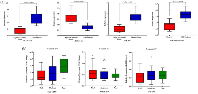
The transcript level of CEACAM6 gene was measured by normalizing to GAPDH as a reference gene, which was performed in the same run. The analysis of Real-Time data demonstrated that the colon tumor cells expressed CEACAM6 4.7-fold higher than the adjacent normal colon tissues (Fig. 1a, p < 0.05). From the status of tumor differentiation point of view, the CEACAM6 transcript was increased by 26% in the poor differentiated samples than well differentiated (Fig. 1b).

The expression profile of CEACAM6, HOXA-AS3, & miR-29a in CRC patients. (a) The transcript level of CEACAM6, HOXA-AS3, and miR-29a across colon cancer and the adjacent tissues demonstrated. High expression of CEACAM6 and low expression of HOXA-AS3 were significantly observed in colon tumor tissues (p < 0.05). Also, miR-29a highly expressed in both tissue and serum (p < 0.05). (b) The transcript level of CEACAM6, HOXA-AS3, and miR-29a according to tumor differentiation status, well, moderate, and poor. The expression pattern of CEACAM6 was only significantly changes (p < 0.05).
HOXA-AS3 expression in colorectal cancer tissues
The quantification analysis of expression data indicated that the colon tumors transcribed lower amount of HOXA-AS3 than the adjacent normal tissues (Fig. 1a, p < 0.05). While, no significant differences between expression of HOXA-AS3 and the status of tumor differentiation, as shown in Fig. 1b (p > 0.05).
Correlation of CEACAM6 and HOXA-AS3 expression with clinicopathological features in CRC
To investigate the potential clinical relevance of CEACAM6 and HOXA-AS3 expression, we examined their correlation with various clinicopathological parameters in CRC patients (Table 4). Our analysis revealed that CEACAM6 expression was significantly higher in tumors with poor differentiation compared to well-differentiated tumors (p = 0.050), indicating its potential role in tumor aggressiveness. Furthermore, patients with advanced-stage tumors (TNM stage III) exhibited significantly elevated CEACAM6 levels compared to those in stages I/II (p = 0.019).
In contrast, HOXA-AS3 expression did not show a statistically significant correlation with tumor differentiation, TNM stage, or other clinicopathological variables (p > 0.05). Although its downregulation was observed in tumor tissues, no clear association with disease severity was established. These findings suggest that while CEACAM6 may serve as a potential marker of tumor progression, the role of HOXA-AS3 in CRC requires further investigation.
miR-29a expression in colorectal cancer patients
The comparison of miR29a transcript level in colon tumor tissue than normal adjacent tissue indicated that tumor tissue was significantly expressed higher value of this miRNA (p < 0.05). The same result was found in the serum of patients (Fig. 1a). However, its transcript level was not differentially expressed based on the tumor differentiation status (Fig. 1b; p > 0.05).
Correlation of miR-29a expression in tissue and serum with clinicopathological features in CRC
The relationship between miR-29a expression in both tumor tissue and serum with clinicopathological characteristics was assessed (Table 5). Notably, serum levels of miR-29a were significantly lower in patients with lymphovascular invasion (p = 0.028), suggesting a potential link between reduced miR-29a expression and increased metastatic potential. Additionally, miR-29a expression in both tissue and serum was significantly higher in patients with early-stage CRC (TNM stage I/II) compared to those with stage III tumors (p = 0.026 and p = 0.043, respectively).
Despite these associations, no significant correlation was found between miR-29a levels and other clinicopathological parameters, such as tumor differentiation, tumor size, smoking, alcohol consumption, or diabetes status (p > 0.05). Importantly, a significant positive correlation was observed between miR-29a expression in tissue and serum samples (p = 0.038), reinforcing its potential as a minimally invasive biomarker for CRC.
Table 6 presents the correlation analysis of miR-29a expression with CEACAM6 and HOXA-AS3. The data indicate that while miR-29a expression shows no significant correlation with CEACAM6 and HOXA-AS3 (p > 0.05), its strong positive correlation between tissue and serum levels further highlights its potential as a liquid biopsy biomarker.
IL-6 expression in colorectal cancer patients
Figure 2 shows the expression pattern of Il-6 in colon cancer patients. The expression level of the Il-6 gene in serum was significantly elevated in patients with colon cancer compared to normal individuals (p < 0.05).

The expression profile of Il-6 in CRC patients. The serum expression levels of Il-6 significantly increased in CRC patients (p < 0.05).
Diagnostic performance of miR-29a in CRC: ROC curve analysis
To evaluate the diagnostic utility of miR-29a, a Receiver Operating Characteristic (ROC) curve analysis was performed. As depicted in Fig. 3, the analysis demonstrated a high area under the curve (AUC) of 0.918 (p < 0.001), indicating excellent diagnostic accuracy. These results suggest that miR-29a exhibits strong sensitivity and specificity in distinguishing CRC patients from healthy individuals.
Given that circulating miR-29a levels in serum correlated with tumor tissue expression, our findings support the feasibility of using miR-29a as a non-invasive biomarker for early CRC detection. Further validation in larger cohorts is warranted to establish its clinical applicability.
In silico analysis of CEACAM6, HOXA-AS3, and IL-6 expression in CRC
Bioinformatics analysis were employed to elucidate the molecular mechanisms underlying the regulation of CEACAM6, HOXA-AS3, and Il-6. Data from The Cancer Genome Atlas (TCGA) accessed via GEPIA revealed that CEACAM6 was significantly upregulated in colon adenocarcinoma (COAD) compared to adjacent normal tissue (Fig. 4a). In contrast, the expression levels of HOXA-AS3 and IL-6 were moderately lower in the same COAD samples. Notably, the elevated CEACAM6 mRNA levels were significantly correlated with increased Il-6 expression in COAD (Fig. 4b).
Further analysis extended to other cancer types revealed that CEACAM6 was also upregulated in several cancers including breast cancer (BRCA), cervical cancer (CESC), cholangiocarcinoma (CHOL), lung adenocarcinoma (LUAD), pancreatic adenocarcinoma (PAAD), rectal adenocarcinoma (READ), stomach adenocarcinoma (STAD), testicular germ cell tumors (TGCT), and thyroid cancer (THCA) (Fig. 5).
Conversely, CEACAM6 expression was significantly reduced in cancers such as diffuse large B-cell lymphoma (DLBC), head and neck squamous cell carcinoma (HNSC), lung squamous cell carcinoma (LUSC), skin cutaneous melanoma (SKCM), and thymoma (THYM) compared to normal tissues.
As depicted in Fig. 5, HOXA-AS3 was found to be overexpressed only in acute myeloid leukemia (LAML) relative to non-tumor tissues. IL-6 expression was notably high in cancers such as DLBC, esophageal carcinoma (ESCA), glioblastoma multiforme (GBM), PAAD, and TGCT. Conversely, lower levels of IL-6 were observed in bladder cancer (BLCA), BRCA, kidney chromophobe (KICH), LAML, LUAD, LUSC, and THCA.

Diagnostic accuracy of miR29a in CRC patients. The receiver operating characteristic (ROC) curve analysis revealed an area under the ROC curve (AUC) of 0.918, indicating high diagnostic accuracy. miR29a demonstrated significant specificity and sensitivity in identifying the condition.

In silico expression profile of CEACAM6, HOXA-AS3, & Il-6 in COAD. (a) Expression profile of CEACAM6, HOXA-AS3, & Il-6 in COAD. (b) Pearson correlation of CEACAM6 and HOXA-AS3, CEACAM6 and IL-6, along with HOXA-AS3 and IL-6 genes.

Expression profile of CEACAM6 in different cancer types. Data retrieved from the Gene Expression Profiling Interactive Analysis (GEPIA) database.
In silico profiling of miR-29a in colorectal cancer
According to the dbDEMC database, upregulation of miR29a clearly indicated in colon cancer tissues (Table 7).
Among twenty experiments, only three of them showed the low expression of miR29a in colon cancer vs. normal tissue. Interestingly, the result indicates the higher expression of this miRNA in exosomes and blood samples, suggesting that it could be potentially introduced as a biomarker in colon cancer.
Molecular interactions of HOXA-AS3 with CEACAM6 and IL-6
To explore the molecular interactions of HOXA-AS3, bioinformatics analyses were conducted using the RNAInter and EVlncRNA databases. Our findings revealed that HOXA-AS3 directly interacts with CEACAM6 and IL-6 through specific binding regions, suggesting a potential regulatory role in CRC progression (Fig. 6a).

Interaction analysis of HOXA-AS3 with CEACAM6 and Il-6. (a) The junction point and energy interaction of HOXA-AS3 with CEACAM6 and IL-6 identified. Data obtained from the RNAInter database. (b) The interaction of HOXA-AS3 with other mRNAs was identified through the EVlncRNA database.
Additionally, analysis of HOXA-AS3 interactions with other mRNAs identified key targets involved in RNA metabolism, including EIF4A3, IGF2BP2, FMR1, FUS, CAPRIN1, and PUM2 (Fig. 6b). These interactions suggest that HOXA-AS3 may modulate CRC pathogenesis by influencing post-transcriptional gene regulation and RNA stability. While experimental validation is necessary, these insights provide a foundation for future studies investigating the role of HOXA-AS3 as a therapeutic target in CRC.
Discussion
Colorectal cancer (CRC) remains a leading cause of cancer-related mortality, necessitating the identification of reliable biomarkers for early diagnosis and prognosis1. This study highlights the significant roles of CEACAM6, HOXA-AS3, and miR-29a in CRC pathogenesis, emphasizing their potential clinical applications. The integration of molecular profiling and bioinformatics validation has provided a comprehensive perspective on their involvement in CRC progression.
Our findings indicate that CEACAM6 is significantly upregulated in CRC tissues, particularly in poorly differentiated tumors and advanced disease stages. These results are consistent with the role of CEACAM6 as a member of immunoglobulin superfamily implicated in various cancers, specifically CRC14,27. The overexpression of CEACAM6 is associated with tumor invasion and migration, and it is considered a potential therapeutic target due to its specific membrane expression in tumor cells28. Previous research highlights CEACAM6’s role in enhancing cell proliferation, migration, and resistance to anoikis, making it a promising candidate for targeted immunotherapy29,30.
In this study, the elevated serum levels of Il-6 observed in patients with colon cancer. The correlation between CEACAM6 and Il-6 suggests a potential interplay between these molecules in CRC progression. Elevated Il-6 levels have been linked to tumor growth and metastasis, which aligns with our findings and supports the notion that Il-6 and CEACAM6 may jointly contribute to CRC pathogenesis31,32. Additionally, the correlation of pro-inflammatory cytokines, specifically Il-6, with the progression, detection, and monitoring of CRC has been well-documented31,33. It has also been reported its role in the STAT3 pathway in CRC34.
The observed downregulation of HOXA-AS3 in CRC adds to the growing body of evidence highlighting the role of long non-coding RNAs (lncRNAs) in cancer. HOXA-AS3, a member of the HOX gene cluster, is known to regulate various cellular processes, including cell proliferation, migration, and drug resistance35,36. This suggests its involvement in modulating cancer-related processes through interactions with multiple mRNAs and miRNAs17,37. Our study found a significant reduction in HOXA-AS3 expression in CRC tissues, aligning with data from GEPIA, although the difference was not statistically significant38. The in silico analysis supports these findings, revealing potential regulatory interactions with CEACAM6 and Il-6, which may influence CRC pathogenesis. While our data did not establish a direct association between HOXA-AS3 expression and tumor differentiation or stage, recent research has reported its correlations with several clinicopathological features, including tumor size and metastasis17. These findings underscore the potential of HOXA-AS3 as a diagnostic or prognostic marker in CRC.
Furthermore, our results revealed that elevated expression of miR29a in CRC tissues and serum reinforces its potential as a biomarker. Recent studies have demonstrated its contribution to CRC diagnosis and prognosis24,35. Specifically, the observed decrease in miR29a levels in patients with lymphovascular invasion and advanced stages aligns with its role in suppressing cellular metastatic behaviors in hepatocellular carcinoma by targeting LOX, LOXL2, and VEGFA20. Additionally, miR29a has been identified as a tumor suppressor that regulates the PTEN/Akt/ GSK3β and Wnt/β-catenin signaling pathways, which are involved in colon cancer progression21. In contrast, miR29a overexpression has been shown to inhibit NSCLC cell growth and tumor progression, acting as a tumor suppressor by targeting CEACAM6. It has also been reported that mir29a targets LASP1 and CDC42, thereby regulating the proliferation, migration, and invasion of lung adenocarcinoma cells39.
The association of miR29a with clinicopathological factors in CRC, coupled with the correlation between miR29a levels in serum and tissue samples, underscores its significant diagnostic accuracy, as indicated by a high area under the curve. Additionally, in silico analyses revealing its higher expression in exosomes further support the potential utility of miR29a in non-invasive diagnostic approaches. Therefore, the elevated expression levels of miR29a in CRC indicate that it could serve as a valuable biomarker for early detection and monitoring of disease progression.
Our interaction analysis using the RNAInter database identified potential binding sites and favorable energy interactions between HOXA-AS3 and the mRNAs of CEACAM6 and IL-6. These bioinformatics predictions suggest that HOXA-AS3 may directly or indirectly regulate the expression of these genes. However, such in silico findings require experimental validation to confirm the physical interactions and functional relevance. Therefore, future studies recommend to employ experimental approaches such as RNA immunoprecipitation or RNA pulldown assays to experimentally verify the interaction between HOXA-AS3 and CEACAM6/IL-6 transcripts. Confirming these interactions will provide deeper insight into the regulatory mechanisms of HOXA-AS3 in colorectal cancer pathogenesis.
In conclusion, our study highlights the significant roles of CEACAM6, HOXA-AS3, and miR-29a in CRC, providing insights into their potential as diagnostic and prognostic biomarkers. The integration of experimental and bioinformatics data enhances our understanding of their involvement in CRC and offers a foundation for future research aimed at exploiting these molecules for therapeutic purposes. Further investigation into the mechanisms regulating these biomarkers and their interactions could lead to novel strategies for CRC management.
Conclusions
This study underscores the significance of CEACAM6, HOXA-AS3, and miR-29a in CRC pathogenesis. Their distinct expression patterns and associations with clinicopathological features suggest potential utility as diagnostic, prognostic, and therapeutic biomarkers. The correlation between CEACAM6 and IL-6 further highlights the role of inflammatory signaling in CRC progression, presenting new avenues for targeted interventions. Future studies should aim to translate these findings into clinical applications, paving the way for improved CRC management and patient outcomes.
Data availability
The applied datasets in this study are available from the corresponding author on reasonable request.
References
-
Sawicki, T. et al. A review of colorectal cancer in terms of epidemiology, risk factors, development, symptoms and diagnosis. Cancers 13(9), 1–23 (2021).
-
Rawla, P., Sunkara, T. & Barsouk, A. Epidemiology of colorectal cancer: incidence, mortality, survival, and risk factors. Przeglad Gastroenterologiczny. 14 (2), 89–103 (2019).
-
Siegel, R.L., Miller, K.D., Wagle, N.S. & Jemal, A. Cancer statistics, 2023. Cancer J. Clin. 73 (1), 17–48 (2023).
-
Kumar, A. et al. Current and emerging therapeutic approaches for colorectal cancer: A comprehensive review. World J. Gastrointest. Surg. 15 (4), 495–519 (2023).
-
Roohinejad, Z. et al. Upregulation of the c-MYC oncogene and adjacent long noncoding RNAs PVT1 and CCAT1 in esophageal squamous cell carcinoma. BMC Cancer. 23 (1), 34 (2023).
-
Bahramian, S. et al. Evaluation of arylsulfatase D (ARSD) and long noncoding RNA ARSD-AS1 gene expression in breast cancer patients and their association with oncogenic transcription factors. J. BUON: Official J. Balkan Union Oncol. 25 (4), 1805–1813 (2020).
-
Taha, S. R. et al. Crosstalk between non-coding RNAs and programmed cell death in colorectal cancer: implications for targeted therapy. Epigenetics Chromatin. 18 (1), 3 (2025).
-
Eldakhakhny, B. et al. Exploring the role of noncoding RNAs in cancer diagnosis, prognosis, and precision medicine. Non-coding RNA Res. 9 (4), 1315–1323 (2024).
-
Bahramian, S. et al. Low expression of LncRNA-CAF attributed to the high expression of HIF1A in esophageal squamous cell carcinoma and gastric cancer patients. Mol. Biol. Rep. 49 (2), 895–905 (2022).
-
Yang, J. et al. CircRNAs in colorectal cancer: potential roles, clinical applications, and natural product-based regulation. Front. Oncol. 15, 1525779 (2025).
-
Rizeq, B., Zakaria, Z. & Ouhtit, A. Towards understanding the mechanisms of actions of carcinoembryonic antigen-related cell adhesion molecule 6 in cancer progression. Cancer Sci. 109 (1), 33–42 (2018).
-
Johnson, B. & Mahadevan, D. Emerging role and targeting of carcinoembryonic antigen-related cell adhesion molecule 6 (CEACAM6) in human malignancies. Clin. Cancer Drugs. 2 (2), 100–111 (2015).
-
Ru, G-Q. et al. CEACAM6 is a prognostic biomarker and potential therapeutic target for gastric carcinoma. Oncotarget 8 (48), 83673 (2017).
-
Liu, C. et al. CEACAM6 promotes cholangiocarcinoma migration and invasion by inducing epithelial–mesenchymal transition through Inhibition of the SRC/PI3K/AKT signaling pathway. Oncol. Lett. 23 (1), 1–9 (2022).
-
Wu, G. et al. The emerging roles of CEACAM6 in human cancer (review). Int. J. Oncol. 64(3) (2024).
-
Meena, A. S. & Shukla, P. K. Epidermal growth factor receptor signaling in colon cancer. Adv. Cancer Signal. Transduct. Therapy. 1, 51–84 (2020).
-
Yao, Q. et al. The integrated comprehension of LncRNA HOXA-AS3 implication on human diseases. Clin. Transl. Oncol. 24 (12), 2342–2350 (2022).
-
Chong, Z. X., Ho, W. Y. & Yeap, S. K. Tumour-regulatory role of long non-coding RNA HOXA-AS3. Prog. Biophys. Mol. Biol. 189, 13–25 (2024).
-
Wang, A. et al. MicroRNA-29a inhibits cell proliferation and arrests cell cycle by modulating p16 methylation in cervical cancer. Oncol. Lett. 21 (4), 272 (2021).
-
Han, H. S., Son, S-M., Yun, J., Jo, Y. N. & Lee, O-J. MicroRNA-29a suppresses the growth, migration, and invasion of lung adenocarcinoma cells by targeting carcinoembryonic antigen-related cell adhesion molecule 6. FEBS Lett. 588 (20), 3744–3750 (2014).
-
Han, X., Zheng, J., Wang, Y. & Gao, Z. miRNA–29a inhibits colon cancer growth by regulation of the PTEN/Akt/GSK3β and Wnt/β–catenin signaling pathways. Oncol. Lett. 16 (2), 2638–2644 (2018).
-
Wang, L. & Gu, J. Serum microRNA-29a is a promising novel marker for early detection of colorectal liver metastasis. Cancer Epidemiol. 36 (1), e61–e7 (2012).
-
Nguyen, T. T. P., Suman, K. H., Nguyen, T. B., Nguyen, H. T. & Do, D. N. The role of miR-29s in human cancers-An update. Biomedicines 10(9) (2022).
-
Mo, W-Y. & Cao, S-Q. MiR-29a-3p: a potential biomarker and therapeutic target in colorectal cancer. Clin. Transl. Oncol. 25 (3), 563–577 (2023).
-
Schmittgen, T. D. & Livak, K. J. Analyzing real-time PCR data by the comparative CT method. Nat. Protoc. 3 (6), 1101–1108 (2008).
-
Holmer, R., Wätzig, G. H., Tiwari, S., Rose-John, S. & Kalthoff, H. Interleukin-6 trans-signaling increases the expression of carcinoembryonic antigen-related cell adhesion molecules 5 and 6 in colorectal cancer cells. BMC Cancer. 15 (1), 1–12 (2015).
-
Thomas, J. et al. CEACAMS 1, 5, and 6 in disease and cancer: interactions with pathogens. Genes Cancer. 14, 12 (2023).
-
Burgos, M. et al. Prognostic value of the immune target CEACAM6 in cancer: a meta-analysis. Therapeutic Adv. Med. Oncol. 14, 17588359211072621 (2022).
-
Pandey, R. et al. Carcinoembryonic antigen cell adhesion molecule 6 (CEACAM6) in pancreatic ductal adenocarcinoma (PDA): an integrative analysis of a novel therapeutic target. Sci. Rep. 9 (1), 18347 (2019).
-
Kim, E. Y. et al. Overexpression of CEACAM6 activates Src-FAK signaling and inhibits anoikis, through homophilic interactions in lung adenocarcinomas. Transl. Oncol. 20, 101402 (2022).
-
Turano, M., Cammarota, F., Duraturo, F., Izzo, P. & De Rosa, M. A potential role of IL-6/IL-6R in the development and management of colon cancer. Membranes 11 (5), 312 (2021).
-
Zheng, J., Wang, X., Yu, J., Zhan, Z. & Guo, Z. IL-6, TNF-α and IL-12p70 levels in patients with colorectal cancer and their predictive value in anti-vascular therapy. Front. Oncol. 12, 997665 (2022).
-
Florescu, D. N. et al. Correlation of the pro-inflammatory cytokines IL-1β, IL-6, and TNF-α, inflammatory markers, and tumor markers with the diagnosis and prognosis of colorectal cancer. Life 13 (12), 2261 (2023).
-
Lin, Y. et al. Progress in understanding the IL-6/STAT3 pathway in colorectal cancer. OncoTargets Ther. 13, 13023–13032 (2020).
-
Wang, J. et al. Microrna mir-29a inhibits colon cancer progression by downregulating b7-h3 expression: potential molecular targets for colon cancer therapy. Mol. Biotechnol. 63 (9), 849–861 (2021).
-
Degani, N., Lubelsky, Y., Perry, R. B. T., Ainbinder, E. & Ulitsky, I. J. P. Highly conserved and cis-acting LncRNAs produced from paralogous regions in the center of HOXA and HOXB clusters in the endoderm lineage. PLoS Genet. 17 (7), e1009681 (2021).
-
Jiang, Y. et al. Long non-coding RNA HOXA-AS3 facilitates the malignancy in colorectal cancer by miR-4319/SPNS2 axis. J. Physiol. Biochem. 77, 653–666 (2021).
-
Tang, Z. et al. GEPIA: a web server for cancer and normal gene expression profiling and interactive analyses. Nucleic Acids Res. 45 (W1), W98–W102 (2017).
-
Son, S-M. et al. MicroRNA 29a therapy for CEACAM6-expressing lung adenocarcinoma. BMC Cancer. 23 (1), 843 (2023).
Acknowledgements
The experiments were conducted in Molecular Genetic laboratory of the Golestan University of Medical Sciences. We appreciatively acknowledge all laboratory staffs who helped us in this study.
Funding
The present investigation was financially supported by the Golestan University of Medical Sciences (grant number 110114).
Ethics declarations
Competing interests
The authors declare no competing interests.
Ethics statement
This research is ethically approved (IR.GOUMS.REC.1398.165) by the Ethic committee of the Golestan University of Medical Sciences.
Informed consent
Written informed consent was obtained from all participants prior to sample collection.
Additional information
Publisher’s note
Springer Nature remains neutral with regard to jurisdictional claims in published maps and institutional affiliations.
Rights and permissions
Open Access This article is licensed under a Creative Commons Attribution-NonCommercial-NoDerivatives 4.0 International License, which permits any non-commercial use, sharing, distribution and reproduction in any medium or format, as long as you give appropriate credit to the original author(s) and the source, provide a link to the Creative Commons licence, and indicate if you modified the licensed material. You do not have permission under this licence to share adapted material derived from this article or parts of it. The images or other third party material in this article are included in the article’s Creative Commons licence, unless indicated otherwise in a credit line to the material. If material is not included in the article’s Creative Commons licence and your intended use is not permitted by statutory regulation or exceeds the permitted use, you will need to obtain permission directly from the copyright holder. To view a copy of this licence, visit http://creativecommons.org/licenses/by-nc-nd/4.0/.
About this article

Cite this article
Bahramian, S., Akbar, S., Nikoo, M.H.R. et al. Molecular profiling and bioinformatics validation of CEACAM6, HOXA-AS3 and miR29a in colorectal cancer. Sci Rep 15, 37281 (2025). https://doi.org/10.1038/s41598-025-21185-1
-
Received:
-
Accepted:
-
Published:
-
DOI: https://doi.org/10.1038/s41598-025-21185-1