
Introduction
Tomato plants (Solanum lycopersicum L.) are among the most widely cultivated and economically significant crops worldwide1, valued not only for their economic significance but also as a model plant because of their diploid genome, compact size, and recent sequencing2. Recent studies have highlighted the economic and nutritional importance of the tomato, as well as factors affecting its growth, fruit development, and susceptibility to phytopathogens3,4,5. They are also prized as model plants. Since they are the second most widely grown vegetable in the world, price stability is a major socioeconomic concern because changes in their prices have an immediate effect on both farmers and consumers6. As a key component of many processed foods, it also plays a significant role in the food and agricultural industries7. Minerals, vitamins, amino acids, and important fatty acids like leucine, threonine, valine, histidine, lysine, and arginine are all abundant in them. Tomatoes also contain phytosterols like β-sitosterol and stigmasterol, carotenoids like lycopene and β-carotene, and monounsaturated fatty acids like linoleic and linolenic acids8.
Tomatoes are particularly vulnerable, as more than 200 pathogens are known to infect them during both cultivation and post-harvest stages, leading to major yield reductions9,10. Despite their nutritional benefits, tomatoes are susceptible to a variety of diseases caused by fungi, bacteria, viruses, and nematodes, which can affect them even before fruit development begins1. Plant pathogens collectively threaten agricultural productivity, affecting up to 70–80% of crops globally, with climate change further increasing disease incidence and severity11,12. Fungal pathogens, in particular, pose a significant threat to both the quality and quantity of tomato yields. Among these, Fusarium wilt, caused by the soil-borne fungus Fusarium oxysporum, is considered one of the most destructive diseases affecting tomato plants13. Early blight, caused by Alternaria solani, has been reported to result in up to an 80% loss in tomato production14. Additionally, Sclerotinia stem rot, caused by Sclerotinia sclerotiorum, is another significant disease detrimental to tomato crops15.
To combat these challenges and enhance tomato production, large quantities of fungicides are routinely applied1. However, the excessive use of these chemicals poses risks to human health and the environment16, with adverse effects that depend on toxicity levels, contamination rates, and exposure duration17. In agriculture, carbendazim, a systemic fungicide, is widely used to control plant fungal diseases like Alternaria alternata-caused tomato blight disease. However, when used extensively and repeatedly, carbendazim can have both acute and delayed toxic effects on humans, invertebrates, aquatic life, and soil microorganisms18. Similarly, mancozeb is a common fungicide that has been authorized for use in agriculture in numerous nations. It has an environmental impact and bioaccumulates in biological fluids and tissues19. Agricultural scientists are currently faced with the pressing challenge of developing eco-friendly alternatives for effective disease management. This new approach is crucial for controlling the spread of fungal diseases, improving crop yields, and ultimately supporting global economic growth20.
Nanotechnology presents a promising strategy for managing plant diseases, offering benefits such as enhanced efficacy, decreased resource consumption, and reduced environmental toxicity21. By mitigating the negative impacts of conventional fungicides, nanotechnology can improve plant health while safeguarding the environment22.
Nanoparticles (NPs) are defined as particles that range in size from 1 to 100 nanometers in diameter23. They are distinguished by their high surface-to-volume ratio, sub-micron size, and improved targeting capabilities, providing benefits such as biocompatibility, lower toxicity, and enhanced stability24. Recent research has shown that it is possible to synthesize nanoparticles with improved antifungal activity and regulated size, shape, and stability25. Various methods, including physical, chemical, and biological techniques, can be employed to synthesize NPs with specific size, shape, and stability characteristics26. However, the production of nanoparticles through physical and chemical methods can be costly and may pose environmental risks27. In contrast, biological synthesis, or green synthesis, utilizes plant extracts and microorganisms to produce NPs28. This approach is simpler, cost-effective, and free from toxic chemicals, making it increasingly important in recent years29.
Plant extracts are particularly favorable for synthesizing NPs due to their rapid growth, non-pathogenic properties, and ability to facilitate eco-friendly conditions in a single step30. Several recent studies have also demonstrated that the use of various plant extracts to produce NPs shows that plant-based synthesis is both efficient and environmentally friendly31,32,33.In green synthesis, plant extracts change the physicochemical characteristics of metal ions, such as size, shape, surface charge, and dispersibility, in addition to reducing them to nanoparticles and serving as stabilizing and capping agents34. These improved characteristics make NPs more stable, improve their bioavailability, and improve their interaction with phytopathogens, making them more efficient and environmentally friendly agents for managing plant diseases35,36. The current study focuses on specific plant species used as biogenic agents for nanoparticle fabrication, concentrating on Acacia tortilis subsp. raddiana seeds (A. raddiana). This tree is well-adapted to survive the harsh climatic conditions typical of arid and desert regions37. A. raddiana is noted for its production of a wide variety of secondary bioactive metabolites, including phenolic acids, flavonoids, and procyanidins, each of which may have potential therapeutic applications38. Recent research has emphasized its considerable therapeutic potential, showcasing significant cytotoxicity along with potent anti-inflammatory, antibacterial39, and antifungal properties40.
In recent decades, silver nanoparticles (AgNPs) have garnered significant attention for their antimicrobial properties and protective capabilities41. AgNPs are known for their antifungal and antibacterial activities, making them effective in managing various plant diseases42. Previous research has demonstrated the stability and efficacy of AgNPs synthesized from different plant extracts in combating several fungal pathogens that affect tomatoes, particularly Alternaria solani1, Fusarium equiseti, F. venenatum, and Sclerotinia sclerotiorum43. Moreover, iron oxide nanoparticles (FeONPs) emerging as a promising solution for inhibiting fungal infections and enhancing plant growth parameters44. Recent studies have shown that biogenic FeONPs synthesized from various plant extracts exhibit significant antifungal activity against pathogens such as Fusarium oxysporum f. sp. lycopersici45. and Alternaria alternata46. Furthermore, bimetallic nanoparticles (BNPs), created by combining two different metals, have attracted interest due to their unique optical, electronic, magnetic, and catalytic properties, which differ from those of monometallic nanoparticles47. BNPs have offered enhanced properties compared to their monometallic counterparts. They exhibit improved catalytic and antimicrobial activities due to their increased surface area, which enhances adsorption capacity and overall efficacy48,49. BNPs also display promising antimicrobial properties50,51. Previous studies have reported the successful synthesis of BNPs from various plant extracts to combat tomato pathogenic fungi, including Alternaria sp., Sclerotinia sclerotiorum, Fusarium equiseti, and Fusarium venenatum43, as well as Aspergillus sp52.
The present study seeks to establish a sustainable method for synthesizing silver nanoparticles (AgNPs), iron oxide nanoparticles (FeONPs), and silver-iron oxide nanoparticles (Ag-FeONPs). Additionally, it aims to evaluate the effectiveness of these synthesized materials as nano-fungicides against various phytopathogenic fungi, including Alternaria sp., Sclerotinia sclerotiorum, Fusarium equiseti, and Fusarium venenatum.
Materials and methods
Materials
Seeds of Acacia tortilis subsp. raddiana were collected from the nursery of the Royal Commission for Riyadh City (RCRC) in Riyadh, Saudi Arabia. Silver nitrate, hydrated ferrous chloride, and ferric chloride (hexahydrate), along with potato-dextrose agar (PDA), were obtained from the Laboratory of Princess Nourah bint Abdulrahman University in Riyadh, Saudi Arabia. Organic cherry tomatoes (Solanum lycopersicum var. cerasiforme) were purchased from the local market in Riyadh. Additionally, the following pathogens were isolated from infected tomatoes (Solanum lycopersicum L.) at the Health Sciences Research Center of Princess Nourah bint Abdulrahman University: Alternaria sp. (ON876489), Sclerotinia sclerotiorum (ON876495), Fusarium venenatum (ON876497), and F. equiseti (ON876499).
Preparation of plant extract
Initially, the seeds of A. raddiana were washed with distilled water and dried overnight at 60 °C. They were subsequently ground into a fine powder. Then, 5 g of the powdered A. raddiana were combined with 100 mL of distilled water and boiled for 15 min at 90 °C. The resulting extract was filtered using filter paper with a pore diameter of 20 μm to eliminate any impurities and was stored at room temperature for future use.
Fabrication of monometallic nanoparticles
Silver nanoparticles (AgNPs)
Silver nanoparticles (AgNPs) were synthesized by mixing 90 mL of a 0.01 M aqueous silver nitrate (AgNO3) solution with 10 mL of the plant extract in an Erlenmeyer flask. The mixture was heated at 90 °C for 15 min. A noticeable color change indicated the formation of AgNPs. After the mixture cooled to room temperature, it was centrifuged at 5,550 rpm for 30 min. The resulting pellets were washed twice with distilled water, dried at room temperature, and then stored for further analysis.
Iron oxide nanoparticles (FeONPs)
Iron oxide nanoparticles (FeONPs) were synthesized by combining 50 mL of separately prepared 0.01 M solutions of ferric chloride (FeCl3) and ferrous chloride hexahydrate (FeCl2) in a 2:1 ratio. This mixture was then combined with 50 mL of plant extract at a 1:1 ratio in an Erlenmeyer flask and heated for 15 min at 90 °C. A color change indicated the successful formation of FeONPs. After cooling, the solutions were centrifuged at 5,550 rpm for 30 min, washed twice with distilled water, dried, and stored for subsequent analysis.
Fabrication of silver-iron oxide bimetallic nanoparticles (Ag-FeONPs)
To synthesize bimetallic nanoparticles, 1.6 g of AgNO3 was dissolved in 100 mL of the extract at a temperature of 70 °C. Once the salt was completely dissolved, 0.3 g of FeCl2 and 0.6 g of FeCl3 were added to the mixture. The solution was then heated to 90 °C for one hour. After this duration, the nanoparticles were centrifuged at 5,550 rpm for one hour. The resulting pellets were washed twice with distilled water, dried at room temperature, and stored for further study.
Characterization of nanoparticles
A variety of analytical techniques were utilized to characterize the green-synthesized nanomaterials derived from A. raddiana seed extract. UV-visible spectral analysis was carried out using an Evolution 201 UV-visible spectrophotometer (Thermo Fisher Scientific, Waltham, MA, USA), covering a wavelength range from 200 to 500 nm, to confirm nanoparticle formation and analyze their optical properties.Fourier-transform infrared spectroscopy (FT-IR) was performed with a Perkin-Elmer instrument (Waltham, MA, USA) to identify the functional groups in the phytoconstituents responsible for the reduction and stabilization of the nanoparticles, with a scanning range of 500 to 4,000 cm⁻¹. The size and zeta potential of the nanoparticles were measured using Dynamic Light Scattering (DLS) with a Zetasizer Nano device (NANO ZSP; Malvern Instruments Ltd., Serial Number: MAL1118778, version 7.11, Malvern, UK) to determine particle size distribution and assess colloidal stability. Scanning Electron Microscopy (SEM) (JEOL, Tokyo, Japan), combined with Energy Dispersive X-ray (EDX) analysis and elemental mapping,, was employed to examine surface morphology, particle size, and elemental composition. Additionally, Transmission Electron Microscopy (TEM) (JEOL, Tokyo, Japan) was used to provide detailed information on the size and shape of the nanoparticles.
Identification of secondary metabolites in A. raddiana seeds extract using gas chromatography-mass spectrometry (GC-MS)
A total of five grams of Acacia tortilis subsp. raddiana seeds were extracted using 80 mL of methanol as the solvent. The mixture was kept in a dark environment at room temperature for 24 h, after which it was filtered using 20 μm pore filter paper to isolate the extract. The filtered extracts were then stored for further analysis via Gas Chromatography-Mass Spectrometry (GC-MS). The GC-MS analysis was performed using the Agilent Technologies 7890B Gas Chromatograph coupled with the Agilent 7000D Mass Spectrometer Detector (Santa Clara, CA, USA). High-purity helium was used as the carrier gas, with an ionization energy set at 70 eV and a scanning mass-to-charge (m/z) range of 30 to 500. The temperatures for the transfer line, source, and quadrupole were maintained at 245 °C, 230 °C, and 150 °C, respectively. The analysis utilized an Agilent HP-5MS 5% phenyl methyl siloxane capillary column (30 m × 0.25 mm × 0.25 μm film thickness) with the following temperature program: 60 °C for 1.5 min, ramping up to 160 °C over 3.5 min, and then increasing to 290 °C for 20 min. Injections were conducted in splitless mode with a volume of 10 µL, and the injector temperature was maintained at 280 °C.
Fungal isolation
The collected infected tomato samples underwent an accurate washing process with tap water. Small pieces of infected tomato, approximately 2–3 mm in length, were extracted from the junction of the diseased portion with the aid of an alcohol sterilized sharp blade. These samples were subsequently transferred aseptically to Petri plates containing sterile potato dextrose agar (PDA) medium. The inoculated plates were then incubated at 25 °C for 1 week, with regular monitoring for the growth of colonies. The cultures obtained and subjected to purification using a single spore culture technique to isolate the strains, following the method described by Nizamani et al.53.
Assessment of the antifungal activity of nanoparticles
The antifungal activities of biosynthesized AgNPs, FeONPs, and Ag-FeONPs were evaluated against tomato fungal pathogens using the agar dilution method. Concentrations of 15 mg/mL for each nanoparticle were prepared and added to separate Petri dishes, followed by the incorporation of 9 mL of sterilized potato dextrose agar before it solidified. This methodology was adapted from Khatami et al.54. The negative control consisted of PDA media without any treatment. Aseptic inoculation was carried out by placing 9 mm diameter samples from each fungal strain, sourced from a 7-day-old culture, at the center of the solidified agar plates. The plates were then incubated for 4 days at 25 °C.
Statistical analysis
Statistical analysis was performed using a two-way analysis of variance (ANOVA) to determine significant differences among the study factors, with a significance level established at P ≤ 0.001. Additionally, the least significant differences were calculated. The analysis was conducted with GraphPad Prism version 10.0.2.232 (GraphPad Software, La Jolla, CA, USA). The FTIR and UV-Vis spectra were generated using OriginPro 2023b.
Result
Synthesis of nanoparticles using A. raddiana seed extract and their characterization
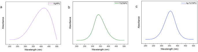
The UV-Vis spectrophotometric analysis was conducted as a preliminary investigation into the synthesis of nanoparticles. A notable color change was observed in the mixture of the plant extract and the metal salt solution. The results from UV-visible spectroscopy indicated the presence of absorbance peaks for the phyto-fabricated nanoparticles within the wavelength range of 200–500 nm, as depicted in Fig. 1. Specifically, the measurements revealed absorption peaks at 428 nm for AgNPs, 331 nm for FeONPs, and 359 nm for Ag-FeONPs.

UV analysis of the (a) AgNPs, (b) FeONPs, and (c) Ag-FeONPs fabricated from A. raddiana seed extract.
The FTIR spectra of the A. raddiana seed extract and NPs synthesized from it showed absorption peaks at similar positions, indicating the presence of various functional groups. Notably, the spectra displayed significant absorption peaks at 3338 cm⁻¹ and 1636 cm⁻¹, as illustrated in Fig. 2.

The FTIR spectra for the seed extract of A. raddiana and the phyto-fabricated nanoparticles.
The mean size of AgNPs is 248 nm, with a polydispersity index (PDI) of 0.22, as shown in Fig. 3, and their zeta potential, which is indicated at -45.15 mV.

The size of distribution for the AgNPs fabricated by A. raddiana seed extract.
In Fig. 4, the mean size of FeONPs is presented, with an average size of 213 nm and a PDI of 0.10, and their zeta potential value is shown at 0.8663 mV.

The size of distribution for the FeONPs fabricated by A. raddiana seed extract.
For the Ag-FeONPs, Fig. 5 indicates an average size of 208.8 nm, with a PDI of 0.22, and their zeta potential, which is measured at -32.85 mV.

The size of distribution for the Ag-FeONPs fabricated by A. raddiana seed extract.
Additionally, SEM analysis was performed to assess the surface morphology and dispersion of the nanoparticles. The SEM images showed that the AgNPs have irregular spherical shapes with a rough surface and exhibit significant aggregation, as illustrated in Fig. 6a. EDX analysis indicated that the silver element displays its highest peaks at 0.6 keV, 3 keV, and 3.4 keV, as shown in Fig. 6b. Furthermore, the results of the elemental mapping indicate the presence of silver, carbon (C), and oxygen (O). Among these elements, silver is the most abundantly distributed, making up 40.6% of the total composition, while carbon accounts for 37.7% and oxygen comprises 21.6%, as presented in Fig. 6c.

(A) SEM image; (B) EDX pattern with elemental composition; (C) element mapping of AgNPs.
Figure 7a illustrates the surface structure of FeONPs, which exhibits a spherical and irregular shape with a smooth surface, showing signs of agglomeration. The EDX analysis revealed peaks for the iron (Fe) element at 0.8 keV, 6.4 keV, and 7 keV, as shown in Fig. 7b. Additionally, Fig. 7c displays the element mapping analysis of the elements, indicating the presence of Fe, C, and O.

(A) SEM image; (B) EDX pattern with elemental composition; (C) element mapping of FeONPs.
The SEM image of the bimetallic Ag-FeONPs, presented in Fig. 8a, show that they are spherical in shape with irregular surfaces and a rough texture, along with a tendency to agglomerate. Figure 8b indicates that the EDX analysis detected peaks at 0.8 keV and 3 keV, providing strong evidence for the presence of Ag, while peaks at 0.8 keV, 6.5 keV, and 7 keV confirm the presence of Fe. The results from elemental mapping reveal that the Ag-FeONPs consist of 2.9% Fe, 85% Ag, 3% C, and 8% O, as shown in Fig. 8c.

(A) SEM image; (B) EDX pattern with elemental composition; (C) element mapping of Ag-FeONPs.
Transmission Electron Microscopy (TEM) was employed to analyze the morphology and size of both mono and bimetallic nanoparticles. In alignment with the SEM results, the AgNPs display an irregular spherical shape with an average size of 28.09 nm, as depicted in Fig. 9a. Similarly, the FeONPs also present an irregular spherical shape and exhibit some agglomeration, with an average size of 73.154 nm, as illustrated in Fig. 9b. The Ag-FeONPs are spherical in shape, with an average size of 21.36 nm, as shown in Fig. 9c.

The TEM images of (A) AgNPs, (B) FeONPs, and (C) Ag-FeONPs.
Identification of bioactive compounds in A. raddiana seed extract by GC-MS
The GC-MS analysis of the methanol extract from A. raddiana seeds identified a total of 507 bioactive compounds belonging to various chemical groups, including fatty acids, esters, phenolic compounds, and alkanes. The detected compounds include Phenol, 4-ethenyl-2,6-dimethoxy, Hexadecanoic acid, methyl ester, n-Hexadecanoic acid ,9,12-Octadecadienoic acid (Z, Z)-, methyl ester, 9-Octadecenoic acid, methyl ester, (E), cis-Vaccenic acid, Bis(2-ethylhexyl) phthalate, and 9,12-Octadecadienoic acid (Z, Z)-, 2-hydroxy-1 (hydroxymethyl)ethyl ester. These findings, summarized in Table 1, shed light on the chemical profile of the seed extract, while the GC-MS spectrum of A. raddiana is presented in Fig. 10.

Total ion chromatogram and GC-MS spectrum of A. raddiana compounds.
Antifungal activity of fabricated NPs
The antifungal activity of Ag, FeO, and FeO-Ag nanoparticles synthesized from A. raddiana seed extract was successfully evaluated against the phytopathogenic fungi Alternaria sp., S. sclerotiorum, F. equiseti, and F. venenatum, as illustrated in Fig. 11. The analysis revealed significant differences among the fungal strains, treatment effects, and interactions, with a P-value ≤ 0.0001. AgNPs exhibited a strong inhibitory effect on various fungal strains, achieving complete suppression of Alternaria sp. (100% inhibition), a 65% reduction in the growth of S. sclerotiorum, a 20% inhibition of F. venenatum, and a 32% decrease in the growth of F. equiseti. In addition, FeONPs showed effectiveness by inhibiting Alternaria sp. by 49%, S. sclerotiorum by 36%, F. venenatum by 40%, and F. equiseti by 13%. Furthermore, the combination of these nanoparticles resulted in suppression rates of 100% for Alternaria sp., 65% for S. sclerotiorum, 40% for F. venenatum, and 32% for F. equiseti.
According to Tukey’s post hoc comparison, groups labeled with different letters were significantly different, whereas those sharing the same letter did not differ statistically. In the control group, F.venenatum and F. equiseti showed comparable and relatively lower growth, while S.clerotiorum and Alternaria exhibited the highest growth. Under AgNPs and Ag–FeONPs treatments, S. sclerotiorum, F. venenatum, and F. equiseti showed statistically similar growth (a), indicating lower sensitivity to these treatments, whereas Alternaria was completely inhibited, reflecting strong antifungal activity. In the FeONPs-treated group, S. sclerotiorum maintained the highest growth (a), while F. venenatum displayed the lowest growth (d), suggesting that FeONPs were more effective against F. venenatum. Overall, AgNPs and Ag–FeONPs showed the strongest inhibitory effects, completely suppressing Alternaria.

Antifungal activity of synthesized A. raddiana nanoparticles against tomato pathogens. Different letters above the bars indicate statistically significant differences (Tukey’s HSD, p < 0.05).
Discussion
This study utilized aqueous A. raddiana seed extract as a reducing and capping agent to synthesize silver, iron oxide, and their combined nanoparticles. The resulting nanoparticles exhibited surface plasmon resonance (SPR) bands in the range of 331–450 nm, as detected through UV spectroscopy.
This spectral range indicates that the nanoparticles are well-dispersed in solution and reflects their unique characteristics55. Previous research has demonstrated that the shell extract of Adansonia digitata was employed to fabricate AgNPs, FeONPs, and Ag-FeONPs, which exhibited SPR bands at 418 nm, 338 nm, and 352 nm, respectively43. These findings align closely with the results obtained in the present study.
FTIR spectroscopy was employed to identify the biomolecules in the seed extract that are responsible for reducing and stabilizing the nanoparticles56. Both the extracts and nanoparticles exhibited similar prominent peaks at 3338 cm⁻¹ and 1636 cm⁻¹. These peaks are primarily linked to functional groups, with the peak at 3338 cm⁻¹ corresponding to O-H (hydroxyl) stretching attributed to secondary amines57, and the peak at 1636 cm⁻¹ associated with the N-H bond of primary amines58. In comparison, AgNPs synthesized from green tea leaf extract displayed absorption peaks at 3689.8 and 1631 cm⁻¹59, which are similar to those found in our AgNPs study. In a recent study conducted by Ramzan et al.60, it was shown that AgNPs synthesized by Zingiber officinale extract exhibited a peak at 3320 cm⁻¹. Furthermore, a study by Aldahasi et al.43 reported that the peaks of FeONPs derived from Adansonia digitata fruit shell extract at 1639 cm⁻¹ and 3327 cm⁻¹ closely resemble the peaks associated with FeONPs and Ag-FeONPs in our findings. The DLS technique was employed to measure the hydrodynamic sizes and polydispersity index (PDI) of plant-based nanoparticles, confirming their presence in the nano-range. A study by Purbowati et al.61 reported an average size of 121.22 nm for AgNPs. Similarly62, indicated that FeONPs had an average size of 194.5 nm. Furthermore, Velidandi et al.63 noted a mean size distribution of 171.4 nm for Ag-FeONPs, which aligns with our findings. Our results demonstrated that the nanoparticles fabricated in this study showed relatively uniform size distribution, as the PDI values were below 0.564. Additionally, the observation of a negative zeta potential suggests that the nanoparticles are stable and play a crucial role in preventing particle agglomeration65,66.These negative values may be attributed to the phytochemicals involved, highlighting their importance in the capping and stabilization of the nanoparticles43. Previous studies have indicated that the synthesized nanoparticles exhibit a negative charge67,68.
In the present investigation, SEM analysis revealed that the synthesized AgNPs exhibited a spherical morphology, regardless of the plant species used. This observation aligns with previous studies that reported similar characteristics for AgNPs synthesized from Rubus ellipticus69. A recent study by Manimaran et al.70 on the synthesis of FeO NPs using the leaf extract of Pleurotus citrinopileatus confirmed the formation of spherical nanoparticles, and another study reported spherical FeO NPs synthesized using Pterolobium hexapetalum leaf extract71, which is consistent with our findings. Furthermore, an irregular spherical shape was noted in the silver-iron nanoparticles synthesized from Salvia officinalis72.
TEM analysis was employed to accurately assess the size, shape, and distribution of the nanoparticles. This analysis characterized AgNPs, FeONPs, and Ag-FeONPs derived from Citrus reticulata Blanco peel extract, Chromolaena odorata extract, and Salvia officinalis, respectively. The results of these analyses confirmed the spherical shape of the nanoparticles72,73,74. Moreover, the AgNPs exhibited sizes ranging from 10 to 50 nm75.
Results from the GC-MS analysis indicated that A. raddiana contains several compounds with potential antifungal activity against plant pathogens. Notably, hexadecanoic acid methyl ester and n-hexadecanoic acid are recognized for their antimicrobial properties, particularly in inhibiting the growth of fungal pathogens by disrupting cell membrane integrity. For example, hexadecanoic acid has demonstrated antifungal activity against Fusarium oxysporum76. Additionally, the compounds 9,12-octadecadienoic acid (Z, Z)-methyl ester and 9-octadecenoic acid (E)-methyl ester, both derivatives of unsaturated fatty acids, have been associated with antifungal effects, showing inhibitory activity against Alternaria alternata77. Furthermore, cis-vaccenic acid, a monounsaturated fatty acid, has exhibited antifungal properties against various pathogens78. A study by Khamis et al.79 identified cis-vaccenic acid in the methanolic extract of Indian hawthorn leaves, which displayed significant antifungal activity against Fusarium verticillioides. Dimethoxy, a phenolic compound, is also noted for its antimicrobial properties that inhibit fungal growth, demonstrating activity against Colletotrichum gloeosporioides and C. lindemuthianum80.
Furthermore, the present study explored the antifungal activity of the biosynthesized AgNPs, FeONPs, and Ag-FeONPs against the phytopathogenic fungi Alternaria sp., S. sclerotiorum, F. venenatum, and F. equiseti. The antifungal effectiveness of AgNPs was evaluated against multiple fungal species, consistent with findings from Khan et al.81, Aldahasi et al.43, and Mostafa et al.82, who also reported the effectiveness of AgNPs against fungi such as Fusarium oxysporum, Alternaria sp., Sclerotinia sclerotiorum, Fusarium venenatum, and Alternaria solani. Likewise, the notable antifungal potential of FeONPs supports the work of46, emphasizing their efficacy against Alternaria alternata. Additionally, Ag-FeONPs exhibited broad-spectrum activity against all tested fungal strains, indicating that bimetallic nanoparticles possess significantly enhanced antifungal properties compared to their monometallic counterparts83. According to Aldahasi et al.43, bimetallic nanoparticles, particularly Ag-FeONPs, demonstrate superior efficacy against Alternaria sp., Fusarium equiseti, and Fusarium venenatum. Different mechanisms by which NPs can combat fungal cells in different ways were responsible for the antifungal effect. They first enter the cell by adhering to the fungal cell wall. Once inside, they can interfere with vital cell components, such as DNA, proteins, and fats, impairing the cell’s ability to function normally. This stress can prevent the fungi from growing and spreading, potentially causing DNA damage and even cell death. These actions enable nanoparticles to exhibit potent antifungal properties against a range of fungal species84,85,86.
Conclusion
This study successfully demonstrated the potential of Acacia tortilis subsp. raddiana seed extract for the eco-friendly green synthesis of AgNPs, FeONPs, and Ag-FeONPs. The resulting nanoparticles exhibited well-defined structural and morphological properties, as confirmed by comprehensive characterization techniques, and showed notable antifungal activity against key tomato pathogens. Specifically, AgNPs and Ag-FeONPs completely inhibited Alternaria sp. (100%) and significantly suppressed Sclerotinia sclerotiorum, Fusarium venenatum, and F. equiseti, highlighting their superior efficacy compared to FeONPs. GC-MS analysis of the seed extract revealed multiple bioactive compounds likely contributing to the synthesis and antifungal performance of the nanoparticles. In conclusion, these findings underline the promise of biogenic AgNPs and bimetallic Ag-FeONPs as sustainable and environmentally friendly alternatives to conventional fungicides for controlling tomato fungal diseases. Future studies should focus on optimizing their synthesis for large-scale agricultural applications and evaluating their long-term environmental safety and effectiveness under field conditions.
Data availability
The datasets used and/or analyzed during the current study are available from the corresponding author on reasonable request.
References
-
Ansari, M. et al. Green synthesized silver nanoparticles: A novel approach for the enhanced growth and yield of tomato against early blight disease. Microorganisms 11 (2023).
-
Sato, S. et al. The tomato genome sequence provides insights into fleshy fruit evolution. Nature 485 ,7400,635(2012).
-
Sunera et al. Evaluating the efficacy of endophytic bacteria in controlling rice sheath blight: in vitro and in vivo studies. Microb. Pathog. 197, 107084 (2024).
-
Sunera et al. Characterization and phytostimulatory activity of bacteria isolated from tomato (Lycopersicon esculentum Mill.) rhizosphere. Microb Pathog 140,103966 (2020).
-
Naeem, M. et al. The solanum melongena COP1LIKE manipulates fruit ripening and flowering time in tomato (Solanum lycopersicum). Plant Growth Regul 96 (2022).
-
Pathak, P. D., Mandavgane, S. A. & Kulkarni, B. D. Tomato utilization: Techno-economic and social aspects 15. In Biorefinery: A Sustainable Approach for the Production of Biomaterials, Biochemicals and Biofuels (2023). https://doi.org/10.1007/978-981-19-7481-6_15
-
Guo, L. et al. Biochar improves soil-tomato plant, tomato production, and economic benefits under reduced nitrogen application in Northwestern China. Plants 10 (2021).
-
Ali, M. Y. et al. Nutritional composition and bioactive compounds in tomatoes and their impact on human health and disease: A review. Foods 10 (2021).
-
Samaras, A., Roumeliotis, E., Ntasiou, P. & Karaoglanidis, G. Bacillus subtilis mbi600 promotes growth of tomato plants and induces systemic resistance contributing to the control of soilborne pathogens. Plants 10 (2021).
-
Singh, V. K., Singh, A. K. & Kumar, A. Disease management of tomato through PGPB: Current trends and future perspective. 3 Biotech 7 Preprint (2017). https://doi.org/10.1007/s13205-017-0896-1
-
Campos, M. D. et al. Defense strategies: The role of transcription factors in tomato–pathogen interaction. Biology 11 Preprint (2022). https://doi.org/10.3390/biology11020235
-
Nazarov, P. A., Baleev, D. N., Ivanova, M. I., Sokolova, L. M. & Karakozova, M. V. Infectious plant diseases: Etiology, current status, problems and prospects in plant protection. Acta Nat. 12, 46–59. Preprint (2020). https://doi.org/10.32607/actanaturae.11026
-
Abdelaziz, A. M., Kalaba, M. H., Hashem, A. H., Sharaf, M. H. & Attia, M. S. Biostimulation of tomato growth and biocontrol of fusarium wilt disease using certain endophytic fungi. Bot. Stud. 63 (2022).
-
Pandey, S. et al. Early blight disease management by herbal nanoemulsion in Solanum lycopersicum with bio-protective manner. Ind. Crops Prod. 150 (2020).
-
Mazumdar, P. Sclerotinia stem rot in tomato: A review on biology, pathogenicity, disease management and future research priorities. J. Plant Dis. Protect. 128, 1403–1431. Preprint (2021). https://doi.org/10.1007/s41348-021-00509-z
-
Salazar, B. et al. Bacillus spp. as bio-factories for antifungal secondary metabolites: Innovation beyond whole organism formulations. Microb. Ecol. Preprint (2022). https://doi.org/10.1007/s00248-022-02044-2
-
Zubrod, J. P. et al. Fungicides: An overlooked pesticide class? Environ Sci. Technol 53 (2019).
-
Singh, S. et al. Toxicity, monitoring and biodegradation of the fungicide carbendazim. Environ. Chem. Lett. 14. Preprint (2016). https://doi.org/10.1007/s10311-016-0566-2
-
Bianchi, S. et al. Association between female reproductive health and mancozeb: systematic review of experimental models. Int. J. Environ. Res. Public Health. 17. Preprint. https://doi.org/10.3390/ijerph17072580 (2020).
-
Anand, G. & Rajeshkumar, K. C. Challenges and threats posed by plant pathogenic fungi on agricultural productivity and economy. (2022). https://doi.org/10.1007/978-981-16-8877-5_23
-
Fu, L., Wang, Z., Dhankher, O. P. & Xing, B. Nanotechnology as a new sustainable approach for controlling crop diseases and increasing agricultural production. J. Exp. Bot. 71. Preprint (2020). https://doi.org/10.1093/jxb/erz314
-
Kutawa, A. B. et al. Trends in nanotechnology and its potentialities to control plant pathogenic fungi: A review. Biology 10 Preprint (2021). https://doi.org/10.3390/biology10090881
-
Mohajerani, A. et al. Nanoparticles in construction materials and other applications, and implications of nanoparticle use. Materials 12 Preprint (2019). https://doi.org/10.3390/ma12193052
-
Gavas, S., Quazi, S. & Karpiński, T. M. Nanoparticles for cancer therapy: Current progress and challenges. Nanoscale Res. Lett. 16 Preprint (2021). https://doi.org/10.1186/s11671-021-03628-6
-
Saqib, S. et al. Organometallic assembling of chitosan-iron oxide nanoparticles with their antifungal evaluation against rhizopus oryzae. Appl. Organomet. Chem. 33 (2019).
-
Singh, A. et al. Green synthesis of metallic nanoparticles as effective alternatives to treat antibiotics resistant bacterial infections: A review. Biotechnol. Rep. 25 (2020).
-
Khan, F. et al. Green nanotechnology: Plant-mediated nanoparticle synthesis and application. Nanomaterials 12. Preprint (2022). https://doi.org/10.3390/nano12040673
-
Ijaz, I., Gilani, E., Nazir, A. & Bukhari, A. Detail review on chemical, physical and green synthesis, classification, characterizations and applications of nanoparticles. Green Chem. Lett. Rev. 13. Preprint (2020). https://doi.org/10.1080/17518253.2020.1802517
-
Pal, G., Rai, P. & Pandey, A. Green synthesis of nanoparticles: A greener approach for a cleaner future. In Green Synthesis, Characterization and Applications of Nanoparticles (2018). https://doi.org/10.1016/B978-0-08-102579-6.00001-0
-
Mustapha, T., Misni, N., Ithnin, N. R., Daskum, A. M. & Unyah, N. Z. A Review on plants and microorganisms mediated synthesis of silver nanoparticles, role of plants metabolites and applications. Int. J. Environ. Res. Public Health 19 Preprint (2022). https://doi.org/10.3390/ijerph19020674
-
Manimegalai, G. et al. Acorus calamus-mediated CuFe2O4/reduced graphene oxide (AcL-CF-G) nanocomposite and its versatile biomedical and environmental remediation applications. J. Ind. Eng. Chem. 147, 278–292 (2025).
-
Prem, P. et al. Valeriana Jatamansi root extract a potent source for biosynthesis of silver nanoparticles and their biomedical applications, and photocatalytic decomposition. Green Chem. Lett. Rev 17 (2024).
-
Elangovan, M. et al. Sunlight-driven photocatalytic and anticancer properties of biogenic synthesized gold nanoparticles (AuNPs) employing Polygala elongata. J. King Saud Univ. Sci. 36 (2024).
-
Villagrán, Z. et al. Plant-based extracts as reducing, capping, and stabilizing agents for the green synthesis of inorganic nanoparticles. Resources 13, 70 (2024).
-
Irshad, M. A. et al. Exploring the antifungal activities of green nanoparticles for sustainable agriculture: a research update. Chem. Biol. Technol. Agric. 11, 133 (2024).
-
Saqib, S. et al. Postharvest disease Inhibition in fruit by synthesis and characterization of chitosan iron oxide nanoparticles. Biocatal. Agric. Biotechnol. 28 (2020).
-
Hnini, M., Taha, K. & Aurag, J. Botany, associated microbiota, traditional medicinal uses, and phytochemistry of Vachellia tortilis subsp. raddiana (Savi): A systematic review. J Agric. Food Res 12 (2023).
-
Elshamy, S., Handoussa, H., El-Shazly, M., Mohammed, E. D. & Kuhnert, N. Metabolomic profiling and quantification of polyphenols from leaves of seven Acacia species by UHPLC-QTOF-ESI-MS. Fitoterapia 172 (2024).
-
Ziani, B. E. C. et al. Phenolic profiling, biological activities and in silico studies of Acacia tortilis (Forssk.) Hayne ssp. raddiana extracts. Food Biosci. 36 (2020).
-
Singh, R., Choudhary, A. & Ram, R. Pharmacological assessment of the heartwood of Acacia raddiana Willd for antifungal potential. In Materials Today: Proceedings. Vol. 62 (2022).
-
Ansari, M. et al. Evaluation of in vitro and in vivo antifungal activity of green synthesized silver nanoparticles against early blight in tomato. Horticulturae 9 (2023).
-
Hernández-Díaz, J. A. et al. Plant-mediated synthesis of nanoparticles and their antimicrobial activity against phytopathogens. J. Sci. Food Agric. 101 Preprint (2021). https://doi.org/10.1002/jsfa.10767
-
Aldahasi, R. M., Shami, A. & Mohammed, A. E. Bimetallic nanoparticles and biochar produced by Adansonia digitata shell and their effect against tomato pathogenic fungi. PeerJ 12 (2024).
-
Ashraf, H. et al. Antifungal potential of green synthesized magnetite nanoparticles black coffee–magnetite nanoparticles against wilt infection by ameliorating enzymatic activity and gene expression in Solanum lycopersicum L. Front. Microbiol. 13 (2022).
-
Ashraf, H. et al. Sustainable synthesis of microwave-assisted ionps using Spinacia oleracea L. for control of fungal wilt by modulating the defense system in tomato plants. J Nanobiotechnol. 20 (2022).
-
Yassin, M. T., Al-Otibi, F. O., Al-Askar, A. A. & Alharbi, R. I. Green synthesis, characterization, and antifungal efficiency of biogenic iron oxide nanoparticles. Appl. Sci. (Switzerland) 13 (2023).
-
Loza, K., Heggen, M. & Epple, M. Synthesis structure, properties, and applications of bimetallic nanoparticles of noble metals. Adv. Funct. Mater. 30 Preprint (2020). https://doi.org/10.1002/adfm.201909260
-
Sharma, G. et al. Novel development of nanoparticles to bimetallic nanoparticles and their composites: A review. J. King Saud Univ. Sci. 31 Preprint (2019). https://doi.org/10.1016/j.jksus.2017.06.012
-
Sumbal et al. Synthesis, characterization and biological activities of monometallic and bimetallic nanoparticles using Mirabilis jalapa leaf extract. Biotechnol. Rep. 22 (2019).
-
Padilla-Cruz, A. L. et al. Synthesis and design of Ag–Fe bimetallic nanoparticles as antimicrobial synergistic combination therapies against clinically relevant pathogens. Sci .Rep. 11 (2021).
-
Thirumoorthy, G. et al. Phytofabricated bimetallic synthesis of silver-copper nanoparticles using Aerva lanata extract to evaluate their potential cytotoxic and antimicrobial activities. Sci. Rep. 14 (2024).
-
Hashem, A. H. et al. Sustainable biosynthesized bimetallic ZnO@SeO nanoparticles from pomegranate peel extracts: antibacterial, antifungal and anticancer activities. RSC Adv. 13, 22918–22927 (2023).
-
Nizamani, S. et al. Isolation and identification of the fungi causing tomato fruit rot disease in the vicinity of tandojam, Sindh. Agric. Sci. Digest. 41 (2021).
-
Khatami, M., Sharifi, I., Nobre, M. A. L., Zafarnia, N. & Aflatoonian, M. R. Waste-grass-mediated green synthesis of silver nanoparticles and evaluation of their anticancer, antifungal and antibacterial activity. Green. Chem. Lett. Rev. 11, 125–134 (2018).
-
Yamada, M. et al. Synthesis of silver nanoparticles by atmospheric-pressure pulsed discharge plasma in a slug flow system. Jpn. J. Appl. Phys. 58 (2019).
-
Marslin, G. et al. Secondary metabolites in the green synthesis of metallic nanoparticles. Materials 11 Preprint (2018). https://doi.org/10.3390/ma11060940
-
Javed, R. et al. Phytochemical-mediated biosynthesis of silver nanoparticles from strobilanthes glutinosus: exploring biological applications. Micromachines (Basel) 14 (2023).
-
Muthukumar, H., Palanirajan, S. K., Shanmugam, M. K. & Gummadi, S. N. Plant extract mediated synthesis enhanced the functional properties of silver ferrite nanoparticles over chemical mediated synthesis. Biotechnol. Rep. 26 (2020).
-
Widatalla, H. A. et al. Green synthesis of silver nanoparticles using green tea leaf extract, characterization and evaluation of antimicrobial activity. Nanoscale Adv. 4 (2022).
-
Ramzan, M., Abusalah, M. A. H. A., Ahmed, N., Yean, C. Y. & Zeshan, B. Green synthesis and characterization of silver nanoparticles using Zingiber officinale extracts to investigate their antibacterial potential. Int. J. Nanomed. 19, 13319–13338 Preprint (2024). https://doi.org/10.2147/IJN.S475656
-
Purbowati, R. et al. Green synthesis of one-dimensional silver nanoparticles using Quercus infectoria gall extract. Case Stud. Chem. Environ. Eng. 9 (2024).
-
Umair, M. Green nanotechnology mediated silver and iron oxide nanoparticles: Potential antimicrobials. Agrobiol. Rec. 10, 35–41 (2022).
-
Velidandi, A., Sarvepalli, M., Gandam, P. K. & Baadhe, R. R. Silver/silver chloride and gold bimetallic nanoparticles: Green synthesis using Azadirachta indica aqueous leaf extract, characterization, antibacterial, catalytic, and recyclability studies. Inorg Chem. Commun 155 (2023).
-
Danaei, M. et al. Impact of particle size and polydispersity index on the clinical applications of lipidic nanocarrier systems. Pharmaceutics 10 Preprint (2018). https://doi.org/10.3390/pharmaceutics10020057
-
Kumavat, S. R. & Mishra, S. Green synthesis of silver nanoparticles using Borago officinalis leaves extract and screening its antimicrobial and antifungal activity. Int. Nano Lett. 11, 355–370 (2021).
-
Bhagat, M., Anand, R., Datt, R., Gupta, V. & Arya, S. Green synthesis of silver nanoparticles using aqueous extract of Rosa brunonii Lindl and their morphological, biological and photocatalytic characterizations. J. Inorg. Organomet. Polym. Mater. 29, 1039–1047 (2019).
-
Rajput, S., Kumar, D. & Agrawal, V. Green synthesis of silver nanoparticles using Indian Belladonna extract and their potential antioxidant, anti-inflammatory, anticancer and larvicidal activities. Plant. Cell. Rep. 39, 921–939 (2020).
-
Periakaruppan, R. et al. Utilization of tea resources with the production of superparamagnetic biogenic iron oxide nanoparticles and an assessment of their antioxidant activities. J Clean. Prod 278 (2021).
-
Khanal, L. N. et al. Green synthesis of silver nanoparticles from root extracts of Rubus ellipticus Sm. and comparison of antioxidant and antibacterial activity. J. Nanomater. (2022).
-
Manimaran, K. et al. Biological synthesis and characterization of iron oxide (FeO) nanoparticles using Pleurotus citrinopileatus extract and its biomedical applications. Biomass Convers. Biorefin. 14 (2024).
-
Loganathan, S., Govindasamy, M., Habila, M. A. & Manimaran, K. Green synthesis of iron oxide nanoparticles using Pterolobium hexapetalum (roth) Santapau & Wagh leaves extract and their biological applications. Biocatal Agric. Biotechnol 54 (2023).
-
Malik, M. A., Alshehri, A. A. & Patel, R. Facile one-pot green synthesis of Ag-Fe bimetallic nanoparticles and their catalytic capability for 4-nitrophenol reduction. J. Mater. Res. Technol. 12, 455–470 (2021).
-
Jaast, S. & Grewal, A. Green synthesis of silver nanoparticles, characterization and evaluation of their photocatalytic dye degradation activity. Curr. Res. Green Sustain. Chem. 4 (2021).
-
Nnadozie, E. C. & Ajibade, P. A. Green synthesis and characterization of magnetite (Fe3O4) nanoparticles using Chromolaena odorata root extract for smart nanocomposite. Mater Lett. 263 (2020).
-
Loganathan, S. et al. Synthesis of silver nanoparticles (AgNPs) using Pterolobium hexapetalum (Roth) Santapau & Wagh and its investigation of biological activities. Biomass Convers. Biorefin. https://doi.org/10.1007/s13399-023-04934-y (2023).
-
Idris, N., Johannes, E. & Dwyana, Z. Potential of hexadecanoic acid as antimicrobials in bacteria and fungi that cause decay in mustard greens Brassica juncea L. Int. J. Appl. Biol. 6 (2022).
-
Youssef, N. H. et al. Antimycotoxigenic activity of beetroot extracts against altenaria alternata mycotoxins on potato crop. Appl. Sci. (Switzerland) 11 (2021).
-
Guimarães, A. & Venâncio, A. The potential of fatty acids and their derivatives as antifungal agents: A review. Toxins 14 Preprint (2022). https://doi.org/10.3390/toxins14030188
-
Khamis, W. M. et al. Phytochemical analysis and insight into insecticidal and antifungal activities of Indian Hawthorn leaf extract. Sci. Rep. 13 (2023).
-
Martinez, J., Ramírez, C., Gil, J., Quiñones, W. & Durango, D. Antifungal activity against anthracnose-causing species of homopterocarpin derivatives. Heliyon 9 (2023).
-
Khan, M., Khan, A. U., Bogdanchikova, N. & Garibo, D. Antibacterial and antifungal studies of biosynthesized silver nanoparticles against plant parasitic nematode meloidogyne incognita, plant pathogens Ralstonia solanacearum and Fusarium oxysporum. Molecules 26 (2021).
-
Mostafa, Y. S., Alamri, S. A., Alrumman, S. A., Hashem, M. & Baka, Z. A. Green synthesis of silver nanoparticles using pomegranate and orange peel extracts and their antifungal activity against alternaria solani, the causal agent of early blight disease of tomato. Plants 10 (2021).
-
Gaber, S. E., Hashem, A. H., El-Sayyad, G. S. & Attia, M. S. Antifungal activity of myco-synthesized bimetallic ZnO-CuO nanoparticles against fungal plant pathogen Fusarium oxysporum. Biomass Convers. Biorefin. https://doi.org/10.1007/s13399-023-04550-w (2023).
-
Li, L. et al. The antifungal activity and mechanism of silver nanoparticles against four pathogens causing kiwifruit post-harvest rot. Front. Microbiol. 13 (2022).
-
Gurunathan, S., Lee, A. R. & Kim, J. H. Antifungal effect of nanoparticles against COVID-19 linked black fungus: A perspective on biomedical applications. Int. J. Mol. Sci. 23 Preprint (2022). https://doi.org/10.3390/ijms232012526
-
Rana, A. et al. Recent advancements in plant- and microbe-mediated synthesis of metal and metal oxide nanomaterials and their emerging antimicrobial applications. ACS Appl. Nano Mater. 6, 8106–8134 (2023).
Acknowledgements
Princess Nourah bint Abdulrahman University Researchers Supporting Project number (PNURSP2025R740), Princess Nourah bint Abdulrahman University, Riyadh, Saudi Arabia.
Ethics declarations
Competing interests
The authors declare no competing interests.
Additional information
Publisher’s note
Springer Nature remains neutral with regard to jurisdictional claims in published maps and institutional affiliations.
Rights and permissions
Open Access This article is licensed under a Creative Commons Attribution-NonCommercial-NoDerivatives 4.0 International License, which permits any non-commercial use, sharing, distribution and reproduction in any medium or format, as long as you give appropriate credit to the original author(s) and the source, provide a link to the Creative Commons licence, and indicate if you modified the licensed material. You do not have permission under this licence to share adapted material derived from this article or parts of it. The images or other third party material in this article are included in the article’s Creative Commons licence, unless indicated otherwise in a credit line to the material. If material is not included in the article’s Creative Commons licence and your intended use is not permitted by statutory regulation or exceeds the permitted use, you will need to obtain permission directly from the copyright holder. To view a copy of this licence, visit http://creativecommons.org/licenses/by-nc-nd/4.0/.
About this article

Cite this article
Aldahasi, R.M., Mohammed, A.E. Bimetallic nanocomposite produced from Acacia tortilis subsp. raddiana for the mitigation fungal pathogens in tomatoes. Sci Rep 15, 41592 (2025). https://doi.org/10.1038/s41598-025-25419-0
-
Received:
-
Accepted:
-
Published:
-
Version of record:
-
DOI: https://doi.org/10.1038/s41598-025-25419-0