Introduction
In the human retina, long (L-, red) and medium (M-, green) wavelength-sensitive cones comprise ~95% of the total cone population and are concentrated in the fovea, a small central region in the retina responsible for high acuity vision in bright light conditions1,2,3. Their corresponding photo-sensitive proteins, L- and M-opsin, are encoded in the OPN1LW/OPN1MW gene locus on the X-chromosome4. Cone opsins are crucial for initiating the phototransduction cascade and for maintaining the structural integrity of cone outer segments (COS) where phototransduction occurs. Blue cone monochromacy (BCM) is an X-linked blinding disorder associated with mutations within the OPN1LW/OPN1MW gene locus and characterized by severely reduced or complete loss of function in red and green cones5. This disorder results in severely reduced visual acuity ranging from 20/60 to 20/200 and impaired color vision. BCM patients also commonly exhibit infantile nystagmus, photophobia, and myopia6,7.
One common class of mutation identified in BCM patients involves large deletions spanning the locus control region (LCR) of the OPN1LW/OPN1MW gene locus and/or part of both genes, thereby preventing L/M-opsin expression. Previous studies demonstrate that BCM patients with deletion mutations exhibit significantly shortened COS as early as 5 years of age. However, mutant cones within these retinas appear to degenerate gradually throughout patients’ lives, evidenced by progressive foveal thinning tracked by optical coherence tomography (OCT) and Adaptive Optics Scanning Laser Ophthalmoscopy (AOSLO) longitudinally6. A second class of BCM mutations involves missense mutations in both OPN1LW and OPN1MW genes, resulting in the absence or translation of nonfunctional opsin. The most common point mutation, predominantly identified within populations of British origin, is a cysteine to arginine substitution at residue 203 (C203R) in both OPN1LW and OPN1MW8,9,10. Similar to deletion mutants, thinning of the fovea and shortening of COS are seen across the retinas of patients with C203R mutations (ages 5–70), but degeneration is less severe in early life compared to patients with deletion mutations10. Despite C203R patients exhibiting significantly shortened COS, these studies show that they also retain intact cone inner segments (CIS) in the foveal region with gradual degeneration over time, serving as potential targets for restoring normal cone opsin expression and cone function via gene therapy.
Previously, our lab generated mouse models that resemble BCM deletion (Opn1mw-/-/Opn1sw-/-, double knockout or DKO) or C203R (Opn1mwC198R/Opn1sw-/-, C198R) genotypes. We performed AAV-mediated gene supplementation of human OPN1LW cDNA driven by a cone-specific PR2.1 promoter via subretinal injection. In both models, we observed robust rescue of cone function and regeneration of cone outer segment morphology when mice were treated at less than 3 months of age. However, therapy efficacy was significantly reduced when DKO and C198R mice were treated at 5 months or older11,12.
Previous studies have shown that success rates of AAV gene therapy for other diseases primarily affecting cones also exhibit age dependence. Restoration of cone-mediated vision using AAV-based gene supplementation therapy has been reported in mouse, dog, sheep, and rhesus macaque models of CNGA3-, CNGB3-, or PDE6C-related achromatopsia. These studies demonstrated long-term restoration of cone-mediated vision13,14,15,16. However, decreases in therapy efficacy have been observed in aged mice, dogs, and rhesus macaques, underscoring the challenges of treating degenerating cones13,14,16. The potential causes have since been investigated, such as promoter silencing and histone modifications of the AAV transgene17,18,19. Additionally, the decreased rescue efficacy in aged photoreceptors may likely be attributed to accumulated structural and molecular changes as a result of the mutation, leading to eventual photoreceptor death20,21.
In this study, we investigate the long-term efficacy of AAV gene therapy in DKO and C198R mice side-by-side and explore molecular mechanisms limiting the therapeutic window in aged mutant cones. We demonstrate that the therapeutic longevity and treatment window are comparable between DKO and C198R mice. We detect severe morphological abnormalities in aging mutant cones and observe that these cones exhibit significantly reduced transgenic mRNA expression compared to younger cones. Additionally, we identify candidate cone-specific promoters that may enhance transgene expression and improve therapeutic outcomes in older mutant mice. Our findings offer insight into future studies that aim to expand the therapeutic window in translational BCM gene therapy research.
Results
AAV8Y733F-mediated OPN1LW expression improves rescue in C198R mice compared to AAV5
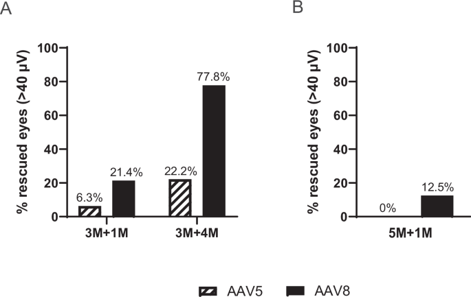
We have previously shown that DKO and C198R mice lack cone-mediated function, and that AAV5-mediated expression of OPN1LW driven by the cone-specific PR2.1 promoter successfully rescues cone function and COS structure11,12. To determine whether the AAV8Y733F capsid, known for facilitating high photoreceptor transduction22, could mediate better rescue compared to AAV5, we treated C198R mice at 3 and 5 months of age with either AAV8Y733F or AAV5 expressing OPN1LW cDNA under the PR2.1 promoter. Electroretinograms (ERGs) were performed at 1 and 4 months post-injection in 3-month-old injected eyes (3M + 1M and 3M + 4M, respectively), and at 1 month post-injection in 5-month-old injected eyes (5M + 1M).
AAV8Y733F-treated C198R eyes demonstrated significantly higher functional rescue compared to AAV5-treated eyes. Here, we show that 21.4% of AAV8-treated eyes had ERG amplitudes greater than 40 µV in 3M + 1M C198R animals, while only 6.3% of AAV5-treated eyes reached this level. In the 3M + 4M group, 77.8% of AAV8-treated eyes showed ERG amplitudes above 40 µV, compared to 22.2% of AAV5-treated eyes (Fig. 1A). In 5M + 1M C198R animals, 12.5% of AAV8-treated eyes showed ERG amplitudes higher than 40 µV, while zero AAV5-treated eyes reached this level (Fig. 1B).

A C198R mice were treated at 3 months of age and assessed by ERG at 1- (3M + 1M) and 4-months (3M + 4M) post-injection. In 3M + 1M, n = 16 eyes (7 male, 9 female) for AAV5, and n = 14 eyes (8 male, 6 female) for AAV8Y733F. In 3M + 4M, n = 9 eyes (5 male, 4 female) for AAV5, and n = 9 eyes (4 male, 5 female) for AAV8Y733F. B C198R mice were treated at 5 months of age and assessed by ERG at 1 month (5M + 1M) post-injection. n = 20 eyes (9 male, 11 female) for AAV5, n = 16 eyes (7 male, 9 female) for AAV8Y733F. Rescued eyes are defined as having b-wave maximum amplitudes higher than 40 µV.
DKO and C198R mice exhibit comparable therapy rescue and therapeutic windows following AAV8Y733F-mediated gene therapy
Next, we conducted a side-by-side study to directly compare the therapeutic efficacy, treatment window, and rescue longevity in DKO vs. C198R mice using the high-transduction AAV8Y733F vector. Age-matched DKO and C198R mice were injected subretinally at 3, 5, and 7 months of age. Functional rescue was assessed by ERG at 1 month post-injection, with follow-up evaluations at 3-month intervals.
In both DKO and C198R mice treated at 3 months (Fig. 2A; Supplementary Figs. S1, S2), we observed robust ERG rescue at 1 month post-injection (3M + 1M). The average b-wave maximum amplitude at the highest tested light intensity of 1.4 log cd·s/m2 was 23 ± 17 μV for DKO, and 25 ± 16 μV for C198R. Functional rescue peaked at 4 months post-injection (3M + 4M) with b-wave amplitudes of 55 ± 32 μV in DKO mice, and 47 ± 23 μV in C198R. At 7 and 10 months post-injection (3M + 7M, 3M + 10M), functional rescue persisted in both models, albeit with a slight decline compared to the 4-month time point. When ERG was assessed at 7 months post-treatment (3M + 7M), average b-wave amplitudes were 23 ± 13 μV for DKO, and 42 ± 20 μV for C198R. At 10 months post-injection (3M + 10M), average b-wave amplitudes were 13 ± 9 μV for DKO, and 35 ± 13 μV for C198R. Rescue efficiency appeared to be better maintained in C198R eyes compared to DKO eyes, though the difference was not statistically significant. The maximum amplitudes of ERG b-waves in treated DKO and C198R cones were significantly higher than those in untreated controls but remained significantly lower compared to wildtype (WT) controls (Fig. 2A).

A Mice were treated at 3 months old, and their photopic visual responses were measured at 1 (3M + 1M), 4 (3M + 4M), and 7 (3M + 7M) months post-injection. For 3M + 1M, n = 24 eyes (10 male, 14 female) for DKO (pink bar), and n = 15 eyes (6 male, 9 female) for C198R (blue bar). In 3M + 4M mice, n = 11 eyes (7 male, 4 female) for DKO, and n = 10 eyes (4 male, 6 female) for C198R. In 3M + 7M mice, n = 10 eyes (6 male, 4 female) for DKO, and n = 10 eyes (4 male, 6 female) for C198R. In 3M + 10M mice, n = 5 eyes (2 male, 3 female) for DKO, and n = 7 eyes (3 male, 4 female) for C198R. n = 8 eyes (4 male, 4 female) for WT (10-month-old), DKO untreated (1-month-old), and C198R untreated (one-month-old). All treated groups are significantly lower than WT (*P < 0.05) but significantly higher than untreated controls (*P < 0.05). Data shown are average ± SD, 2-way ANOVA. B In 5M + 1M mice, n = 21 eyes (10 male, 11 female) for DKO, and n = 15 eyes (8 male, 7 female) for C198R. In 5M + 4M mice, n = 5 eyes (2 male, 3 female) for DKO and n = 5 eyes (2 male, 3 female) for C198R. Data shown are average ± SD. No comparisons between DKO and C198R at matched timepoints achieved statistical significance, 1-way ANOVA. C In 7M + 1M mice, n = 13 eyes (5 male, 8 female) for DKO and n = 11 eyes (6 male, 5 female) for C198R. Data shown are average ± SD. No comparisons achieved statistical significance, non-paired t-test.
In mice treated at 5 months of age, both DKO and C198R models showed similar levels of rescue at 1 and 4 months post-injection (5M + 1M, 5M + 4M; Fig. 2B, Supplementary Fig. S1B). In 5M + 1M mice, average b-wave amplitudes were 18 ± 9 μV and 23 ± 16 μV for DKO and C198R eyes, respectively. In 5M + 4M mice, average b-wave amplitudes were 19 ± 14 μV for DKO and 31 ± 13 μV for C198R eyes. Rescue appeared to be maintained slightly better in 5M + 4M C198R eyes, though the difference was not statistically significant.
Rescue efficiency of eyes treated at 7 months of age proved to be much lower than in those treated at 3 months, with b-wave amplitudes of 11 ± 6 μV and 14 ± 9 μV in DKO and C198R eyes, respectively (Fig. 2C, Supplementary Fig. S1). These results are consistent with our previous findings that gene therapy is less effective in older mice11,12
Next, we performed immunohistochemistry (IHC) on treated eyes to confirm functional rescue when treated at 3 or 5 months is accompanied by structural rescue. COS morphology and cone phototransduction proteins were analyzed in retinal cross-sections from 3M + 10M and 5M + 1M treated DKO and C198R mice, staining for OPN1LW/MW (L/M-opsin), PDE6H (cone phosphodiesterase 6 γ-subunit), and GNAT2 (cone transducin α-subunit). In WT retinas, OPN1MW, GNAT2, and PDE6H were abundantly expressed and localized primarily to COS (Fig. 3A, B, top rows). In contrast, untreated DKO and C198R retinas showed no expression of OPN1MW or GNAT2, and minimal PDE6H expression was detected but mislocalized to the CIS (Fig. 3A, B, second and third rows). In contrast, all treated eyes showed AAV-mediated OPN1LW expression that was correctly localized to the COS (Fig. 3A, B, bottom four rows). Additionally, treatment partially restored PDE6H and GNAT2. Eyes treated at 3M + 10M displayed more abundant expression of these three proteins compared to 5M + 1M treated eyes, consistent with the observed stronger ERG b-wave amplitudes in 3-month-old treated mice.

A OPN1LW/MW (magenta) and PDE6H (green) expression and localization in DKO and C198R eyes treated at 3 months of age and analyzed at 10 months post-injection (3M + 10M), and DKO and C198R eyes treated at 5 months of age and analyzed at 1 month post-injection (5M + 1M). B OPN1LW/MW (magenta) and GNAT2 (green) expression and localization in DKO and C198R eyes treated at 3 months of age and analyzed at 10 months post-injection (3M + 10M), and DKO and C198R eyes treated at 5 months of age and analyzed at 1 month post-injection (5M + 1M). Scale bars = 20 µm.
Examination of cone ultrastructure before and after gene therapy
To closely examine cone ultrastructural changes before and after treatment, we performed transmission electron microscopy (TEM) on retinas from 1M + 1M treated DKO and C198R mice, as well as untreated controls. Identifiable structures in WT cones include organized membrane discs in the COS, the connecting cilia (CC), the basal body (BB) which is localized between the COS and CIS at the base of the axoneme, as well as mitochondria concentrated towards the apical end of the CIS (Fig. 4A). In untreated 1-month-old DKO and C198R cones, COS were either absent or significantly shortened. A majority of cones at this age still contained intact CC and BB, along with mitochondria of typical size and concentration. However, some cones exhibited degenerating mitochondria that had begun to migrate toward the middle or base of the CIS (Fig. 4B, D). With early intervention, 1M + 1M treated DKO and C198R cones showed varying degrees of COS regeneration; some cones showed well-organized membrane discs, while others remained disorganized and contained abnormal vacuoles. Additionally, both healthy and abnormal mitochondria were observed in the treated cones (Fig. 4C, E).

A WT cone featuring normal cone outer segment disc formation (COS, blue dashed line), cone inner segment (CIS, magenta line), basal body (BB), connecting cilium (CC), and mitochondria (M). Untreated 1-month old DKO (B) and C198R (D) cones displaying disorganized, shortened, or missing COS (blue dashed line). Some demonstrate normal CIS, intact CC, and mild CIS (magenta line) degeneration with abnormal mitochondria (M#). DKO (C) and C198R (E) cones treated at 1 month of age and analyzed at 1 month post-injection, demonstrating elaboration of COS (COS#) and mixed presence of normal (M) or abnormal (M#) mitochondria. Scale bars = 2 μm.
Disease mechanisms underlying reduced therapy efficacy in aged mutant cones
We have previously shown and were able to confirm in the current study that gene therapy is less effective in DKO and C198R mice older than 5 months, likely due to progressive structural and molecular changes caused by the absence of M-opsin11,12. To gain a better understanding of the ultrastructural changes in aged cones, we next examined untreated DKO and C198R cones at 5 months of age using TEM, an age which we previously observed and currently reaffirm shows significantly reduced therapeutic rescue11,12. While we identified some cones with intact CC and BB, we also observed cones with more apparent signs of degeneration. These degenerating cones exhibited mislocalized BB and mitochondria towards the middle and basal regions of the CIS (Fig. 5).

A DKO and B C198R cones show more severe CIS defects; many contain partially or highly mislocalized mitochondria (yellow or orange M, respectively) and partially or highly mislocalized BB (yellow arrows). Blue dashed line = COS region. Magenta line = CIS region. Scale bars = 2 μm.
We have also previously observed fewer cones express AAV-transduced L-opsin in DKO and C198R mice when treated at 5 months and older11,12. This is a phenomenon not associated with natural aging in mouse cones, as IHC of 2-, 8-, and 14-month-old WT eyes injected with a cone-specific GFP reporter vector (AAV8Y733F-IRBPe/PR0.5-GFP) demonstrate strong GFP expression at 1–2 months post-treatment (Supplementary Fig. S3). To better quantify the percentage of cones expressing L-opsin and other COS proteins in BCM eyes treated at young (3 months old) versus older (5 months old) ages, we performed IHC on whole retina mounts from 3M + 1M and 7M + 1M treated DKO and C198R mice, staining cones for the matrix sheath with peanut agglutinin (PNA), as well as AAV-mediated L-opsin and GNAT2 (Fig. 6A; Supplementary Fig. S4). We found that 3M + 1M treated DKO and C198R retinas displayed similar numbers of viable PNA-positive (PNA+) cones, with 40% vs. 48% of PNA+ cones expressing L-opsin and 28% vs. 46% PNA+ cones expressing GNAT2 in DKO vs. C198R, respectively (Fig. 6B). Importantly, in 7M + 1M treated eyes, viable cones expressing L-opsin and GNAT2 were much lower compared to 3M + 1M cones. However, both DKO and C198R retinas again showed comparable numbers of viable cones and similar percentages of L-opsin+ and GNAT2+ cones, with 6.5% vs. 6.3% of PNA+ cones expressing L-opsin and 12.2% vs. 15.8% of PNA+ cones expressing GNAT2 in DKO vs. C198R treated eyes, respectively (Fig. 6B).

A Representative images of flat mounts stained for PNA (blue), OPN1LW/MW (magenta), and GNAT2 (green). Scale bars = 10 µm. B Quantification of cone cells positive for PNA, OPN1LW, and GNAT2 in 3M + 1M and 7M + 1M DKO and C198R mice. n = 3 retinas for each age and treatment group (DKO and C198R 3M + 1M: 1 male, 2 female; DKO and C198R 7M + 1M: 2 male, 1 female). 7–10 total retinal areas were counted per group. Error bars = SD. No comparisons between DKO and C198R at matched timepoints achieved statistical significance, non-paired t-test.
To investigate if the significantly reduced L-opsin+ cones in mice treated at older ages are associated with decreased mRNA levels of AAV-mediated OPN1LW expression, we measured the mRNA levels in 1M + 1M vs. 4M + 1M treated DKO and C198R eyes by qRT-PCR. The primers were confirmed to specifically amplify AAV-mediated human OPN1LW, rather than endogenous mouse Opn1mw (Supplementary Fig. S5). qRT-PCR showed that OPN1LW mRNA levels in 4M + 1M DKO and C198R mice were reduced to 45% and 68% compared to 1M + 1M DKO and C198R mice, respectively (Fig. 7). These results suggest that AAV-mediated OPN1LW is less efficiently expressed in these mice when treatment is initiated at 4 months of age.

A For DKO, n = 3 mice (1M + 1M and 4M + 1M: 2 male, 1 female) ×3 technical replicates. B For C198R, n = 3-4 mice (1M + 1M: 1 male, 2 female; 4M + 1M: 2 male, 2 female) ×3 technical replicates. Data shown are all biological and technical replicates. Technical replicates per each mouse were averaged before deriving statistics. Error bars = SD. No comparisons between timepoints achieved statistical significance, non-paired t-test.
Cone-specific promoters with robust activity in degenerating DKO and C198R cones
Another possible reason gene therapy is less effective in aged cones lies in the PR2.1 promoter, derived from the promoter region of the human OPN1LW/OPN1MW gene locus23, which may be downregulated in degenerating cones. To identify potential candidate promoters that may efficiently drive expression of the OPN1LW transgene in older mice, we investigated cone-specific genes whose expression remain stable or even increase in degenerating cones. We analyzed mRNA levels of seven cone phototransduction pathway genes that are abundantly expressed in normal COS, including Opn1mw, Cngb3 (cone cyclic nucleotide-gated channel β subunit), Cnga3 (cone CNG α subunit), Pde6c (cone PDE6 α’ subunit), Pde6h, Arr3 (cone arrestin), and Gnat2 from single-cell RNA-sequencing (scRNAseq) data of 1- and 4-month-old DKO and C198R retinas. Among these genes, Cngb3 and Pde6c maintained their expression levels in 4-month-old DKO and C198R cones compared to 1-month-old cones (Fig. 8A; Supplementary Fig. S6). To confirm these findings, we performed qRT-PCR in 1- and 4-month-old DKO, C198R, and WT retinas using Pde6c and Cngb3 primers. qRT-PCR showed that 1- and 4-month-old DKO and C198R mice had similar mRNA levels for both Pde6c and Cngb3. Moreover, they exhibit higher mRNA levels compared to 1-month-old WT controls for Pde6c and Cngb3 (Fig. 8B). These results suggest that the Pde6c and Cngb3 promoters may provide an alternative for robust transgene expression in aged mutant cones.

A scRNA seq plots showing Pde6c and Cngb3 transcript levels in DKO 1- and 4-month-old, C198R 1- and 4-month-old, and WT 1- and 4-month-old retinas. n = 4 mice (1 male + 1 female pooled, ×2 biological replicates) per group. B qRT-PCR showing mRNA levels of Cngb3 and Pde6c in DKO 1- and 4-month-old, C198R 1- and 4-month-old, and WT 1-month-old retinas. n = 2–3 mice (DKO 1M and 4M: 2 male, 1 female; C198R and WT 1M and 4M: 2 female, 1 male) ×3 technical replicates. Data points shown are all biological and technical replicates. Technical replicates per each mouse were averaged before deriving statistics. Error bars = SD. No comparisons achieved statistical significance, 1-way ANOVA.
Discussion
Our study presents a side-by-side comparison of AAV-mediated gene therapy in DKO and C198R mouse models, representing the two most prevalent causes of BCM. We evaluated therapeutic efficacy, window of treatment, and longevity while also investigating the mechanisms that limit treatment effectiveness in aged cones. Our results reveal comparable therapeutic windows and treatment longevity between DKO and C198R mice. To improve therapeutic outcomes in aged cones, we provided evidence that combining the enhanced transduction facilitated by the AAV8Y733F capsid with a cone-specific promoter (Pde6c or Cngb3) could offer promising strategies for therapeutic advancement.
Age-related decreases in therapy efficacy in diseases primarily affecting cones has been observed in mice, dogs, and rhesus macaques13,14,16. Given the variable outcomes in clinical trials targeting cone diseases associated with CNGB3, CNGA3, ABCA4 (ATP-binding cassette subfamily A member 4), CEP290 (centrosomal protein of 290 kDa), and GUCY2D (guanylate cyclase 2D, retinal)24, addressing the therapeutic window is critical to understanding these inconsistencies. Reduced therapy efficacy in aged cones may result from one or more of several factors: 1) reduced transduction efficiency due to fewer AAV particles entering cells, 2) impaired endosomal uptake and release of AAV particles, 3) decreased overall cellular transcription/translation caused by molecular changes in aged cones, 4) targeted degradation of transgene mRNA or protein, and 5) structural deterioration in aging cones.
Quantification of PNA+OPN1LW+ cones in mice treated at a young (<3 months) vs. older (>5 months) ages revealed that significantly fewer older cones successfully expressed the transgene compared to younger cones. Additionally, we showed that AAV-mediated OPN1LW mRNA is lower in mice treated at older ages. These findings suggest that some limitations in therapy efficacy likely arise prior to protein translation. Possible contributing factors could therefore include a reduced capacity of aged cones to be transduced by AAVs, decreased efficiency in viral endocytosis, increased endosomal degradations, or reduced efficiency of the PR2.1 promoter. These mechanisms are not mutually exclusive.
Furthermore, we observed significant structural abnormalities in CIS in 5-month-old DKO and C198R cones, including degenerating CC, mislocalized BB, and mitochondria migrating toward the base of the CIS. Since M-opsin and other COS proteins are synthesized in the CIS and transported unidirectionally to the COS via the CC, structural deterioration of the CC could disrupt this trafficking process and further contribute to a reduced therapy efficacy in aging cones.
Teasing out the mechanisms underlying reduced therapy efficacy in aged cones requires quantifying molecular and structural changes at the single-cell level, which presents significant technical challenges considering cones are only ~3% of all photoreceptors in mouse retina. One of our future goals focuses on profiling the structural integrity of the CIS and CC at various ages in DKO and C198R mice, categorizing these changes, and correlating them with transcriptomic alterations. Another ongoing effort of our lab involves determining whether hindrances in AAV transduction in aged cones serves as a major factor in the observed low rescue efficacy. Additionally, our current method for evaluating therapy outcomes relies on full-field ERG, which measures a composite outcome by pooling the responses of all cones. However, this approach inherently lacks sensitivity. Moving forward, a visual-guided behavioral test will potentially offer a better method of detecting vision improvement in animals treated at middle or late stages, as the results would be more clinically relevant.
Prior research in human patients demonstrated that individuals harboring the C203R missense mutation experience a slower rate of degeneration compared to those with deletion mutations10. This observation is puzzling, as in vitro studies have shown the C203R mutation causes protein misfolding, retention in the endoplasmic reticulum, and potential toxicity to cones25. However, in C198R mice, we found that the mutant opsin was undetectable, indicating that cones can degrade this protein very efficiently in vivo12. Our side-by-side comparison of DKO and C198R mice revealed that C198R mice may have a slightly extended therapeutic window and longevity, although these differences were not statistically significant. Considering the short lifespan of mice and the intrinsic variability of AAV-mediated gene therapy, proving significantly better outcomes in C198R mice remains challenging. Nonetheless, our findings corroborate patient studies6,10, confirming that the C198R mutation does not cause more severe cone degeneration than deletion mutations, and treatment in C198R mice showed similar if not better outcomes than DKO mice. Both C198R and DKO cones exhibited similar abnormalities at older ages, including defects in COS discs, degenerating connecting cilia, and mislocalized centrioles and mitochondria. These findings suggest that both models experience comparable cellular deterioration over time.
Our findings highlight an age-dependent decline in the efficacy of AAV-mediated gene therapy in cones, raising important translational considerations. As older BCM patients demonstrate degeneration of their cones, these findings underscore the need for strategies to enhance long-term treatment outcomes. One potential approach is repeated AAV administration in bilateral eyes, which has previously been suggested to cause minimal adverse effects26,27. Alternative strategies may include 1) using engineered AAV capsids with enhanced tropism or nuclear entry in aged cells, 2) incorporating promoters with higher activity in aged cones or resistance to epigenetic silencing, or 3) combining gene therapy with neuroprotective agents. Future studies will be critical to evaluate these strategies in preclinical models and determine their translational feasibility in BCM patients of varying ages.
A comparison of the cone photoreceptor transcriptome between young and older mutants did not yield meaningful pathway insights from the identified differentially expressed genes. One major limitation was the substantial variability in the number of cone photoreceptors captured across samples. To address this, we performed 100 iterations of sampling and differential gene expression analysis, recording the frequency with which each gene was identified to be significantly differentially expressed. However, due to the low number of cone cells in some datasets, many of the same cells were resampled across iterations, limiting the robustness of this approach. Despite these constraints, we identified about 100 and 60 differentially expressed genes between 1- and 4-month-old cones in the DKO and C198R models, respectively. However, these gene lists did not converge on specific biological pathways or processes that could help explain the reduced therapeutic efficacy in older cones.
In summary, we demonstrated that the AAV8Y733F serotype proves superior in its transduction and gene delivery efficacy to cones compared to AAV5. DKO and C198R mice showed similar therapeutic windows and longevity, and treatment efficacy declined significantly in older mice of both genotypes likely due to factors affecting cone degeneration and reduced transgene expression. Lastly, the cone-specific promoters for Pde6c and Cngb3 demonstrated the potential to enhance therapy outcomes in aged cones due to their robust, upregulated expression compared to younger mice. Additional efforts are required to further understand the molecular changes that occur in aged mutant cones to confer resistance to gene therapy rescue.
Materials and methods
Animals
Opn1mw-/-Opn1sw-/- (double knockout, DKO) mice were generated by crossing Opn1mw-/- (B6J.B6N-Opn1mwtm1a(EUCOMM)Wtsi/WbaeMmmh, Mutant Mouse Resource and Research Center, strain MMRRC_044014-MU, University of Missouri, donated to MMRRC by Wolfgang Baehr, Ph.D., University of Utah) mice with Opn1sw-/- (Jackson Laboratory, strain 032295) mice, both of which are described previously11,28. Opn1mwC198R/Opn1sw-/- (C198R) mice were generated by crossing Opn1mwC198R (Jackson Laboratory, strain 031385) mice with Opn1sw-/- mice, as described previously12. C57BL/6J (WT, Jackson Laboratory, strain 000664) mice from a separate colony were used for controls. All mouse lines are on the C57BL/6J background. Both male and female mice were used in all experiments.
All strain genotyping was outsourced by Transnetyx Genotyping services, using tail tip biopsies. Opn1mw-/- primer sequences were forward CATGAATGTATGTATTTGCCTATGACATC and reverse GTACCCAGTGTTCACTTACACTATAGCC. Opn1sw-/- primer sequences were forward GCCCCTCAACTATATTCTGGTCAAT and reverse GGCGATGAAGACTGTGAAGACA. Opn1mwC198R primer sequences were forward CCCTTCTCTTTAGGTACTGGCCTTA and reverse GTACGAGGTACCGCTGAACAC.
AAV vectors
The AAV construct used in this study contains a PR2.1 promoter that drives expression of human OPN1LW (PR2.1-OPN1LW; Supplementary Table S1)23. This construct was packaged in AAV serotypes 5 and 8Y733F and purified according to previously published methods at the University of Florida Ocular Gene Therapy Core29.
Subretinal injection
The viral vector for injection was prepared by adding fluorescein dye (0.1% final concentration, Akorn, NDC 59390-199-05) to AAV at a concentration of 1 ×10¹¹ vector genomes per microliter. Preceding injection, mouse eyes were dilated using Tropi-Phen (Phenylephrine HCl 2.5%, Tropicamide 1%) ophthalmic solution (Pine Pharmaceuticals, Tonawanda, NY). Subsequently, mice were anesthetized by an intramuscular (IM) injection of ketamine (80 mg/kg) and xylazine (10 mg/kg) in sterile phosphate-buffered saline (PBS). A 25-gauge needle was then used to create a small hole at the coronal edge. A transcorneal subretinal injection was administered using a 33-gauge blunt-end needle attached to a 5 μl Hamilton syringe containing 1 μl of the prepared AAV. The injection was administered into the subretinal space, and the injection bleb was visualized by the dispersion of fluorescence behind the retina. Immediately following injection, eyes were treated with Neomycin/Polymixin B Sulfates/Bacitracin Zinc ophthalmic ointment (Bausch & Lomb, Inc., Tampa, FL). Lastly, Antisedan (2.5 mg/kg, Orion Corporation, Espoo, Finland) was administered via intraperitoneal injection (IP) to reverse anesthesia.
In our previous study, we showed that negative control subretinal injections of either AAV-GFP or buffer did not restore retinal functions, as expected30. However, all subretinal injections in mice induce retinal detachments and some degree of damage, including outer segment shortening at the injection site. In order not to exaggerate morphological differences before and after treatment, we used uninjected eyes rather than AAV-GFP or buffer injected eyes as the controls for comparison.
Electroretinography
All mouse eyes were dilated using Tropi-Phen drops (Pine Pharmaceuticals, Tonawanda, NY) prior to ERG testing. Following eye dilation, animals were anesthetized with isoflurane (5% in 2.5% oxygen) for 3–5 min and placed onto a heated stage (37 °C) with a nose cone supplying isoflurane (1.5% in 2.5% oxygen) throughout testing. Eyes were lubricated with GenTeal gel (0.3% Hypromellose), and silver wire electrodes were positioned above the corneal surface. A ground electrode was placed in the animals’ tails and a reference electrode was placed subcutaneously between the ears. ERG recordings were performed using the UTAS Visual Diagnostic System, which included the BigShot Ganzfeld, UBA-4200 amplifier and interface, and EMWIN software (version 9.0.0, LKC Technologies, Gaithersburg, MD, USA). Following a 5-minute light adaption period, photopic (cone) responses were measured using a 30 cd/m2 white background light. Cone-mediated ERG responses were recorded at increasing light intensities (0.4, 0.7, 0.9, 1.4 log cd·s/m2) following stimulation with long (627 nm, treated eyes) or medium (430 nm, untreated eyes) wavelength light.
Retinal whole mount preparation and cone quantification
Mice were humanely euthanized directly before collections by CO2 asphyxiation and cervical dislocation. The Change-a-Tip Deluxe Cautery tool (Braintree Scientific, Braintree, MA) was used to mark the dorsal position of the eye above the coronal line and before the eye was enucleated. Next, a 16-gauge needle was used to poke a hole along the edge of the cornea and the eye was placed in a 24-well plate containing 4% paraformaldehyde (PFA) in 1X PBS for a 60-minute incubation period at room temperature. After incubation, a radial cut was made at the dorsal position towards the optic nerve. The cornea was excised by cutting around the coronal line and the lens was removed. The neural retina was then dissected away from the eyecup and placed into a 94-well plate containing 4% PFA. The retinas were washed in 1X PBS for 30 s and were then blocked in 3% bovine serum albumin (BSA) with 0.3% Triton-X-100 in 1X PBS for 2 h at room temperature. Next, retinal tissue was labeled with biotinylated peanut agglutinin (PNA) (Vector Laboratories, 1:500 dilution), L/M-opsin antibody (Kerafast EJH006, 1:500), and GNAT2 (Invitrogen, PA5-24553, 1:500) diluted in 1% BSA in 1X PBS overnight at 4 °C. The following day, retinas were washed three times for 15 min in 0.05% Tween-20 in 1X PBS and incubated overnight with Fluorescein Avidin D (Vector Laboratories, 1:500), Donkey anti-Rabbit IgG Alexa Fluor™ 488 (Thermo Fisher, A-21206, 1:500) for GNAT2, and Goat anti-Chicken IgY Alexa Fluor™ Plus 594 (Thermo Fisher, A32759, 1:500) for L/M opsin. Retinas were washed, and three additional radial cuts were made at the ventral, temporal, and nasal edges of the retina. The retinal whole mount was then flattened under a coverslip and mounted using ProLong Gold Antifade Mountant (Thermo Fisher, Waltham, MA). Whole mounts were imaged using a Nikon C2 confocal microscope and processed using ImageJ FIJI software. Cells positive for PNA, GNAT2, and L/M-opsin were counted using ImageJ software. Three retinas from each group were processed, imaged, and counted.
Immunohistochemistry and frozen retinal cross-section preparation
Mice were humanely euthanized directly before collections via CO2 asphyxiation and cervical dislocation. The Change-a-Tip Deluxe Cautery tool (Braintree Scientific, Braintree, MA) was used to mark the dorsal position above the coronal line before the eye was enucleated. Next, a 16-gauge needle was used to poke a hole along the edge of the cornea, and the eye was incubated in 4% paraformaldehyde (PFA) in 1X PBS for 2 h at room temperature. The cornea was then removed by cutting around the coronal line. Next, the eyes were washed three times in 1X PBS for 10 min and placed in 20% sucrose in 1X PBS overnight at 4 °C. The next day, the eyes were incubated in a 50/50 mixture of 20% sucrose in 1X PBS and Tissue-Tek O.C.T compound (Sakura Finetek USA, Inc., Torrance, CA) for 1 h at 4 °C. After incubation, the eyes were positioned in a cryomold filled with Tissue-Tek® O.C.T. and flash frozen in a dry ice and ethanol bath. Before staining the retinal tissue, 16 µm cross-sections were cut using a Leica CM1850 Cryostat and placed on Superfrost plus slides (Fisher Scientific). A hydrophobic PAP pen was used to draw a barrier around the tissue on the slides and then washed with 1X PBS to eliminate O.C.T. compound and to hydrate the retinal cross-sections. Tissue sections were then incubated in a blocking buffer containing 3% bovine serum albumin (BSA) and 0.3% Triton-X-100 for 1 h at room temperature. Then, the primary antibodies for OPN1LW/MW (Sigma-Aldrich, AB5405, 1:1000), PDE6H (Proteintech, 18151-1-AP, 1:500), and GNAT2 (Invitrogen, PA5-24553, 1:1000 dilution) were diluted in 1% BSA in 1X PBS and incubated overnight at 4 °C. The following day, cross-sections were washed three times for 7 min with 0.1% Triton-X-100 in 1X PBS, followed by one 7-min wash with 1X PBS. After washing, the secondary antibodies Goat anti-Chicken IgY Alexa Fluor™ Plus 594 (Thermo Fisher, A32759, 1:500) and Donkey anti-Rabbit IgG Alexa Fluor™ 488 (Thermo Fisher, A-21206, 1:500) with DAPI (Thermo Fisher, 1:1000) diluted in 1X PBS were added and incubated for 2 h at room temperature. Following additional washes in 0.1% Triton-X-100 in 1X PBS, coverslips were mounted using ProLong Gold Antifade Mountant (Thermo Fisher, Waltham, MA). Retinal cross-sections were imaged using a Nikon C2 confocal microscope and processed using ImageJ FIJI software.
Ultrastructural analysis of cone photoreceptors
Retinal samples were prepared and imaged using previously published procedures31,32,33. Briefly, the eyes were enucleated and fixed in a solution containing 2% paraformaldehyde and 2.5% glutaraldehyde in 100 mM cacodylate buffer (pH 7.4). A small incision was made at the edge of the cornea using a 20 G needle. The eyes were then incubated in a glass vial containing the fixative for 30–60 min at room temperature. Subsequently, the eyes were transferred to a petri dish with a drop of 7% sucrose in 200 mM cacodylate buffer (pH 7.4). After removing the cornea and lens, the eyecups were placed back into the fixative-filled glass vial and incubated for two days.
After fixation, the eyecups were sectioned into smaller trapezoid-shaped pieces and treated with 2% osmium tetroxide in 0.1 M cacodylate buffer, followed by incubation with 1% uranyl acetate. The fixed tissue was then dehydrated through a graded ethanol series and embedded in Polybed 812 resin (Polysciences, Inc.). Thin sections were mounted on grids, post-stained with 3% Reynold’s lead citrate, and imaged using a JEOL 1010 transmission electron microscope at 80 kV.
RNA extraction, cDNA synthesis, and quantitative RT-PCR
Immediately after CO2 asphyxiation and cervical dislocation of the mice, retinas were collected by cutting an opening across the cornea with a sterile razor and wrinkling the retina out of the eye with forceps. Both retinas from a given animal were flash frozen in dry ice together as one sample. Total RNA was extracted from retinal samples using the Quick-RNA MicroPrep system (Zymo Research, R1050) according to the manufacturer’s protocol. Concentration and purification quality were measured by the Nanodrop ND-1000 Spectrophotometer. First-strand cDNA was synthesized using the iScript cDNA Synthesis kit (Bio-Rad, 1708891), and qRT-PCR was performed with iQ SYBR Green Supermix (Bio-Rad, 1708882), loading ~120 ng cDNA per reaction. 40 PCR cycles were run with an annealing temperature of 57 °C. Fold change was calculated using the 2−ΔΔCT method. Actin as an endogenous control for all experiments.
Primers used to amplify OPN1LW transcripts in treated mice are: CTGCATCATCCCACTCGCT (forward) and GCTTTGCCACCGCTCG (reverse). ΔΔCT values were generated by normalizing 4M + 1M treated replicates to 1M + 1M treated.
To amplify Pde6c transcripts, primer sequences were GGATAGTTGGCTGGGTCGCT (forward) and TGCCATGACCACAGCAAGGA (reverse). Cngb3 primer sequences were GCCAACCATAGCACAGGGAG (forward) and TGCTCGACATTCAGGGTCAG (reverse). Actin primers were used as internal controls and data normalization. Actin primers are: ACCAACTGGGACGACATGGAGAA (forward) and CATGGCTGGGGTGTTGAAGGT (reverse); Both sets of ΔΔCT values were generated by normalizing all DKO and C198R replicates to WT controls.
Transcriptomics analysis
Single-cell suspensions were prepared with pooled retinas from one- and four-month-old male and female from each genotype according to the protocol by Fadl and colleagues to preserve cone viability34. Single-cell libraries were prepared using the 10× Chromium Platform following the manufacturer’s instructions at the WVU Flow Cytometry & Single Cell Core Facility. Sequencing was performed at the Admera Health (South Plainfiled, NJ). scRNA-seq FASTQ reads were aligned to the GRCm38 (mm10) reference genome and cellranger count (version 6.1.2) was used to perform alignment, filtering, barcode counting, and UMI (Unique Molecular Identifier) counting. Seurat (version 4.2.1) was used for downstream analysis35. All sample data are combined into a merged Seurat object. Normalize Data was used for normalization and set normalization method as ‘CLR’ (centered log ratio transformation). The cell cycle phase score was calculated by CellCycleScoring. FindVariableFeatures was used to calculate a subset of features that exihibit high cell-to-cell variation in the dataset. Focusing on these genes in downstream analysis helps to highlight biological signal in single-cell datasets. The top 2000 features were selected. Downstream principal component analysis (PCA) was then performed on the scaled data, use the determined variable features as the input. Data were scaled and centered by ScaleData function and regress out the cell cycle’s effect. RunPCA was used for principal component analysis (PCA) dimensionality reduction. RunUMAP was used for dimensional reduction and visualization via uniform manifold approximation and projection (UMAP). FindNeighbours was used to compute the nearest neighbors for the object. FindClusters was used to identify clusters of cells by a shared nearest neighbors modularity optimization based on original Louvain clustering algorithms. Each subclusters features were plotted to confirm quality. After filter out the subclusters which have a significant low level of RNA fragments, further quality control was based on each library’s features with these settings: 500 < nCount_RNA (total number of RNA fragments) < 20,000, 500 < nFeature_RNA (number of genes) < 6000, percent.mt < 30%, percent.ribo < 15%. Percent.mt (RNA reads from mitochondrial genes) and percent.ribo (reads from ribosomal genes) were calculated by the PercentageFeatureSet function. After quality control, subclusters were identified based on the same subclusters identification procedures. Cone photoreceptor cells were annotated through marker genes, including Arr3, Cnga3, Cngb3, Gnat2, Opn1mw, and Pde6c. Then this specific population was extracted for analysis (Supplementary Table S2).
Statistics and reproducibility
All data are presented as the mean ± SD unless otherwise noted, and figure legends contain details about sample size. Minimal sample size was determined per each experiment for 0.8 power and default alpha level of 0.05. All statistical analyses were carried out using GraphPad Prism 9 software by non-paired, two-tailed Welch’s t test (2 groups), ordinary 1-way ANOVA with Turkey ad hoc test (more than two groups), or two-way ANOVA with Turkey ad hoc test for multiple comparisons, unless noted differently. Significance is indicated as *P ≤ 0.05, ns not significant. All information required to reproduce this data has been included in the current paper and supplementary information.
Ethics statement
All mice were bred and maintained under a 12 h/12 h light/dark cycle. Food and water were available ad libitum. All animal handling, housing, and experimental procedures were executed according to the protocol #2102039943, which has been approved by the Institutional Animal Care and Use Committee (IACUC) of West Virginia University. We have complied with all relevant ethical regulations for animal use.
Reporting summary
Further information on research design is available in the Nature Portfolio Reporting Summary linked to this article.
Data availability
All data supporting this study are available within the paper and its supplementary information. Supplementary Data 1 represents the numerical source data for graphs in Figs. 1, 2, 6, 7 and 8. All raw sequenced data are publicly available at Gene Expression Omnibus under accession number GSE289547. All other data and information can be provided upon request to the corresponding author.
References
-
Ahnelt, P. K. The photoreceptor mosaic. Eye 12, 531–540 (1998).
-
Roorda, A., Metha, A. B., Lennie, P. & Williams, D. R. Packing arrangement of the three cone classes in primate retina. Vis. Res. 41, 1291–1306 (2001).
-
Wells-Gray, E. M., Choi, S. S., Bries, A. & Doble, N. Variation in rod and cone density from the fovea to the mid-periphery in healthy human retinas using adaptive optics scanning laser ophthalmoscopy. Eye 30, 1135–1143 (2016).
-
Vollrath, D., Nathans, J. & Davis, R. W. Tandem array of human visual pigment genes at Xq28. Science 240, 1669–1672 (1988).
-
Nathans, J. et al. Molecular genetics of human blue cone monochromacy. Science 245, 831–838 (1989).
-
Cideciyan, A. V. et al. Human cone visual pigment deletions spare sufficient photoreceptors to warrant gene therapy. Hum. Gene Ther. 24, 993–1006 (2013).
-
Luo, X. et al. Blue cone monochromacy: visual function and efficacy outcome measures for clinical trials. PLoS ONE 10, e0125700 (2015).
-
Nathans, J. et al. Genetic heterogeneity among blue-cone monochromats. Am. J. Hum. Genet. 53, 987–1000 (1993).
-
Gardner, J. C. et al. Blue cone monochromacy: causative mutations and associated phenotypes. Mol. Vis. 15, 876–884 (2009).
-
Sumaroka, A. et al. Blue cone monochromacy caused by the C203R missense mutation or large deletion mutations. Invest. Ophthalmol. Vis. Sci. 59, 5762 (2018).
-
Ma, X. et al. Gene therapy in Opn1mw-/-/Opn1sw-/- mice and implications for blue cone monochromacy patients with deletion mutations. Hum. Gene Ther. 33, 708–718 (2022).
-
Sechrest, E. R. et al. Structural and functional rescue of cones carrying the most common cone opsin C203R missense mutation. JCI Insight 9, e172834 (2024).
-
Carvalho, L. S. et al. Long-term and age-dependent restoration of visual function in a mouse model of CNGB3-associated achromatopsia following gene therapy. Hum. Mol. Genet. 20, 3161–3175 (2011).
-
Komáromy, A. M. et al. Gene therapy rescues cone function in congenital achromatopsia. Hum. Mol. Genet. 19, 2581–2593 (2010).
-
Banin, E. et al. Gene augmentation therapy restores retinal function and visual behavior in a sheep model of CNGA3 achromatopsia. Mol. Ther. 23, 1423–1433 (2015).
-
Moshiri, A. et al. AAV-mediated gene therapy for PDE6C achromatopsia: progress and challenges. Investigative Ophthalmol. Vis. Sci. 65, 4267 (2024).
-
Qiao, C. et al. Liver-specific microRNA-122 target sequences incorporated in AAV vectors efficiently inhibits transgene expression in the liver. Gene Ther. 18, 403–410 (2011).
-
Das, A. et al. Epigenetic silencing of recombinant adeno-associated virus genomes by NP220 and the HUSH complex. J. Virol. 96, e02039 (2022).
-
Handyside, B. et al. Vector genome loss and epigenetic modifications mediate decline in transgene expression of AAV5 vectors produced in mammalian and insect cells. Mol. Ther. 30, 3570–3586 (2022).
-
Cideciyan, A. V. et al. Human retinal gene therapy for Leber congenital amaurosis shows advancing retinal degeneration despite enduring visual improvement. Proc. Natl. Acad. Sci. 110, E517–E525 (2013).
-
Cepko, C. L. & Vandenberghe, L. H. Retinal gene therapy coming of age. Hum. Gene Ther. 24, 242–244 (2013).
-
Petrs-Silva, H. et al. High-efficiency transduction of the mouse retina by tyrosine-mutant AAV serotype vectors. Mol. Ther. 17, 463–471 (2009).
-
Komáromy, A. et al. Targeting gene expression to cones with human cone opsin promoters in recombinant AAV. Gene Ther. 15, 1049–1055 (2008).
-
Jacobson, S. G. et al. Improvement and decline in vision with gene therapy in childhood blindness. N. Engl. J. Med. 372, 1920–1926 (2015).
-
Kazmi, M. A., Sakmar, T. P. & Ostrer, H. Mutation of a conserved cysteine in the X-linked cone opsins causes color vision deficiencies by disrupting protein folding and stability. Invest. Ophthalmol. Vis. Sci. 38, 1074–1081 (1997).
-
Bennett, J. et al. AAV2 gene therapy readministration in three adults with congenital blindness. Sci. Transl. Med. 4, 120ra15 (2012).
-
Bennett, J. et al. Safety and durability of effect of contralateral-eye administration of AAV2 gene therapy in patients with childhood-onset blindness caused by RPE65 mutations: a follow-on phase 1 trial. Lancet 388, 661–672 (2016).
-
Daniele, L. L. et al. A mouse M-opsin monochromat: retinal cone photoreceptors have increased M-opsin expression when S-opsin is knocked out. Vis. Res. 51, 447–458 (2011).
-
Zolotukhin, S. et al. Production and purification of serotype 1, 2, and 5 recombinant adeno-associated viral vectors. Methods 28, 158–167 (2002).
-
Zhang, Y. et al. Gene-based therapy in a mouse model of blue cone monochromacy. Sci. Rep. 7, 6690 (2017).
-
Moakedi, F. et al. Prenylation is essential for the enrichment of cone phosphodiesterase-6 (PDE6) in outer segments and efficient cone phototransduction. Hum. Mol. Genet. 32, 2735–2750 (2023).
-
Goldberg, A. F. X. et al. An intramembrane glutamic acid governs peripherin/rds function for photoreceptor disk morphogenesis. Invest. Ophthalmol. Vis. Sci. 48, 2975–2986 (2007).
-
Kirschman, L. T. et al. The Leber congenital amaurosis protein, AIPL1, is needed for the viability and functioning of cone photoreceptor cells. Hum. Mol. Genet. 19, 1076–1087 (2010).
-
Fadl, B. R. et al. An optimized protocol for retina single-cell RNA sequencing. Mol. Vis. 26, 705–717 (2020).
-
Hao, Y. et al. Integrated analysis of multimodal single-cell data. Cell 184, 3573–3587.e29 (2021).
Acknowledgements
This work was supported by National Institutes of Health (R01 EY030056 to W.T. Deng, R01 EY08123, EY014800-039003 to W. Baehr, and R01 EY025536, R21 EY036144 to P. Stoilov), West Virginia University startup fund, NIH NIGMS P20GM144230 Visual Sciences COBRE grant to WVU and an unrestricted challenge grant from Research To Prevent Blindness (RPB) to the Ophthalmology department to WVU, BCM Families Foundation, unrestricted grants to the University of Utah Department of Ophthalmology from Research to Prevent Blindness (RPB), and the West Virginia Lions and Lions Club International Foundation. Data analysis support through the WVU Bioinformatics Core is supported by NIGMS P20 GM103434 and NIGMS U54 GM104942 (to G. Hu). Overall Core support: TME CoBRE GM121322, WVCTS grant #GM104942 and WV-INBRE grant #GM103434.
Ethics declarations
Competing interests
The authors declare no competing interests.
Peer review
Peer review information
Communications Biology thanks the anonymous reviewers for their contribution to the peer review of this work. Primary Handling Editors: Huan Bao & Rosie Bunton-Stasyshyn. A peer review file is available.
Additional information
Publisher’s note Springer Nature remains neutral with regard to jurisdictional claims in published maps and institutional affiliations.
Supplementary information
Rights and permissions
Open Access This article is licensed under a Creative Commons Attribution-NonCommercial-NoDerivatives 4.0 International License, which permits any non-commercial use, sharing, distribution and reproduction in any medium or format, as long as you give appropriate credit to the original author(s) and the source, provide a link to the Creative Commons licence, and indicate if you modified the licensed material. You do not have permission under this licence to share adapted material derived from this article or parts of it. The images or other third party material in this article are included in the article’s Creative Commons licence, unless indicated otherwise in a credit line to the material. If material is not included in the article’s Creative Commons licence and your intended use is not permitted by statutory regulation or exceeds the permitted use, you will need to obtain permission directly from the copyright holder. To view a copy of this licence, visit http://creativecommons.org/licenses/by-nc-nd/4.0/.
About this article

Cite this article
Brothers, B.A., Sechrest, E.R., Ma, L. et al. Molecular mechanisms limiting the AAV gene therapy treatment window in mouse models of blue cone monochromacy. Commun Biol 8, 1654 (2025). https://doi.org/10.1038/s42003-025-09045-0
-
Received:
-
Accepted:
-
Published:
-
Version of record:
-
DOI: https://doi.org/10.1038/s42003-025-09045-0