
Introduction
Diabetic foot ulcer (DFU) is one of the most severe and challenging complications of diabetes mellitus, a metabolic disease characterized by prolonged hyperglycemia due to impairments in insulin secretion, insulin activity or both. It is broadly classified into Type 1 diabetes, which is caused by the destruction of β cells of the pancreas, leading to insulin deficiency and type 2 diabetes, which is characterized by insulin resistance1. The prolonged hyperglycemia is known to cause oxidative stress mediated nerve cell dysfunction and death ultimately leading to peripheral neuropathy, loss of sensations in the lower extremity. This sensory loss alters gait, biomechanical loading and walking patterns resulting in persistent repetitive tissue injury and eventually skin ulcerations2. Concurrently, hyperglycemia triggers vascular dysfunction – characterized by tissue hypoxia and elevated reactive oxygen species – and prolonged systemic inflammation – marked by M1 macrophage polarization and increased proinflammatory cytokines. Together, these changes predispose the body for a reduced acute inflammatory response, impairing macrophage repolarization and delaying effective tissue repair following injury2,3,4.
As a result, DFU are associated with high rates of amputation, significant morbidity, diminished quality of life, economic burden, and even death. Around 15–25% of patients with diabetes suffer from diabetic foot ulcers in their lifetime, with type 2 diabetic patients at a higher risk of amputation5,6. Several risk factors, including neuropathy, peripheral vascular disease, decreased arterial perfusion, foot deformities, trauma, and secondary infections, further contribute to DFU development7.
Treatment strategies for DFU include debridement, offloading, antibiotics, and the use of different types of wound dressings8. The current standard approach, surgical debridement, can delay wound healing as nonviable tissue is debrided down to the bleeding tissue, resulting in unintended damage to normal tissues9,10. Offloading devices although intended to reduce the pressure, may negatively impact the patient’s static and dynamic balance11. Furthermore, the reduced efficacy of standard antibiotics against the colonization of multidrug-resistant bacterial strains in the wounded area complicates the treatment and often lead to lower limb amputations12,13. Moreover, the frequent hospitalizations and specialized treatments add to the economic burden. These drawbacks significantly limit treatment approaches for DFUs.
Polymer based scaffolds have emerged as a promising class of wound dressing materials. Among these polymers are natural polymers, such as chitosan, known for its numerous biological properties, such as biocompatibility, antimicrobial activity, biodegradability, and nontoxicity14. However, the application of chitosan has been limited by its low solubility in water due to its rigid crystalline structure. To overcome this limitation, carboxy methyl chitosan (CMCh) was created by the carboxymethylation of chitosan. CMCh possesses superior qualities to those of chitosan, such as increased moisture retention, water solubility, biodegradability, biocompatibility, antioxidant properties, and antimicrobial activity, making CMCh an apt choice as a polymer for scaffold preparation15,16,17.
In addition to polymer-based scaffolds, plant derived extracts have gained immense traction for wound healing due to their diverse biological properties, high efficacy, and low side effects. Green Tea extract (GTE) (Camellia sinensis) is a phytoextract with numerous wound-healing properties, such as antimicrobial, anti-inflammatory, antioxidant, and antidiabetic effects. GTE mainly contains phenolic compounds such as (+)-catechin, (-)-epicatechin, (+)-gallocatechin, (-)-epicatechin gallate, (-)-epigallocatechin, and (-)-epigallocatechin gallate, which possess biological properties that aid in wound healing18,19. Another such phytoextract is Acacia catechu extract (ACE) that contains tannins, flavonoids, and phenolic compounds such as catechin, epicatechin, epigallocatechin, procyanidin, quercetin, and taxifolin imparting it immunomodulatory, antimicrobial, antioxidant, antidiabetic, and anti-inflammatory qualities20. These properties of GTE and ACE phytoextracts can make them a suitable active ingredient for DFU treatment.
Despite advances in polymer-based scaffolds and phytoextracts as active compound, there is limited research on integrating active phytoextracts within a single biodegradable polymer scaffold. Even if so, there are minimal studies that explore embedding GTE and ACE onto a scaffold for wound healing that is validated through in-vitro and in-vivo studies. To address this current gap, the present study aims to develop novel CMCh – based nanoscaffolds, impregnated with GTE and ACE, intending to exploit their synergistic effect on DFU healing.
The study hypothesizes that the combined bioactivities of the phytoextracts would enhance DFU healing through improved antimicrobial efficacy, comprehensive ECM remodeling and anti-inflammatory activity. The nanoscaffolds were characterized for their physical and chemical properties. In addition, the antimicrobial activity in various bacterial strains and wound healing properties were evaluated in streptozotocin induced diabetic rat foot ulcer model. This comprehensive approach provides robust validation of the therapeutic potential of these novel nanoscaffolds as an effective DFU treatment and establish a new direction for phytoextract loaded biomaterials in chronic wound management.
Materials and methods
Materials
Carboxymethyl chitosan polymer (batch no: CM2023002) was purchased from Aura Biotechnologies Private Limited, Chennai. The polymer had a degree of substitution of 84.2% and a viscosity of 46 CPs. GTE and ACE were gift samples from Green Chem Private Limited, Bengaluru. Total polyphenol and catechins in GTE were 98.4% and 83.2%, respectively. Whereas, Epicatechin and total catechin in ACE were 16.5% and 70.3%, respectively. Streptozotocin (STZ) (Catalog no. 14653, CAS 18883-66-4) was purchased from Sisco Research Laboratories Pvt. Ltd., India. STZ and stored at − 20 °C, in a desiccated, light-protected environment. It was kept in its original amber vial or wrapped in aluminum foil to prevent photodegradation when aliquoted. Mucoadhesive tape, gauze, and disposable syringes were procured from a local pharmacy.
New Zealand white rabbits were obtained from the animal house of the Department of Pharmacology, Ramaiah University of Applied Sciences. Male Albino Wistar rats were procured from Chromed Biosciences Private Limited, Tumkur, India and allocated into five groups, with six rats in each group (Table 1). They were housed in cages, acclimated one week prior to experimentation, and had access to a standard pellet diet and water.
Fabrication of scaffolds
Preparation of the phytoextracts solution
100 mg of phytoextracts were weighed using a digital analytical weighing balance and were triturated for 5 min using a motor and pestle. The triturated sample was dissolved in 100 ml of distilled water to prepare a solution with a concentration of 1000 µg/ml. This solution was subjected to sonication using a probe sonicator for 30 min with a pulse of 8 s, a frequency of 20 kHz, and an amplitude of 50% with solutions maintained in 250 mL glass beakers containing 100 mL extract solution. The solution temperature was maintained at 35 ± 1 °C and the temperature control was rigorously maintained using an ice bath at 4 °C throughout the 30 min sonication process with intermittent cooling periods to prevent thermal degradation of bioactive compounds.
Preparation of carboxymethyl chitosan scaffold
A 2% CMCh dispersion was prepared, and 2 mL of the nanophytoextracts solution was added to every 10 mL of the 2% CMCh dispersion and magnetically stirred for 1 h at room temperature. The extract loading was 1% w/w (200 mg of phytoextract / 200 mg CMCh) and a 4% vanillin crosslinking solution in acetone was added to the CMCh and nanophytoextracts mixture. The Vanillin: CMCh ratio was maintained at 1:5 w/w to ensure adequate crosslinking. The final mixture was microwaved in a Microwave Oven (Glass vessel type) for 1 min at 400 W and mixed at a constant 200 RPM for 1 h to ensure proper crosslinking. The final mixture was poured into a 6 cm diameter Petri dish and frozen for 24 h. After the overnight freezing period (− 5 °C to − 40 °C at 0.5 °C/min), the frozen samples were immediately transferred to the lyophilizer, where they were subjected to freeze-drying conditions with the condenser temperature maintained at -50 °C to -80 °C and vacuum pressure below 100 mTorr. The lyophilization process continued for a full day (24 h) and then the scaffold was carefully peeled out.
Before any in vitro and in vivo testing, scaffolds were sterilized by exposure to UV light for 30 min on each side under a laminar airflow cabinet. Additionally, samples were immersed in 70% ethanol for 30 min, followed by rinsing with sterile phosphate-buffered saline (PBS) to remove traces of ethanol and air-dried under sterile conditions. Sterility was maintained throughout by handling within a class II biosafety cabinet and storing in sterile containers until use. After fabrication, the scaffolds were thoroughly processed to ensure complete evaporation of acetone. Initially, scaffolds were air-dried in a fume hood for 24 h to allow primary evaporation of acetone. Subsequently, they were placed in a vacuum desiccator at 37–40 °C for 24–48 h to remove any trace residual solvent. However, complete removal of acetone was confirmed indirectly by weight constancy and absence of acetone odour.
Evaluation of nanoscaffolds
In vitro swelling studies using a 1x PBS solution at pH 7.4
A 7.4 pH PBS solution was prepared, and the dry weight of the scaffolds was noted. The scaffolds of 2 cm diameter cut out and placed in petri plates containing PBS solution and incubated for 24 h at room temperature. The wet weight of the scaffolds was noted every 4 h, and the swelling ratio of the scaffold was calculated using the following formula:
({text{Swelling ratio }}=(({{text{W}}_{({text{wet}})}}-{{text{W}}_{({text{dry}})}})/{{text{W}}_{({text{dry}})}}) times {text{1}}00)
where W(wet) is the weight of the scaffold after moisture absorption, and W(dry) is the dry weight of the scaffold weighed before being placed in a 1X PBS solution. The studies were carried out with n = 3 per group and results reported as mean ± SD.
Antibacterial assay
An antibacterial assay was performed using the microorganisms Pseudomonas aeruginosa, Escherichia coli, Staphylococcus aureus, Enterococcus faecalis, Acinetobacter baumannii, and Proteus mirabilis, which were purchased from the Microbial Type Culture Collection and Gene Bank (MTCC), Chandigarh, India. The bacterial strains were inoculated into a nutrient agar plate and subcultured at 37 °C for 24 h. The medium was prepared by following the standard protocol using Muller–Hinton medium (Cat No: M173, Himedia). The plates containing the Muller‒Hinton medium were swabbed with the subcultured pathogenic bacterial strains. The sterile disks, which were loaded with the scaffold at a concentration of 100 µg/ml, were placed on the surface of the Muller–Hinton medium, along with 5 µg ciprofloxacin disks as a standard control and distilled water-loaded disks as a negative control, which were incubated at 37 °C for 24 h. The zone of inhibition was noted after 24 h.
Characterization of nanoscaffolds
ATR-IR
ATR-IR analysis was performed at the Faculty of Pharmacy, Ramaiah University of Applied Science, Bengaluru, India, using ALPHA (Bruker, Germany) with ATR as the sampling method for analyzing the major functional groups present. The samples were placed directly in contact with the crystal and subjected to IR irradiation and the generated spectrum was noted. The spectra of FTIR was recorded using ATR method in the scan range of 4000–400 cm-1. The resolution was 4 cm-1 with averaged scans being 32 per sample. The background correction was carried out using clean, empty ATR crystal surface without sample, which serves to acquire environmental contributions. Then, the background was subtracted from the sample spectra resulting in identification of functional groups at the molecular level.
X-ray diffraction
XRD analysis was performed at the Center for Advanced Material Technology (CMAT) at M.S. Ramaiah Institute of Technology, Bengaluru, India. XRD was performed using a D8 ADVANCE manufactured by Bruker, Germany. XRD was performed with a 2Ø angle range of 0–90°. The XRD measurements employed Cu Kα radiation (λ = 1.5406 Å) generated at 40 kV and 40 mA. Data were collected over a 2θ range of 0° to 90°, with a step size of 0.02° and a scan rate of 1° per minute. The samples were prepared by gently powdering the scaffolds and mounting them flat on the sample holder to ensure uniform surface exposure. This method was used to evaluate changes in crystallinity upon scaffold formation in comparison to plain CMCh.
Zeta potential analysis and dynamic light scattering (DLS)
Zeta potential and DLS analyses were performed at the Center for Nanotechnology and Nanoscience Engineering (CeNSE), which was established under the Indian nanoelectronic user’s program at the Indian Institute of Science (IISc), Bengaluru. Zeta potential and DLS were performed on the synthesized nanophytoextract solutions to obtain the zeta potential and particle size, respectively, via ZetaPALS and Brookhaven. The analysis was carried out with a scattering angle of 90°, measuring temperature of 25 °C and deionized water as the diluent.
The samples were filtered through a 0.45 μm syringe filter prior to DLS, each sample was measured in triplicate (three runs per measurement) and reported as mean ± SD.
Scanning electron microscopy (SEM)
SEM was performed at the Center for Nanotechnology and Nanoscience Engineering (CeNSE) at the Indian Institute of Science (IISc), Bengaluru. SEM analysis was performed on the fabricated CMCh scaffolds to analyze the structure, morphology and pore size of the scaffolds via Ultra55 (Zeiss). The images were acquired at 100 μm–1 μm with a magnification ranging from 100x to 500x to capture detailed scaffold morphology. The accelerating voltage was set between 5 kV and 15 kV, and the working distance varied from 5 mm to 10 mm according to the imaging requirements. Surface topography was recorded using the Everhart-Thornley secondary electron detector. The fabricated CMCh scaffolds were freeze-dried to preserve their porous architecture, mounted on aluminium stubs with conductive carbon tape, and sputter-coated with a thin (~ 5 nm) layer of gold to prevent charging effects during imaging. Pore size was measured from the SEM micrographs using ImageJ software by analyzing multiple representative images per sample. Pore diameters were quantified by manual tracing or automated thresholding methods, and the mean pore size with standard deviation was reported.
Thermogravimetric analysis (TGA) and differential scanning calorimetry (DSC)
TGA was performed at the Center for Nanotechnology and Nanoscience Engineering (CeNSE) at the Indian Institute of Science (IISc), Bengaluru. TGA and DSC analyses were performed to analyze the thermal degradation properties of the scaffolds over a temperature range of 25–900 °C, heating rate of 10 °C/min, in a nitrogen atmosphere, with a sample mass of 5-10 mg and use of alumina crucibles.
In vivo studies
Dermal irritation studies
The dermal irritation test was performed after obtaining ethical clearance from the Institutional Animal Ethical Committee, Faculty of Pharmacy, Ramaiah University of Applied Sciences (Ref no: XXIX/MSRFPH/RIT/UG-20/15.02.2024). The fabricated scaffolds were tested for dermal irritation on female New Zealand white rabbits as per the OECD guidelines 404. The animals selected for the study were female New Zealand white rabbits that were 4–5 months old and weighed approximately 1–2.5 kg, (n = 3). The dorsal area of each rabbit was shaved 24 h prior to the study, and 0.5 g of scaffold material was applied to a 6 cm² area of skin under a gauze patch, secured with non-irritating tape. Skin reactions were observed and scored at 1 h, 4 h, and subsequently at 24, 48, and 72 h post-application for signs of erythema and edema. Scoring was done using the OECD 404 scale (0–4) by a blinded observer. The patch was removed after 4 h of exposure as per guideline recommendations. No severe adverse reactions were observed in any of the animals. Female rabbits were selected based on standardized practice for dermal irritation testing and because sex-related differences in skin response were not anticipated for this scaffold material.
Wound healing studies
Wound healing studies were performed in albino Wistar rats with diabetic foot ulcers, and the study was approved by the Institutional Animal Ethical Committee, Faculty of Pharmacy, Ramaiah University of Applied Sciences (Ref no: XXIX/MSRFPH/RIT/UG-20/15.02.2024). Induction of diabetes foot ulcers and testing of the wound closure rate of CMCh scaffolds were performed on the Albino Wistar strain, which was 8–12 weeks old and had a body weight of approximately 200–250 g. The blood glucose levels of the rats were measured prior to the experiment. The streptozotocin (STZ) powder was weighed and dissolved in ice cold 0.1 M citrate buffer (pH 4.5) to prepare a solution of 45 mg of STZ/kg body weight. The freshly prepares solution was protected from light using foil wrap and kept on ice until injection. The prepared solution was given to the rats as a single-dose intraperitoneal injection and was used within 10–15 min of preparation to avoid decomposition. The blood glucose levels of the rats were checked after 24 h, 48 h and 72 h using a digital glucometer. Rats with fasting blood glucose levels ≥ 250 mg/dL on two consecutive readings after 72 h were considered diabetic. To prevent early hypoglycemia, rats received 5% glucose solution orally for the first 24 h post-STZ injection.
Once diabetes was confirmed in the rats, they underwent anesthesia by administration of ketamine and xylazine at a concentration of 80 mg/kg body weight and 10 mg/kg body weight respectively in accordance with previous reports. The dorsal foot was sterilized using surgical spirit, and an excision wound was created on the foot using a plastic template. The wound was left to develop into an ulcer by the pressure exerted on the foot by the rat. The scaffolds were cut to the wound dimension and placed directly on the ulcer. To ensure that the scaffolds remained in the wound site, an extra secondary dressing was applied using a sterile mucoadhesive tape.
The diameter of the ulcer was noted before dressing as day 0, and the diameter of the ulcer was noted on subsequent days using a vernier caliper until the ulcer healed completely. The rate of wound closure was calculated as described below.
({text{Rate of wound healing }}={text{ }}({text{A}}{{text{W}}_{text{o}}}-{text{A}}{{text{W}}_{text{t}}})/{text{A}}{{text{W}}_{text{o}}} times {text{1}}00)
where AWo is the initial wound and AWt is wound size at time t.
The wound dressing was changed as needed, and the treatment was continued for 28 days. To ensure the comfort of the animal, 20 mg/kg body weight of painkiller (diclofenac sodium injection) was administered for 3 day. The animals were randomly divided into five groups (n = 6 rats per group), as summarized in Table 1, to test the fabricated scaffolds of various combinations. The healthy rats which were 8–12 weeks old with no pre-existing illness were included for the studies whereas the rats that exhibited severe illness unrelated to the experimental protocol or failure to develop the required diabetes induction failure were excluded from the studies. To ensure animal welfare, humane endpoints were defined as: sudden weight loss > 20%, persistent hypoglycemia symptoms, severe infection or unmanageable pain at the wound site, lack of mobility or response to stimuli. Animals showing such signs were humanely euthanized using approved overdose anesthesia (ketamine/xylazine).
Histopathology studies
At the end of the wound healing period (day 28), the rats were euthanized using an overdose of ketamine (300 mg/kg) and xylazine (30 mg/kg). The skin tissue, including the wound area and surrounding margin, was excised using sterile instruments. Tissues were immersed in 10% neutral buffered formalin within 10 min of excision and fixed for 24 h at room temperature. The Samples were oriented longitudinally during embedding to include the full wound depth and adjacent dermal tissue. After paraffin embedding, 5 μm thick sections were prepared using a rotary microtome. Slides were stained with (i) Hematoxylin and Eosin (H&E) to evaluate general tissue morphology, inflammatory response, re-epithelialization, and granulation tissue and (ii) Masson’s Trichrome to assess collagen fiber deposition and ECM remodeling.Histological evaluation was performed by a trained pathologist at Herbal Research Pvt. Ltd., Bengaluru, using standard light microscopy at 10× and 40× magnification.
Statistical analysis
All qualitative findings are derived from observations made across at least three independent experimental replicates. The images exhibited represent 8–10 images captured in different areas of each sample/culture plate. All the quantitative data are presented as the means ± SDs. Statistical analysis of the data was performed using Prism (GraphPad). For quantitative variables, two-tailed unpaired Student´s t test with or without Welch’s correction and two-tailed paired Student´s t test was performed for the wound closure rate. The significant difference in the rate of wound closure among the different groups was determined by performing One-way ANOVA. A P value < 0.05 was considered statistically significant.
Results
DLS and zeta analysis of the nano sized dispersed phytoextracts
DLS analysis indicated size reduction of extract particles following sonication: the GTE extract decreased from 17.41 μm to 411.5 nm, and the ACE extract decreased from 661.3 nm to 229.9 nm. The zeta potential values for the nanosized phytoextracts in a water dispersion (pH 7) were − 18.25 mV and − 22.87 mV for GTE and ACE, respectively (Table 2) indicating that GTE and ACE nanoparticles are anionic in nature. As per previous study conducted by Wu et al. 2025, self-assembled nanoparticles of similar size ranges having a negative zeta potential were reported to be stable and well-integrated within the nanostructures of chitosan hydrogel and noted to have an increased uptake by M1 macrophages and cause the microtissue remodeling towards a pro healing immunomodulatory phase, we hypothesize similar effect to be exerted by GTE and ACE particles in this study21.
Moisture absorption properties of the nanoscaffolds
The nanoscaffolds placed in a 1x PBS solution initially absorbed the buffer in an efficient manner, but the scaffolds started degrading after 24 h. In the first 4 h interval, the swelling ratio of the scaffold was 280 ± 35%, whereas at the end of 24 h, the swelling ratio was 400 ± 35%. In addition, after 40 h, the swelling ratio was 290 ± 35%. These results point to a reliable yet time limited scaffold for wound healing, providing a dependable material for absorbing wound exudate22,23. Such behavior aligns with previous reports showing that porous nanoscaffolds promote high water absorption, promoting homeostasis and wound healing. Concurrently, the scaffold had a regulated swelling, and which is imperative to retain the scaffold’s mechanical properties24,25,26.
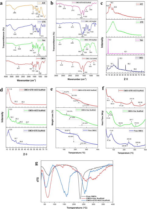
Chemical structure analysis by ATR-IR
The ATR-IR spectra of the pure components and composite scaffolds were analyzed to confirm crosslinking interactions and the incorporation of phytoextracts within the carboxymethyl chitosan (CMCh) matrix. The spectra analysis of pure compounds is as follows.
The IR spectra of pure CMCh, exhibited a broad-wide absorption peak in the range of 3500 cm− 1 to 2877 cm− 1, indicating –OH stretching and -CH2 stretching. NH deformation vibration and C = O stretching are indicated by a 1627 cm− 1 peak27. Additionally, the C-O stretching vibration peak at 1062 cm− 1 can be attributed to the presence of a carboxylate group (Fig. 1a). The vibrational absorption peak observed at 1551 cm− 1 is indicative of the presence of the characteristic carboxylate moiety (-COOH) in carboxymethyl chitosan and the peaks at 1551 and 1062 cm− 1 are indicative of primary amide and glycosidic bond stretching. These spectra peaks implicate the presence of the characteristic saccharide structure and of CMCh28.
Pure vanillin powder exhibited characteristic peaks at 3150 cm− 1, 2838 cm− 1 and 1663 cm− 1, which correspond to the -OH and methyl groups of vanillin and the C = O stretching vibration of the aldehyde group, respectively29. The sharp peaks at 1577 and 1435 cm− 1 and at 1127 cm− 1 are assigned to the stretching vibrations of the benzene ring and C-O-C stretching, respectively30,31. (Fig. 1a). GTE showed a characteristic peak at 3230 cm− 1, which represents the -OH group; a strong intensity peak at 1606 cm− 1 that corresponds to aromatic C = C ring quadrant stretch vibrations; and a strong peak at 1010 cm− 1, which represents the C-O-C group. At 1207 cm− 1, a band corresponding to the deformation of the hydroxyl group in aromatic alcohol is also detected. These agree with spectra reported previously32,33. (Fig. 1a). ACE displayed characteristic peaks at 3230 cm− 1, 1617 cm− 1, 1454 cm− 1, 1363 cm− 1, 1264 cm− 1, 1135 cm− 1, and 1040 cm− 1. The broad peak at 3230 cm− 1 represents the -OH bond, and the sharp peak at 1617 cm− 1 indicates the C = O (amide) bond. The peaks at 1454 cm− 1 and 1363 cm− 1 are attributed to alkanes, those at 1454 cm− 1 are attributed to N-H, those at 1264 and 1040 cm− 1 indicate alkyl halides, and those at 1135 cm− 1 peak are attributed to aliphatic amines34, as represented in Fig. 1a.
Compared with those of the individual components, the ATR-IR spectra of various composite scaffolds crosslinked with the organic crosslinker vanillin (CMCh + vanillin, CMCh + GTE + ACE, CMCh + ACE, CMCh + GTE) revealed significant interactions and bonding. A detailed analysis of each composite is discussed below. The IR spectra of the CMCh + Van scaffold, which is devoid of phytoextracts, were obtained to provide insights into the crosslinking reaction between CMCh and vanillin.
The new peaks at 1530 and 801 cm− 1 are related to the benzene ring and OH of vanillin and are the result of cross-linking reactions, such as bonding between the carbonyl groups of vanillin and the amino groups of CMCh. The 1678 cm− 1 peak corresponding to the distinctive stretching vibration of C = N can be attributed to the Schiff base reaction between the aldehyde group of vanillin and the amino group of CMCh35 (Fig. 1b). The Schiff base reaction consumed the amino group, so the stretching vibration peak at 1627 cm− 1 disappeared, which is proof of crosslinking by the modification of CMCh28. The -OH peak of CMCh shifted from 3500 to 3105 cm− 1, with a significant reduction in intensity, which could be due to hydrogen bonding interactions between CMCh and vanillin during cross-linking. The peaks at 1043 cm− 1 and 2311 cm− 1 represent shifts in the ether group and methyl group from 1127 to 2838 cm− 1 of pure vanillin, respectively, with a decreased intensity, indicating the presence of free vanillin in the scaffold. The peak at 1043 cm− 1 is also attributed to -O-C-O vibration absorption, which is the result of the acetalization reaction. As the intensity of this peak is significantly lower than that of the imine bond, it can be inferred that crosslinking occurred via the creation of a Schiff base and that a small amount of free vanillin was present in the samples. This observed crosslinking behavior corroborates earlier findings on vanillin-mediated modification of CMCh, further supporting the proposed mechanism31,35,36.

Comparison of ATR-IR spectra, XRD patterns, and thermal degradation studies of various scaffolds and their individual components. (a) ATR-IR spectra of phytoextracts (Acacia catechu extract [ACE] and green tea extract [GTE]) and polymers (vanillin and CMCh), the characteristic peaks in the graph confirm the presence of the key functional groups found in these individual components. (b) ATR-IR spectra of the CMCh + GTE + ACE scaffold, CMCh + ACE scaffold, CMCh + GTE scaffold, and placebo scaffold, showing retention of individual component peaks and the appearance of new peaks, indicating the structural integrity of the scaffolds, resulting from the bonding and interactions between the functional groups (c) XRD patterns demonstrating the crystalline and amorphous nature of the phytoextracts, polymers, and developed scaffolds. The sharp peaks for the crystalline vanillin and CMCh, and broad peaks for the phytoextracts indicate its amorphous nature. (d) XRD patterns of the scaffolds, indicating reduced crystallinity of CMCh when phytoextracts are incorporated, leading to an increased amorphous phase. (e) Thermal degradation studies performed to analyze the effects of the incorporation of nanoparticles into the nanoscaffolds revealed three-stage weight loss, indicating decreased thermal stability upon the addition of crosslinkers and nanophytoextracts. (f) DSC graphs of the constituents and nanoscaffolds and (g) dTG thermograms as a function of the rate of weight loss (dTG).
In the CMCh + GTE scaffold, the broad O-H stretching peak shifts to 3233 cm⁻¹ with reduced intensity was noted, suggesting hydrogen bonding with carbonyl groups. The characteristic peaks of GTE molecules are retained, thereby indicating their presence in the scaffold. Aromatic ring vibrations and slight shifts in C-O stretching at 1063 cm⁻¹ indicate the formation of new linkages. The peak at 1526 cm− 1 (NH bending in secondary amines) is observed, depicting that this functional group is involved in cross-linking and that most amine groups are involved in this interaction. The peak at 1663 cm− 1 (C-O stretching in secondary amines) appeared in the cross-linked scaffold. The sharp peak at 1600 cm− 1 corresponds to the vibration of an imine band (C = N). Furthermore, the vanillin-crosslinked scaffold exhibited a sharp peak at 1526 cm− 1 (− COO symmetric stretching). Overall, the sharp, intense peaks indicate an increase in the intensity of intermolecular interactions in the crosslinked scaffold25,37. (Fig. 1b)
The CMCh + ACE scaffold shifted the O-H stretching peak to 3265 cm⁻¹, suggesting enhanced hydrogen bonding involving hydroxyl groups from Acacia catechu and CMCh. The carbonyl group interactions at 1658 cm⁻¹ and 1593 cm⁻¹ are noted. The appearance of new peaks at 1522 cm⁻¹ and 1429 cm⁻¹ indicates the involvement of aromatic ring vibrations from Acacia catechu interacting with CMCh. The characteristic peaks of ACE were retained, with small shifts in wavelength and intensity at 1276, 1142, and 1052 cm− 1, which can be attributed to the encapsulation of the nano Acacia catechu molecules in the scaffold. (Fig. 1b)
In the CMCh + GTE + ACE scaffold, O-H stretching shifted from 3230 cm⁻¹ (Acacia catechu and GTE) to 2916 cm⁻¹, suggesting enhanced hydrogen bonding involving the hydroxyl groups of ACE, GTE and CMCh. The presence of peaks at 1710 cm⁻¹ and 1681 cm⁻¹ indicates carbonyl group interactions between CMCh and vanillin. The appearance of new peaks at 2312 cm⁻¹ and 1314 cm⁻¹ indicates interactions likely from the Acacia catechu and Green Tea components. The peak at 1069 cm⁻¹ suggests slight shifts due to interactions involving the C-O groups, indicating new linkages.
X-ray diffraction of the scaffolds revealed that CMCh, ACE, and GTE existed in an amorphous state
The XRD spectrum of CMCh showed peaks at 11.6°, 19.1°, 33.3°, 48.2°, and 60.2°. CMCh displayed a peak at an angle of 2θ = 33.3°, which is similar to the characteristic peak (20–40°) of CMCh as reported in literature38,39. Vanillin showed a sharp peak at 13.2°. The phytoextract ACE displayed an amorphous nature with a few broad peaks at 14.8°, 19.7°, and 29.8°; GTE had an amorphous nature, which can be inferred from Fig. 1c.
The molecular and structural stability of the composites was validated when the overall XRD data of the individual components were compared with those of the scaffolds. The composite scaffolds, CMCh + GTE, CMCh + ACE and CMCh + GTE + ACE, existed in an amorphous state with a few diffraction peaks and a broad band between 30° and 75° (Fig. 1d). The presence of the peaks of increased intensity is a result of increased ordered structure formation during crosslinking, and its amorphous structure indicates the suitability of the material as a good biosorbent40. The peaks shifting and the differences in the 2θ and d-spacing values in the nanocomposite scaffold suggest alterations in the crystallinity patterns among the constituent polymers/components. The incorporation of the phytoextracts into CMCh almost completely collapsed the crystalline region in the original CMCh, leaving more amorphous regions.
Scanning electron microscopic analysis of the scaffolds revealed a significantly porous architecture with substantial interconnectivity
Three-dimensional scaffolds require high porosity with interconnected pore networks conducive for wound healing. The morphological characteristics of pores, including, size and shape, significantly influence cellular attachment and proliferation, and overall efficiency of tissue regeneration. In our study, a porous architecture was established through freeze‒drying methodology. Figure 2 illustrates the SEM images of the various scaffold formulations. SEM micrographs of the nanoscaffolds revealed pore sizes ranging from 110 μm to 143 μm. Compared with the scaffold devoid of the phytoextracts (127.23 ± 31.6 μm), the CMCh + GTE + ACE scaffold presented a porous structure with a smaller pore size (131.41 ± 43.27 μm) (Fig. 2a and c). Scaffolds bearing these pore sizes have shown enhanced nutrient and oxygen transport throughout the scaffold matrix but are too big for cellular infiltration41,42,43. In addition, regular round micropores ranging from 3 to 10 μm in size were found on the wall of the scaffold. These microporous structures can potentially permit fibroblast attachment and proliferation. Furthermore, a distribution of mineral crystals coating the scaffold framework was evident in the CMCh + GTE + ACE scaffold composition. Another study indicates that pore size lesser than 160 μm is conducive for fibroblast attachment and proliferation and facilitate cellular interaction and cross talk with host tissue44. These pore sizes better mimic the extracellular matrix. The generated scaffolds are macroporous and whether they can allow cellular infiltration and attachment is ambiguous due to the discrepancies in literature.

SEM and EDS analysis of fabricated scaffolds and their incorporation with phytoextracts. (a) SEM analysis of scaffolds without phytoextracts, showing a pore size of 127.23 ± 31.6 (m. (b) SEM analysis of scaffolds incorporated with phytoextracts, showing a slightly larger pore size of 131.41 ± 43.27 (m, indicating the incorporation of nanophytoextracts into the scaffold structure. (c) SEM of nanophytoextract molecules, demonstrating nanoscale particle sizes that retained their structure after ultrasonication, facilitating incorporation into the porous scaffolds. (d) EDS analysis of scaffolds without phytoextracts, showing dominant peaks for carbon (C), oxygen (O), and nitrogen (N), with trace elements of chlorine (Cl), sulfur (S), and sodium (Na) likely from the crosslinking salt. (e) EDS analysis of scaffolds with phytoextracts, showing additional minor peaks for calcium (Ca) and iron (Fe), suggesting the presence of these trace elements in the phytoextracts. (f) EDS analysis of phytoextract molecules, revealing prominent peaks for carbon (C), oxygen (O), and nitrogen (N), indicating purity and minimal impurities in the nanoparticles. (g) The table presents the weight and atomic percentages of detected elements in the samples.
Energy-dispersive X-ray (EDS) was also performed on these samples to identify the elemental composition of the nano scaffolds (Fig. 2d and g). The nanoparticles are composed of C, N and O only, whereas the nanoscaffold devoid of phytoextracts (CMCh + Van scaffold) contains Cl along with C, N and O. It indicated that the nitrogen and chlorine contents increased in the fabricated CMCh + GTE + ACE nanoscaffold and could be attributed to the incorporation of the nanoparticles into the scaffold during the fabrication process. As indicated, the carbon, oxygen, chlorine, nitrogen, and sodium percentages were 39.14%, 35.13%, 10.75%, 8.86% and 5.57%, respectively, in the CMCh + GTE + ACE sample. On the other hand, the presence of an atomic percentage of nitrogen in the CMCh + Van scaffold and CMCh + GTE + ACE composite scaffold confirmed the formation of Schiff’s base, which was supported by ATR-IR analysis. These findings also fortify the proposed crosslinking mechanism of the composite scaffolds.
Thermogravimetric analysis and differential scanning colorimetric analysis
Thermogravimetric analysis was performed on the samples to determine their thermal degradation properties. A graph of weight against temperature was plotted to determine the weight loss profile across the temperature range. These weight losses can be correlated with the DSC data to determine the cause of weight loss. The thermograms of pure CMCh indicated a three-stage weight loss. The weight loss was 29.81%, 40.59%, and 25.16% at temperatures ranging from 47.3 to 118 °C, 180–289 °C, and 307–400 °C, respectively, in each of the stages45. (Fig. 1e − 1 g). The initial stage is associated with the loss of adsorbed water, the second stage accounts for bonded water loss, and the third stage is due to CMCh decomposition. At a Tm of 231.9 °C, the second-stage weight loss of 40.59% occurred, which can be attributed to polysaccharide decomposition, thereby indicating lower polymer thermal stability. These observations are also in agreement with the formation of an endothermic peak at 229.5 °C by DSC and validated in literature45. (Figure 1f g)
The CMCh + Van scaffold displayed a three-stage thermal degradation, with the first stage occurring at a Tm of 64.9 °C and a weight loss of 20.31%. This was most likely related to the release of physically absorbed and loosely bonded water molecules, as chitosan is highly hygroscopic and can be easily hydrated. This loss could also be due to the structural differences in the chitosan polymer upon crosslinking with vanillin. As supported by ATR-IR and XRD, crosslinking occurred through the formation of various intermolecular hydrogen bonds, thereby altering the hydration capacity of the polymer46.
The thermal stability of CMCh + GTE + ACE falls below that of pure CMCh. The nanoscaffolds displayed a multistage thermal decomposition at a faster rate than the nanophytoextract-devoid scaffold. The incorporation of GTE nanoparticles and ACE nanoparticles into chitosan decreased the thermal stability of the CMCh polymer. The scaffold showed a moisture loss at slightly lower temperatures in comparison to the CMCh and CMCh + Van scaffolds, indicating a weak water-matrix interaction. The onset of thermal decomposition at Tm of 161.2 °C (Endothermic peak at 171.20 °C by DSC graph) for the CMCh + GTE + ACE scaffold is lower than both Pure CMCh and CMCh + Van Scaffold, suggesting decreased thermal resistance. The displayed thermal resistance is adequate for in-vivo use but may hinder its sterilization via high temperature or pressure.
Antibacterial assays of the phytoextracts against bacteria commonly found in diabetic wounds revealed notable zones of Inhibition that were comparable with those of standard antibiotics
A chronic wound is highly infiltrated with various gram-positive and gram-negative bacteria that form biofilms and hinder the process of wound healing. To mimic the microbial complexity of a chronic wound, the nanoscaffolds were tested on agar plates that contained a mixture of S. aureus, A. baumannii, E. faecalis, P. mirabilis, E. coli, and P. aeruginosa, microbes. Table 3 shows that the zone of inhibition (ZOI) for the CMCh + GTE scaffold against the above-mentioned bacterial strains at a combined concentration of 100 µg/ml, was 14.2 mm, that for CMCh + ACE was 15.98, and that for CMCh + GTE + ACE was 17.34. (Fig. 3a, b). Notably, the nanoscaffolds displayed a potency similar to that of ciprofloxacin (control, which had a zone of inhibition of 21.17 mm) and indicated that chronic wound microbes are sensitive to the synthesized nanoscaffolds. This indicated the potency of the synergistic effect of the phytoextracts. This can be attributed to the moderate antibacterial properties of carboxymethyl chitosan and the nanoparticles against both gram positive and gram-negative bacteria, that have been reported extensively in previous reports23 .

Antimicrobial efficacy and biocompatibility assessment of the scaffolds. (a) Zone of inhibition observed against S. aureus, A. baumannii, E. faecalis, P. mellifera, E. coli, P. aeruginosa, and a bacterial mixture after exposure to phytocompounds (100 µg/ml) for 24 h, demonstrating antimicrobial properties comparable to ciprofloxacin. (b) Graphical representation of the zone of inhibition, with data presented as means ± SEMs from triplicate experiments (n = 3, P < 0.05). (c) Dermal irritation test results on New Zealand rabbits after 24 h of observation, showing no signs of edema, erythema, or rashes, confirming the nonirritant nature of the scaffolds.
Dermal irritation tests revealed that the nanoscaffolds were safe for the skin
A dermal irritation study was conducted to evaluate whether the fabricated scaffolds produced any allergic reactions or toxic effects upon topical application. Scaffolds incorporated with green tea extract, Acacia catechu extract, and a combination of both were compared with the control (CMCh scaffold without any phytoextracts) after 24 h for any negative reactions. The rabbits were also monitored for any changes in behavior (excessive licking or scratching), feed and water intake, all of which remained normal.
It was concluded that there were no significant differences between the skin that was exposed to the control and those in contact with the phytoextract-incorporated scaffolds, as there were no signs of erythema, edema, rashes, cracks or peeling on the skin. These results can be observed in Fig. 3c. These findings confirmed that the phytoextract-incorporated CMCh scaffolds were safe and nonirritant and did not produce any adverse effects following topical application. While OECD 404 typically requires observations at 1, 24, 48, and 72 h, we conducted observations for 24 h based on extensive literature demonstrating the safety profiles of individual components (CMCh, GTE, and ACE) in topical applications. These findings agree with other studies that report CMCh, vanillin and phytoextracts to be safe for topical application47,48,49,50,51,52.
In vivo wound healing studies revealed a gradual decrease in wound size and enhanced wound healing
The nanoscaffolds were applied to the wound area in the dorsal foot of the rats for 28 days to assess their effectiveness in accelerating wound healing. The daily behaviors, such as the dietary intake and activity levels, of the rats were assessed and noted to be normal. A single intraperitoneal dose of streptozotocin (STZ) (45 mg/kg) in citrate buffer (pH 4.5) was administered to induce diabetes in the rat groups, and blood glucose levels were checked using a glucometer for confirmation. After confirmation of diabetes, anesthesia was administered using ketamine and xylazine at doses of 80 mg/kg and 10 mg/kg body weight, respectively53. During the study, 2 animals were excluded due to unsuccessful diabetes induction, and no animal deaths were observed due to experimental procedures. Post-procedure analgesia was provided (diclofenac sodium, 20 mg/kg for 3 days) to minimize pain.
A wound 1–2 cm in diameter was created on the dorsal foot, and the experimental scaffold and marketed formulations (silver sulfadiazine) were applied topically to the DFU wound. All wound dressing and scaffold application were performed under anesthesia. The wound contraction ratio was assessed and reported as the percentage reduction in wound size after the wound induction procedure. The apparent changes in the wound contraction area are tabulated in Table 4.
The scaffold-treated group started re-epithelialization within 7 days, which was accounted for by the presence of a healthy pinkish-red color, followed by a healing phase that lasted for 14 days, culminating in a completely healed wound in 28 days. Notably, the control group displayed an increase in wound size, whereas the positive control stabilized the wound size, with neither an increase nor a decrease. The CMCh + GTE + ACE scaffold had the highest wound closure rate (98.90%), followed by that of the marketed formulation, the CMCh + GTE scaffold, and the CMCh + ACE scaffold, with rates of 97.97%, 96.70%, and 95.64%, respectively, in comparison with the control group (69.79%) (Fig. 4a and c; Table 4). Compared with the control group, all the groups presented a wound healing rate with P < 0.001 on days 16 to 28. Statistical significance is indicated by asterisks (****, p < 0.001), confirming that all the treatments significantly outperformed the control. These highly significant differences suggest that these biomaterial-based scaffolds have robust therapeutic effects on accelerating wound healing. The timeline of wound healing reported in our study closely parallels those reported by Hajmiri et al., where they achieved nearly complete wound healing within 15 days using a CMCh hydrogel functionalized with recombinant human growth factor for DFU54. This underscores the use of bioactive as cost effective and biocompatible alternatives to achieve comparable healing efficacy.

Comparative analysis of wound healing efficacy in diabetic Wistar rat model and histopathological evaluation of scaffold treated tissues. (a) Acceleration of wound healing in rats treated with CMCh + GTE + ACE, with a wound closure rate of 98.9% by day 28, outperforming the commercial formulation, the CMCh + GTE scaffold, and the CMCh + ACE scaffold at rates of 97.97%, 96.70%, and 95.64%, respectively, in comparison to the control group (69.79%). The synergistic effect of the nanophytoextracts enhanced the wound closure rate, and significantly enhanced wound healing. (b) The figure shows the changes in the wound area (mm²/rat) over time, with a reduction in wound size for all treatment groups, with the CMCh + GTE + ACE scaffold demonstrating the most significant decrease. (c) Depicts the wound closure rates (%), further highlighting the superior performance of the CMCh + GTE + ACE scaffold in accelerating wound healing compared with the other groups. The error bars represent the standard deviation, and **** indicates P < 0.0001; Student’s t test was performed. (d) Histopathological images of healthy and diabetic foot ulcer tissues treated with phytoextract-incorporated nanoscaffolds. (I) Healthy tissue showing vascularization (red arrows) and collagen fibers (black arrows). (II) Untreated tissue showing granulation (green arrow) with follicular congestion (blue arrow) and inflammation (yellow arrow). (III) Tissue treated with the marketed formulation showing keratinocyte (white arrows) and ECM proliferation (orange arrow) with vascularization (red arrows). (IV) Tissue treated with the CMCh + GTE scaffold. Skin showing proliferated keratinocytes (white arrows) resulting in epithelialization with granulation and collagen fibers (black arrows) throughout the dermis area. (V) Tissue treated with the CMCh + ACE scaffold. Skin showing proliferated keratinocytes (white arrows) with ECM (orange arrow) resulting in epithelialization with granulation. (VI) Tissue treated with CMCh + GTE + ACE. Skin showing keratinocyte proliferation (white arrows), extensive vascularization (red arrows) and collagenation (black arrows) highlighting the scaffold’s effectiveness in wound healing.
Histopathological analysis revealed that scaffolds incorporated with phytoextracts enhanced the proliferation of keratinocytes and re-epithelialization
Histopathological examination was conducted to obtain detailed information on how the developed scaffolds affected the wound healing process of diabetic foot ulcers. The histopathological findings of the disease control group revealed that the wound area exhibited keratinocyte hyperplasia, which resulted in abnormal proliferation of keratinocytes. This hyperproliferation of keratinocytes is accompanied by dysregulation of keratinocyte differentiation, resulting in impaired wound healing42,55. These results indicate that blood vessel congestion is likely caused by impaired blood circulation due to hypoxic conditions and inflammation. As shown in Fig. 4d (II), moderate granulation, follicular congestion and inflammation were also observed. However, no collagen fibers were observed in the control group because of the prolonged inflammatory state of the diabetic wounds.
Histopathology evaluations revealed that both the GTE formulation and the ACE formulation promoted moderate keratinocyte proliferation and differentiation, leading to granulation and re-epithelialization. The presence of collagen layers was also observed in both samples, as shown in Fig. 4d (V) and (VI). However, scaffolds incorporated with GTE presented a greater presence of collagen fibers.
A tissue sample that was treated with the scaffold that incorporated both phytoextracts showed superior wound healing activity (Fig. 4d (VI)). Extensive fibroblast proliferation resulted in the generation of a collagenous matrix in the entire dermis area, which was accompanied by extensive vascularization. Compared with the marketed formulation (Fig. 4d (III)), this scaffold showed a superior wound-healing response. This tissue sample was suggestive of moderate keratinocyte proliferation and differentiation despite the hyperglycemic state, that is known to interrupt the natural signaling pathway of fibroblast and keratinocyte proliferation. The increased fibroblast proliferation have been shown to regulate the process of ECM remodeling and allow a faster wound closure rate26,56,57. Compared with the marketed formulation, this tissue sample presented superior wound healing activity.
Discussion
To facilitate wound healing, ideal scaffolds must be highly porous, have a large surface area-to-volume ratio, be flexible enough to acquire the shape of the wound, and shield the wound from any external contamination. Concurrently they must be biodegradable and have degradation kinetics in accordance with the wound healing time and also retain moisture, initiate angiogenesis, and facilitate reepithelialization. The nanoscaffold we synthesized by the freeze-drying method with a composition of 2% CMCh, 4% crosslinker and 1000 µg/mL of the nanophytoextracts were analyzed by various evaluation and characterization methods to meet the above-described characteristics. The nano sized dispersed phytoextracts were anionic in nature and this is critical for enhanced uptake by keratinocytes and fibroblasts. Specifically, the consistent negative zeta potential values (-22.87 mV) suggest their good colloidal stability, preventing aggregation and ensuring uniform distribution within the CMCh nanoscaffold, thereby optimizing the therapeutic efficacy of the scaffold58. We hypothesize the mechanism of uptake of the nanoparticles to be endocytic via electrostatic interactions with the various positive sites at the cellular membrane. Prior work also suggests that the nanoparticles that are bound to the positive sites could also attract more nanoparticles to agglomerate and cause the membrane to bend at the site of adsorption, thereby leading to endocytosis, which our results support qualitatively through increased wound healing rate and ECM remodeling, a mechanistic attribute of the nanoparticles59,60,61.
The ability of the scaffold to absorb moisture is important, as a good scaffold should possess the ability to absorb tissue exudate and remove metabolic waste from the wound site to fasten wound healing. The synthesized scaffolds had a time restricted swelling and degradation capabilities. The scaffolds initially swelled up and held a constant swelling rate but post 24 h, started degrading, gradually impairing the integrity of the scaffold and emphasizing the need to change the wound dressing every 24 h.
The scaffolds were further characterized to evaluate its structure and interactions and their implications in modulating the scaffold’s physico-chemical properties. The ATR-IR analysis confirmed that vanillin-mediated crosslinking successfully stabilized the CMCh scaffold and facilitated the encapsulation of GTE and ACE, thereby providing a chemically integrated system capable of sustained structural and functional performance in wound healing applications. The various interactions namely, hydrogen bonding, aromatic interactions, and possible ether linkages within the composite scaffolds (noticeable as peaks), enhanced its structural integrity (Fig. 1b). The incorporation of the nanophytoextracts decreased the crystallinity of the scaffold, proved by XRD graphs, which led to the creation of a new crystalline phase from the pure amorphous nature of CMCh. The reduced crystallinity is an added advantage to the antibacterial power of nanoscaffolds38, albeit it also increases its degradation rate. Greater amorphous nature of the scaffold makes it greatly susceptible to hydrolysis and enzymatic degradation62. These structural modifications may also be attributed to the differential hydrogen bonding interactions between the components in the scaffold that developed during the freeze‒thawing process.
The SEM images revealed the macroporous structure of the scaffold with limited micropores scattered along the walls of the scaffold to facilitate cellular infiltration and attachment. These highly interconnected heterogenous macropores of 160–210 μm size are susceptible to greater rate of water penetration upon exposure to aqueous environment. The amorphous nature of the composite scaffolds allows water molecules to penetrate the scaffold throughout its volume rather than just surface level. This interpretation is also supported by the TGA and DSC thermograms, that reveal a three-stage weight loss, with rate of moisture loss is greater than the rate of degradation of CMCh at multiple temperatures. The interconnected macropores could facilitate this uniform water diffusion and cause a bulk degradation profile.
Thus, this poses a significant limitation, as the durability of the scaffold for wound healing is questionable. Hence, the scaffold may not be suitable for long-term application in wound healing; this can be overcome by increasing the crosslinking concentration or varying the crosslinker. The current method of utilizing freeze drying is known to cause pore heterogeneity with a rate of freezing dictating the pore size inversely63. By optimizing this parameter of freeze drying along with time and degree of drying we can synthesize scaffolds of better pore size and swelling properties. In this current study we performed crosslinking prior to freeze drying, further studies could be to perform crosslinking on the polymeric scaffold post first freeze drying and subjecting the crosslinked scaffold to another round of freeze drying for better crosslinking. This may improve the mechanical integrity and impart a controlled swelling ability to the scaffold. The addition of plasticizers, such as polyethylene glycol, poly vinyl alcohol, may also help in improving the swelling properties and prevent early degradation64,65. Additionally, the synthesized scaffolds utilize burst method to release phytoextracts, as indicated from bulk degradation of the scaffold; thus, further studies to design controlled release scaffolds are crucial for long-term sustained therapeutic efficacy over prolonged application.
The biological properties of the developed scaffold were evaluated through a series of in-vitro and in-vivo assays to gauge their therapeutic efficacy. The antimicrobial activity is significant in application of wound healing which are colonized by multiple strains. The CMCh + GTE + ACE scaffold showed a comparable zone of inhibition to ciprofloxacin. As indicated by the zeta potential earlier, the nanoparticles are stable and negatively charged, indicating their ability to enhance cellular uptake in gram-positive bacteria, namely, S. aureus and E. faecalis. We hypothesize that the crosslinks that were formed via Schiff’s base gave rise to hydrophobic sides on the CMCh chain allowing for greater attachment of the bacterial peptidoglycan layer /glycoprotein layer to the scaffold. Also, the positive amines of the scaffold could possibly interact with the negatively charged membrane of gram-negative bacteria. The mechanism of action of synergy can be chalked up to be an interplay between the scaffold and the phytoextracts. CMCh scaffolds provides membrane disruption and catechins and tannins from ACE and GTE induce oxidative stress and inhibit microbial enzymatic systems66,67. This theory has been established in a previous study that utilized CMCh/Gelatin scaffold crosslinked with glutaraldehyde for wound healing48. Together these mechanisms provide a broad spectrum antibacterial effect with a satisfactory zone of inhibition against commonly found microbes in DFU, thereby confirming the antimicrobial effect of the nanoscaffolds.
Despite promising results, certain experimental limitations are to be noted. The bacterial mixture used for testing contained multiple strains seeded at a combined concentration of 100 µg/mL, which could result in unequal colony ratios due to different growth rates, oxygen and metabolic preferences, and competitive interaction between the various strains. While this partially mimics the wound microbial complexity, the choice of ciprofloxacin as a may not provide a completely accurate benchmark. Although a broad spectrum antibiotic, ciprofloxacin is selective for gram negative bacteria and exhibits reduced efficacy against gram positive strains as they have shown resistance68. Consequently, the direct comparison of the scaffolds’ ZOI against ciprofloxacin may not be an accurate comparison and can under or overestimate the potency of the scaffold. Thus, the results can be better interpreted as CMCh + GTE + ACE scaffolds provide a broad-spectrum antibacterial effect and is suitable for DFU treatment but requires further optimization in more accurate clinical models.
The scaffolds also demonstrated to be non-toxic and suitable for in-vivo topical applications upon testing on rabbits. Upon in-vivo testing on STZ induced diabetic Albino Wistar rats with a foot ulcer, the scaffolds imparted an accelerated wound closure, with the CMCh + GTE + ACE outperforming the control, silver sulfadiazine, a common antibacterial ointment provided as first step treatment in regimen of DFU. Additionally, the combination formulation had greater efficacy in terms of wound closure than the individual formulations. This potential synergistic effect is not understood on a molecular level in this study, and future work involves the utilization of various molecular and cellular studies to decipher it. The observed synergy effect likely arises from the antioxidant and anti-inflammatory properties of the phytoextracts, which collectively modulate the tissue microenvironment by repolarizing the M0 macrophages to the anti-inflammatory M2 phenotype and simultaneously modulate and restore the ECM that can facilitate re-epithelialization. This is qualitatively evident from the histopathological evaluations of the tissues treated with the scaffolds, as inflammation was not observed in them. ACE is known to enhance various antioxidant enzymes that suppress proinflammatory cytokines69, while the presence of high levels of phenolic compounds in GTE can modulate the oxidative stress and inflammation at the wound site70. Therefore, it can be asserted that the developed scaffolds induced a beneficial wound-healing response, resulting in extensive vascularization, epithelization and remodeling of the ECM.
Conclusions
In this study, we successfully developed a novel CMCh based scaffold incorporating GTE and ACE nano sized dispersions that are known for their antimicrobial, anti-inflammatory, and anti-diabetic properties for the treatment of diabetic foot ulcer. Comprehensive characterization of the fabricated scaffold confirmed successful nanoparticle integration with retention of the major functional groups essential for imparting therapeutic effects. A highly interconnected porous structure (mean pore size: 131.41 ± 43.27 μm) essential for swelling behaviors for wound exudate absorption was developed. The scaffolds exhibited antibacterial activity against DFU associated bacteria and were confirmed to be biocompatible and non-irritant. In-vivo testing in a streptozotocin-induced diabetic rat model demonstrated that the CMCh + GTE + ACE scaffold achieved a 98.90% wound closure by day 28, significantly outperforming the untreated control (69.79%, P < 0.0001) and showing comparable efficacy to marketed formulation (97.97%). The histopathological evaluations indicate favorable tissue regeneration characteristics including epithelialization, collagen deposition and vascularization, suggesting that the scaffold and bioactive provide a conducive microenvironment for healing.
While these results are promising, there are several aspects that warrant further studies and optimization. The scaffold’s limited durability and heterogenous macropores, necessitates further optimization and investigation. The scaffold displayed modest improvement in comparison to existing marketed formulation, thus further refinement of the scaffold is essential for achieving a superior product. This study limits itself with identifying the specific mechanism of action of the scaffold in promoting wound healing and the nature of synergistic interactions – these remain to be systematically and holistically investigated. The current study provides functional evidence of wound healing and establishes CMCh scaffold loaded with phytoextracts as a biocompatible, cost-effective solution for DFU management. The study successfully demonstrated wound healing efficacy and suggest potential for further development. Future research entails optimization of drug concentration, refining the manufacturing technique of scaffold for controlled release of nanoparticles and exploring combinatorial therapies with existing treatments. Addressing these gaps will be essential for advancing this platform toward clinical translation as a bioengineered wound healing solution.
Data availability
Data is provided within the manuscript or supplementary information files.
References
-
Kharroubi, A. T. Diabetes mellitus: the epidemic of the century. World J. Diabetes. 6 (6), 850. https://doi.org/10.4239/wjd.v6.i6.850 (2015).
-
Matoori, S., Veves, A. & Mooney, D. J. Advanced bandages for diabetic wound healing. Sci. Transl Med. 13 (585). https://doi.org/10.1126/scitranslmed.abe4839 (Mar. 2021).
-
Patel, M., Patel, V., Shah, U. & Patel, A. Molecular pathology and therapeutics of the diabetic foot ulcer; Comprehensive reviews. Arch. Physiol. Biochem. 1–8. https://doi.org/10.1080/13813455.2023.2219863 (2023).
-
Pandey, S., Shamim, A., Shaif, M. & Kushwaha, P. Development and evaluation of Resveratrol-loaded liposomes in hydrogel-based wound dressing for diabetic foot ulcer. Naunyn Schmiedebergs Arch. Pharmacol. 396(8), 1811–1825 (2023). https://doi.org/10.1007/s00210-023-02441-5
-
Beulens, J. W. J. et al. Prognostic models for predicting the risk of foot ulcer or amputation in people with type 2 diabetes: A systematic review and external validation study, Diabetologia 64(7), 1550–1562 (2021). https://doi.org/10.1007/s00125-021-05448-w
-
Kendrick, C. et al. Translating clinical delineation of diabetic foot ulcers into machine interpretable segmentation. 1–14 (2025). https://doi.org/10.1007/978-3-031-80871-5_1
-
Noor, S., Zubair, M. & Ahmad, J. Diabetic foot ulcer – A review on pathophysiology, classification and microbial etiology. (Elsevier Ltd, 2015). https://doi.org/10.1016/j.dsx.2015.04.007
-
Afonso, A. C., Oliveira, D., Saavedra, M. J., Borges, A. & Simões, M. Biofilms in diabetic foot ulcers: Impact, risk factors and control strategies. Int. J. Mol. Sci. 22 (15), 8278. https://doi.org/10.3390/ijms22158278 (Jul. 2021).
-
Wang, X., Yuan, C. X., Xu, B. & Yu, Z. Diabetic foot ulcers: Classification, risk factors and management. World J. Diabetes. 13 (12), 1049–1065. https://doi.org/10.4239/wjd.v13.i12.1049 ( 2022).
-
Dai, J., Zhou, Y., Mei, S. & Chen, H. Application of antibiotic bone cement in the treatment of infected diabetic foot ulcers in type 2 diabetes. BMC Musculoskelet. Disord. 24 (1), 135. https://doi.org/10.1186/s12891-023-06244-w ( 2023).
-
Horstink, K. A., van der Woude, L. H. V. & Hijmans, J. M. Effects of offloading devices on static and dynamic balance in patients with diabetic peripheral neuropathy: A systematic review. Rev. Endocr. Metab. Disord. 22( 2), 325–335 (2021). https://doi.org/10.1007/s11154-020-09619-9
-
Baig, M. S. et al. An overview of diabetic foot ulcers and associated problems with special emphasis on treatments with antimicrobials. Life 12(7), 1054 (2022). https://doi.org/10.3390/life12071054
-
Yadav, A. K. et al. Comparative study of various dressing techniques in diabetic foot ulcers in the Indian population: A single-center experience. Int. J. Diabetes Dev. Ctries. 43(5), 647–653 (2023). https://doi.org/10.1007/s13410-022-01163-3
-
Shariatinia, Z. Carboxymethyl chitosan: Properties and biomedical applications. Int. J. Biol. Macromol. 120, 1406–1419. https://doi.org/10.1016/j.ijbiomac.2018.09.131 (Dec. 2018).
-
Shariatinia, Z. Carboxymethyl chitosan: properties and biomedical applications. https://doi.org/10.1016/j.ijbiomac.2018.09.131 (Elsevier B V, 2018).
-
Ahmed, S. B., Mohamed, H. I., Al-Subaie, A. M., Al-Ohali, A. I. & Mahmoud, N. M. R. Investigation of the antimicrobial activity and hematological pattern of nano-chitosan and its nano-copper composite. Sci. Rep. 11 (1), 9540. https://doi.org/10.1038/s41598-021-88907-z (2021).
-
Cheng, L. et al. A microenvironment-modulating dressing with proliferative degradants for the healing of diabetic wounds. Nat. Commun. 15 (1), 9786. https://doi.org/10.1038/s41467-024-54075-7 (2024).
-
Namal Senanayake, S. P. J. Green tea extract: Chemistry, antioxidant properties and food applications – A review. J. Funct. Foods 5(4), 1529–1541 (2013). https://doi.org/10.1016/j.jff.2013.08.011
-
Al-Rawaf, H. A., Gabr, S. A. & Alghadir, A. H. Circulating hypoxia responsive microRNAs (HRMs) and wound healing potentials of green tea in diabetic and nondiabetic rat models. Evid.-Based Complement. Altern. Med. 1–14 (2019). https://doi.org/10.1155/2019/9019253
-
Kumari, M. et al. Nov., Acacia catechu (L.f.) Willd.: A review on bioactive compounds and their health promoting functionalities. Plants 11(22), 3091 (2022). https://doi.org/10.3390/plants11223091
-
Wu, Z. et al. Enhanced diabetic foot ulcer treatment with a chitosan-based thermosensitive hydrogel loaded self-assembled multi-functional nanoparticles for antibacterial and angiogenic effects. Carbohydr. Polym. 347, 122740. https://doi.org/10.1016/j.carbpol.2024.122740 (2025).
-
Vijayan, A. et al. PEG grafted Chitosan scaffold for dual growth factor delivery for enhanced wound healing. Sci. Rep. 9 (1), 19165. https://doi.org/10.1038/s41598-019-55214-7 (Dec. 2019).
-
Youshi, M., Farahpour, M. R. & Tabatabaei, Z. G. Facile fabrication of carboxymethylcellulose/ZnO/g-C3N4 containing nutmeg extract with photocatalytic performance for infected wound healing. Sci. Rep. 13 (1), 18704. https://doi.org/10.1038/s41598-023-45921-7 (2023).
-
Rani, V. V. D. et al. Fabrication of alginate/nanoTiO2 needle composite scaffolds for tissue engineering applications. Carbohydr. Polym. 83 (2), 858–864. https://doi.org/10.1016/j.carbpol.2010.08.065 (2011).
-
Zhu, L. et al. Tannic acid modified keratin/sodium alginate/carboxymethyl chitosan biocomposite hydrogels with good mechanical properties and swelling behavior. Sci. Rep. 14 (1), 12864. https://doi.org/10.1038/s41598-024-63186-6 (2024).
-
Li, H. et al. Qualitative study on diabetic cutaneous wound healing with radiation crosslinked bilayer collagen scaffold in rat model. Sci. Rep. 13 (1), 6399. https://doi.org/10.1038/s41598-023-33372-z (2023).
-
Dayarian, S., Zamani, A., Moheb, A. & Masoomi, M. Physico-mechanical properties of films of chitosan, carboxymethyl chitosan, and their blends. J. Polym. Environ. 22(3), 409–416 (2014). https://doi.org/10.1007/s10924-014-0672-9
-
Zhu, J. et al. Preparation and characterization of vanillin-conjugated chitosan-stabilized emulsions via a Schiff-base reaction. Food Sci. Biotechnol. 32(11), 1489–1499 (2023). https://doi.org/10.1007/s10068-023-01277-2
-
Kamaraj, S. et al. Curcumin drug delivery by vanillin-chitosan coated with calcium ferrite hybrid nanoparticles as carrier. Eur. J. Pharm. Sci. 116, 48–60. https://doi.org/10.1016/j.ejps.2018.01.023 (2018).
-
Shekarforoush, E., Mendes, A., Baj, V., Beeren, S. & Chronakis, I. Electrospun phospholipid fibers as micro-encapsulation and antioxidant matrices. Molecules 22 (10), 1708. https://doi.org/10.3390/molecules22101708 (2017).
-
Zhou, G. et al. Sep., Comparison of chitosan microsphere versus O-carboxymethyl chitosan microsphere for drug delivery systems. J. Bioact. Compat. Polym. 32(5), 469–486 (2017). https://doi.org/10.1177/0883911517690757
-
Christina, S. et al. J. Liq. Chromatogr. Relat. Technol. 25(5), 787–801 (2002). https://doi.org/10.1081/JLC-120003036
-
Ikram, F., Qayoom, A. & Shah, M. R. Synthesis of epicatechin coated silver nanoparticles for selective recognition of gentamicin. Sens. Actuators B Chem. 257, 897–905. https://doi.org/10.1016/j.snb.2017.11.038 (2018).
-
Lakshmi, T., Ramasamy, R. & Thirumalaikumaran, R. Preliminary phytochemical analysis and In vitro antioxidant, FTIR spectroscopy, anti-diabetic activity of Acacia catechu ethanolic seed extract. Pharmacogn. J. 7(6), 356–362 (2015). https://doi.org/10.5530/pj.2015.6.7
-
Zou, Q., Li, J. & Li, Y. Preparation and characterization of vanillin-crosslinked chitosan therapeutic bioactive microcarriers. Int. J. Biol. Macromol. 79, 736–747. https://doi.org/10.1016/j.ijbiomac.2015.05.037 (2015).
-
Czarnecka, E., Nowaczyk, J., Prochoń, M. & Masek, A. Nanoarchitectonics for biodegradable superabsorbent based on carboxymethyl starch and chitosan cross-linked with vanillin. Int. J. Mol. Sci. 23 (10), 5386. https://doi.org/10.3390/ijms23105386 (2022).
-
Westlake, J. R. et al. Vanillin cross-linked chitosan film with controlled release of green tea polyphenols for active food packaging. ACS Food Sci. Technol. 3(10), 1680–1693 (2023). https://doi.org/10.1021/acsfoodscitech.3c00222
-
Olanipekun, E. O., Ayodele, O., Olatunde, O. C. & Olusegun, S. J. Comparative studies of chitosan and carboxymethyl chitosan doped with nickel and copper: Characterization and antibacterial potential. Int. J. Biol. Macromol. 183, 1971–1977. https://doi.org/10.1016/j.ijbiomac.2021.05.162 (2021).
-
Farag, R. K. & Mohamed, R. R. Synthesis and characterization of carboxymethyl chitosan nanogels for swelling studies and antimicrobial activity. Molecules 18(1), 190–203 (2012). https://doi.org/10.3390/molecules18010190
-
Sasirekha, R., Vijayakumar, S., Pazhanisamy, P., Joseph, J. J. & Gomathi, T. Biosorption of copper (II) ion using chitosan nanoparticles/alginate/carboxymethyl cellulose/novel hydroxyapatite biocomposite from aqueous solution. Mater. Today Commun. 35, 106339. https://doi.org/10.1016/j.mtcomm.2023.106339 (2023).
-
Medeiros Borsagli, F. G. L., de Souza, A. J. M. & Paiva, A. E. Ecofriendly multifunctional thiolated carboxymethyl chitosan-based 3D scaffolds with luminescent properties for skin repair and theragnostic of tissue regeneration. Int J Biol Macromol 165, 3051–3064 (2020). https://doi.org/10.1016/j.ijbiomac.2020.10.186
-
Song, L. et al. Peritoneal adhesion prevention with a biodegradable and injectable N,O-carboxymethyl chitosan-aldehyde hyaluronic acid hydrogel in a rat repeated-injury model. Sci. Rep. 6 (1), 37600. https://doi.org/10.1038/srep37600 (2016).
-
Bružauskaitė, I., Bironaitė, D., Bagdonas, E. & Bernotienė, E. Scaffolds and cells for tissue regeneration: different scaffold pore sizes—Different cell effects. Cytotechnology 68 (3), 355–369. https://doi.org/10.1007/s10616-015-9895-4 (2016).
-
Yang, F., Xu, C. Y., Kotaki, M., Wang, S. & Ramakrishna, S. Characterization of neural stem cells on electrospun poly(L-lactic acid) nanofibrous scaffold. J. Biomater. Sci. Polym. Ed. 15 (12), 1483–1497. https://doi.org/10.1163/1568562042459733 (2004).
-
Miranda, M. E. S. et al. Chitosan and N -carboxymethylchitosan: I. The role of N ‐carboxymethylation of chitosan in the thermal stability and dynamic mechanical properties of its films. Polym. Int. 55(8), 961–969 (2006). https://doi.org/10.1002/pi.2060
-
Biernat, M. et al. Effect of selected crosslinking and stabilization methods on the properties of porous Chitosan composites dedicated for medical applications. Polymer (Basel). 15 (11), 2507. https://doi.org/10.3390/polym15112507 (2023).
-
Samuel, S. E., Ghosh, T., Damodar Nayak, A., Deveswaran, R. & Basavaraj, B. V. Exploring self-crosslinked hydrogel: Carboxymethyl chitosan and oxidized alginate for wound healing applications. J. Drug Deliv Sci. Technol. 97, 105746. https://doi.org/10.1016/j.jddst.2024.105746 (2024).
-
Ghosh, T., Rajamanickam, D., Nayak, D. & Srinivasan, B. Physicochemical and in vivo evaluation of crosslinked carboxymethyl chitosan-gelatin scaffolds for wound healing application. Mater. Today Commun. 37, 107307. https://doi.org/10.1016/j.mtcomm.2023.107307 ( 2023).
-
Wang, Y., Cao, H. & Wang, X. Synthesis and characterization of an injectable ε-polylysine/carboxymethyl Chitosan hydrogel used in medical application. Mater. Chem. Phys. 248, 122902. https://doi.org/10.1016/j.matchemphys.2020.122902 (2020).
-
Zhao, T., Li, C., Wang, S. & Song, X. Green tea (Camellia sinensis): A review of its phytochemistry, pharmacology, and toxicology. Molecules 27(12), 3909 (2022). https://doi.org/10.3390/molecules27123909
-
Chang, G. et al. Carboxymethyl chitosan and carboxymethyl cellulose based self-healing hydrogel for accelerating diabetic wound healing. Carbohydr. Polym. 292, 119687. https://doi.org/10.1016/j.carbpol.2022.119687 (2022).
-
Isbrucker, R. A., Edwards, J. A., Wolz, E., Davidovich, A. & Bausch, J. Safety studies on Epigallocatechin gallate (EGCG) preparations. Part 2: Dermal, acute and short-term toxicity studies. Food Chem. Toxicol. 44 (5), 636–650. https://doi.org/10.1016/j.fct.2005.11.003 (2006).
-
Sotoudeh, N. & Namavar, M. R. Optimisation of ketamine-xylazine anaesthetic dose and its association with changes in the dendritic spine of CA1 hippocampus in the young and old male and female Wistar rats, Vet Med Sci 8(6), 2545–2552 (2022). https://doi.org/10.1002/vms3.936
-
Hajimiri, M. et al. Preparation of hydrogel embedded polymer-growth factor conjugated nanoparticles as a diabetic wound dressing. Drug Dev. Ind. Pharm. 42 (5), 707–719. https://doi.org/10.3109/03639045.2015.1075030 (2016).
-
Zingkou, E., Pampalakis, G. & Sotiropoulou, G. Keratinocyte differentiation and proteolytic pathways in skin (patho) physiology. Int J Dev Biol 66(1-2-3), 269–275 (2022). https://doi.org/10.1387/ijdb.210161gs
-
Werner, S., Krieg, T. & Smola, H. Keratinocyte–fibroblast interactions in wound healing. J. Invest. Dermatol. 127 (5), 998–1008. https://doi.org/10.1038/sj.jid.5700786 (2007).
-
Mansoub, N. H. The role of keratinocyte function on the defected diabetic wound healing. Int. J. Burns Trauma. 11 (6), 430–441 (2021).
-
Hoshyar, N., Gray, S., Han, H. & Bao, G. The effect of nanoparticle size on in vivo pharmacokinetics and cellular interaction. Nanomedicine 11(6), 673–692 (2016). https://doi.org/10.2217/nnm.16.5
-
Honary, S. & Zahir, F. Effect of zeta potential on the properties of nano-drug delivery systems – A review (Part 1). Trop. J. Pharm. Res. 12 (2). https://doi.org/10.4314/tjpr.v12i2.19 (2013).
-
Shao, X. et al. Aug., Independent effect of polymeric nanoparticle zeta potential/surface charge, on their cytotoxicity and affinity to cells. Cell Prolif. 48(4), 465–474 (2015). https://doi.org/10.1111/cpr.12192
-
Aliyandi, A., Reker-Smit, C., Bron, R., Zuhorn, I. S. & Salvati, A. Correlating corona composition and cell uptake to identify proteins affecting nanoparticle entry into endothelial cells. ACS Biomater. Sci. Eng. 7(12), 5573–5584 (2021). https://doi.org/10.1021/acsbiomaterials.1c00804
-
Samir, A., Ashour, F. H., Hakim, A. A. A. & Bassyouni, M. Recent advances in biodegradable polymers for sustainable applications. Npj Mater. Degrad. 6 (1), 68. https://doi.org/10.1038/s41529-022-00277-7 (2022).
-
O’Brien, F. J. et al. The effect of pore size on permeability and cell attachment in collagen scaffolds for tissue engineering. Technol. Health Care 15(1), 3–17 (2006). https://doi.org/10.3233/THC-2007-15102
-
Ghosh, T. et al. Vanillin based crosslinked films of CMCh-PVA for wound healing application. J. Drug Deliv Sci. Technol. 83, 104400. https://doi.org/10.1016/j.jddst.2023.104400 (2023).
-
Ahmed, M., Bhat, A. R., Verma, A. K. & Patel, R. Collagen–PVA films plasticized with choline acetate ionic liquid for sustained drug release: UV shielding, mechanical, antioxidant, and antibacterial properties. ACS Appl. Bio Mater. 6(2), 663–673 (2023). https://doi.org/10.1021/acsabm.2c00918
-
Daglia, M. Polyphenols as antimicrobial agents. Curr. Opin. Biotechnol. 23 (2), 174–181. https://doi.org/10.1016/j.copbio.2011.08.007 (2012).
-
Gopal, J., Muthu, M., Paul, D., Kim, D. H. & Chun, S. Bactericidal activity of green tea extracts: The importance of catechin containing nano particles. Sci. Rep. 6 (1), 19710. https://doi.org/10.1038/srep19710 ( 2016).
-
Shariati, A. et al. The resistance mechanisms of bacteria against ciprofloxacin and new approaches for enhancing the efficacy of this antibiotic. Front. Public. Health 10. https://doi.org/10.3389/fpubh.2022.1025633 ( 2022).
-
Stohs, S. J. & Bagchi, D. Antioxidant, anti-inflammatory, and chemoprotective properties of Acacia catechu heartwood extracts. Phytother. Res. 29(6), 818–824 (2015). https://doi.org/10.1002/ptr.5335
-
Lorenzo, J. M. & Munekata, P. E. S. Phenolic compounds of green tea: Health benefits and technological application in food. Asian Pac. J. Trop. Biomed. 6(8), 709–719 (2016). https://doi.org/10.1016/j.apjtb.2016.06.010
Acknowledgements
The authors would like to express our heartfelt gratitude to Dr Basavaraj B V, Head of the Department of Pharmaceutics, and Dr Anbu Jayaraman, Head of the Department of Pharmacology, belonging to the Faculty of Pharmacy, M S Ramaiah University of Applied Sciences, for their unwavering support, guidance, and permission to utilize their resources for the timely completion of our project. All authors would like to thank Dr Rajendran, Green Chem Pvt. Ltd. for providing the free samples of phytoextracts that is heart of the study.
Funding
The current work was funded by RIT SEED Fund Project (Ref No: MSRIT/Admin/1380/24–25).
Ethics declarations
Competing interests
The authors declare no competing interests.
Ethics approval and consent to participate
Institutional Review Board Statement: The Faculty of Pharmacy, Ramaiah University of Applied Sciences’ guide-lines for care and use of laboratory animals were followed, and the animal ethics committee gave its approval to all animal treatment operations. (Ref no: XXIX/MSRFPH/RIT/UG-20/15.02.2024). The study is reported in accordance with ARRIVE guidelines (https://arriveguidelines.org).
Additional information
Publisher’s note
Springer Nature remains neutral with regard to jurisdictional claims in published maps and institutional affiliations.
Rights and permissions
Open Access This article is licensed under a Creative Commons Attribution-NonCommercial-NoDerivatives 4.0 International License, which permits any non-commercial use, sharing, distribution and reproduction in any medium or format, as long as you give appropriate credit to the original author(s) and the source, provide a link to the Creative Commons licence, and indicate if you modified the licensed material. You do not have permission under this licence to share adapted material derived from this article or parts of it. The images or other third party material in this article are included in the article’s Creative Commons licence, unless indicated otherwise in a credit line to the material. If material is not included in the article’s Creative Commons licence and your intended use is not permitted by statutory regulation or exceeds the permitted use, you will need to obtain permission directly from the copyright holder. To view a copy of this licence, visit http://creativecommons.org/licenses/by-nc-nd/4.0/.
About this article

Cite this article
Radhakrishnan, V., Sabu, L., Somayaji, P. et al. Evaluation of wound regeneration potential of nano-phytoextracts loaded carboxymethyl chitosan scaffolds for diabetic foot ulcer. Sci Rep 15, 42809 (2025). https://doi.org/10.1038/s41598-025-26595-9
-
Received:
-
Accepted:
-
Published:
-
Version of record:
-
DOI: https://doi.org/10.1038/s41598-025-26595-9