Introduction
After calving, anatomical and physiological barriers normally prevent bacterial contamination of the bovine reproductive tract. However, pathogenic microorganisms can evade these defensive mechanisms and establish uterine infection, leading to endometritis, a superficial inflammation of the uterine mucosa that typically develops since ~ 3 weeks postpartum. This condition impairs endometrial function, compromises oocyte and embryo quality, and produces deleterious effects that may persist for several months even after apparent clinical recovery1.
In cows affected by endometritis, Escherichia Coli, Trueperella Pyogenes, Streptococcus Alpha-hemolyticus coagulase negative Staphylococcus, Fusobacterium Necrophorum and Porphyromonas Levii are among the most frequently isolated pathogens2. The disease has a marked impact on fertility, reducing pregnancy rates approximately ~ 20%, extending the median calving-to-conception interval by around 30 days, and increasing culling due to conception failure increase by ~ 3%3. It also, decreases conception rates per artificial insemination (AI) (33.4 ± 5.1 vs. 21.6 ± 3.7%), lowers 21-d cycle pregnancy rate % (21 ± 3.4 vs. 12.9 ± 2.3), and prolongs the mean days open interval from 150 to 1744.
Intrauterine antibiotics are routinely used to treat clinical endometritis, which is characterized by purulent vaginal discharge. Cefapirin, a first-generation beta-lactam antibiotic, is among the most widely employed treatments and is typically administered in two intrauterine doses 14-days apart. Application of one or two doses has been reported to increase subsequent pregnancy rates to 28% and 38.8%, respectively5. However, increasing antimicrobial resistance threatens the long-term efficacy of this therapy. E. Coli isolates from cattle have shown resistance to cefapirin6, and extended-spectrum beta-lactamases (ESBL) producing E. Coli strains have been detected in up to 23.4% of bovine endometritis cases7.
Mesenchymal stem/stromal cells (MSCs) are multipotent progenitor cells capable of self-renewal and differentiation into mesodermal cell lineages8,9. They can be isolated from multiple different fetal and adult tissues, including bone marrow (BM-MSCs), adipose tissue (AT-MSCs), umbilical cord (UC-MSCs) and placenta (PL-MSCs)10. MSCs exert regenerative effects through mechanisms that include immunomodulation, anti-apoptosis, angiogenesis, and antimicrobial11, which are mediated by paracrine signaling12. The collection of soluble bioactive molecules secreted by MSC secretome13 has emerged as a promising cell-free therapeutic alternative. Lyophilization of MSC-derived secretome (sMSC) allows concentration and long-term preservation, providing practical advantages for clinical use14.
Compared with cell-based therapies, the use of MSCs-based approaches reduce or eliminate the risk of immune rejection while enabling scalable production of bioactive preparations for a large number of patients15,16,17. In cattle, bovine sMSC offers potential applications in the treatment of several reproductive and inflammatory diseases including endometritis. The aim of this study was to evaluate the antimicrobial activity of bovine fetal sMSCs against E. Coli in vitro and to assess the safety and therapeutic efficacy of an MSCs-based intrauterine treatment for clinical endometritis in dairy cows.
Materials and methods
Ethics
All procedures were approved by the Institutional Committee for the Care and Use of Animals (CICUA) of the University of Chile (certificate 23728–VET–UCH). Experiments were conducted in accordance with national regulations and following ARRIVE guidelines (https://arriveguidelines.org).
Identification of uterine pathogens and antimicrobial resistance
Lactating cows (n = 24) from a commercial dairy located in the Province of Melipilla, Chile, were examined to identify predominant pathogens associated with endometritis and to determine their antibiotic resistance profiles. Cows were housed under confinement and fed a total mixed ration based on corn silage, alfalfa hay and pellet concentrate. Cows with 1–3 lactations and 35.1 ± 5.1 days in milk exhibiting grade 2 endometritis (vulvar discharge with 50% pus and 50% mucus) were selected18. For each animal, a uterine secretion sample was collected from both uterine horns using a trans-cervical guarded swab, placed in Stuart transport medium, refrigerated and transported to the laboratory within four hours. Swabs were enriched in 9 mL tryptone soy broth (Becton, Dickinson and Company, Franklin Lakes, NJ, USA), homogenized and incubated overnight at 42 °C. After homogenization, 25 µL of each culture were plated on MacConkey agar (Becton, Dickinson and Company, Franklin Lakes, NJ, USA) and incubated at 37 °C for 18–24 h. Antimicrobial susceptibility of the isolated strains was determined by minimal inhibitory concentration (MIC) using the VITEK2 system (bioMérieux, Marcy-l’Étoile, France) following the manufacturer’s instructions, and clinical cut-off values of Clinical and Laboratory Standards Institute guidelines.
Isolation and culture of bovine fetal MSCs and fibroblasts
AT-MSCs, BM-MSCs and fibroblasts (FBs) were isolated from male bovine fetuses (n = 9; 7 to 8 months of gestation) obtained from a local abattoir, as previously described19. AT samples from the omentum of three fetuses were pooled, washed twice with PBS and then washed twice with collection media [Dulbecco’s modified Eagle’s low-glucose medium (DMEM, Gibco, Grand Islands, NY, USA) supplemented with 100 IU/mL penicillin, 100 µg/mL streptomycin and 2.5 µg/mL amphotericin B (Hyclone)]. Tissue was digested in 0.5% collagenase I (Sigma-Aldrich, St. Louis MO, USA) (1 mL/g AT) for 45 min, filtered (40 μm) and centrifuged at 400 g for 5 min. The pellet was resuspended in expansion media [Collection media supplemented with 10% fetal bovine serum (FBS)] and cultured in 175 cm2-flasks at 38 °C in humidified atmosphere with 5% CO2. Non-adherent cells were removed after 48 h by replacing the culture medium. BM-MSCs were harvested from BM obtained from the same fetuses used for AT-MSCs collection19. One femur from each fetus was dissected and sectioned at the diaphysis using a saw, then the BM was aspirated from the femoral medullary space using an 18 G needle connected to a syringe containing 2 mL of collection medium. The BM was subsequently centrifuged twice in PBS supplemented with 100 µg/mL amphotericin B, 100 IU/mL penicillin, and 100 µg/mL streptomycin and twice in collection medium at 400 x g for 5 min. The pellet was resuspended in expansion medium and then transferred to 175 cm2-flasks for cell culture. BM-MSCs were incubated at 38 °C in humidified atmosphere with 5% CO2. After 48 h, cells not adhering to the plastic were removed by changing culture medium. Upon reaching 80 to 90% confluence, BM-MSCs were detached using trypsin/EDTA supplemented with 250 µg/mL amphoterecin B, 100 IU/mL penicillin and 100 µg/mL streptomycin. FBs were obtained from epithelium obtained from the same fetuses used for collection of MSC19. Fetal epithelium was obtained from snout area, washed four times with PBS and subsequently fragmented using scalpel and then incubated under constant agitation at 38 °C for 90 min. Subsequently, expansion medium was added in an amount equal to the digestion medium to inactivate the collagenase effect. The digested tissue was filtered using 40 μm pores and centrifuged twice with washing medium at 400 x g for 5 min. The sediment was resuspended in expansion medium and transferred to 75 cm2 cell culture flasks. The pellet was resuspended in expansion medium and then transferred to 175 cm2-flasks for cell culture and incubated at 38 °C in humidified atmosphere with 5% CO2. Upon reaching 80 to 90% confluence, FBs were passaged by trypsin/EDTA removal supplemented with 250 µg/mL amphoterecin B, 100 IU/mL penicillin and 100 µg/mL streptomycin.
Exposure of MSCs to lipopolysaccharide (LPS)
AT-MSCs and BM-MSCs (3–4 passages) were seeded at 1.9 × 10⁵ cells/cm2 in 12-well plates and cultured for 24 h until 80% confluence. Cells were then exposed to 10, 50–100 µg/mL of E. Coli lipopolysaccharide (LPS, O26:B6, L2654, Sigma-Aldrich, Germany) for 48 h. Control wells received vehicle only (distilled water). After incubation, cells were lysed in buffer (Qiagen, CA, USA) containing 1% β-mercaptoethanol and stored at -20 °C for RNA extraction.
qPCR analysis of antimicrobial and Immunomodulatory gene expression
Total RNA was extracted using an RNA easy kit (Qiagen) following manufacturer’s instructions. RNA was quantified using a Qubit RNA HS kit (Life Technologies, NY, USA). Genomic DNA was removed with DNAse I kit (Thermo Scientific., MA, USA). Reverse transcription (RT) reaction was performed with a Brilliant II SYBR Green RT-PCR kit (Agilent Technologies, CA, USA), and cDNA amplification performed with Brilliant SYBR Green Master Mix on a qPCR system (Stratagene, CA, USA). Each reaction tube contained 5 µL of Master mix, 1 µL of forward primer, 1 µL of reverse primer, 1 µL of cDNA sample and 2 µL nuclease-free H2O for a total volume of 10 µL. Cycling and temperature analysis consisted of 1 cycle of increasing the temperature to 95 °C at a rate of 20 °C/s for 10 min for polymerase activation, then 40 cycles of 95 °C for 30 s for cDNA denaturation and then lowering the temperature to 60 °C at a rate of 20 °C/s for 1 min for amplification, recording fluorescence every 0.1 °C. To ensure that amplicons are generated from mRNA and not from genomic DNA amplification, control samples without reverse transcriptase (NRT) were included. Gene expression was recorded as CT values that corresponded to the number of cycles where the fluorescence signal can be detected above a threshold value. The CT averages for each biological replicate were calculated and transformed into relative values denominated quantity (Q) through ∆∆CT formula19. Then, the relative quantification in the expression of target genes for each sample was estimated as the quotient between Q value of the target gene and a normalization factor (NF), which was calculated based on the geometric mean of housekeeping gene Q values. Target genes corresponded to antimicrobial β-Defensin 4 A (bBD4A) and NK lysine 1 (NK1), and immunomodulatory interleukin 6 (IL6), interleukin 10 (IL10), tumor necrosis factor ɑ (TNFɑ) and transforming growth factor β (TGFβ). GAPDH and β-ACTIN were used as housekeeping genes. Primer sequences are shown Table 1.
In vitro antimicrobial assay
Conditioned media (CM) from AT-MSCs, BM-MSCs and FBs were collected after 72 h of culture in serum-free and antibiotics-free medium. Then, CM was filtered (0.2 μm) and stored at -80 °C. Also, media from AT-MSCs, BM-MSCs and FBs co-cultured with E. Coli for 6 h (i.e., preactivated conditioned media, ACM) was obtained. To increase its antimicrobial effect, media was concentrated 10-fold (CCM) using Amicon Ultra tubes (Merk Millipore, Cork, Ireland). Antimicrobial activity against E. Coli ATCC25922 (Manassas VA, USA) was assessed by counting colony forming units (CFU/mL) in CM, CCM, ACM and low-glucose DMEM (negative control) and quantified at 0, 1, 2 and 3 h post-inoculation. Bacterial survival was expressed as percentage as a percentage of the initial (0 h) CFU/mL (100%).
Proteomic analysis of lyophilized sMSC
CM from AT-MSCs was lyophilized at − 50 °C with a vacuum pressure of 4.5 mmHg using a Telstar lyophilizer (LyoQuest, Japan). Peptides (200 ng) were separated within 90 min at a 400 nL/min flow rate on a reversed-phase column (Aurora Series CSI; 25 cm x 75 μm i.d. C18 1.6 μm) (IonOpticks, Australia) with 50 °C using a nano elute liquid chromatography system (Bruker Daltonics, Billerica, Massachusetts, USA). Mobile phases A and B were water and acetonitrile with 0.1% v/v formic acid, respectively. The percentage of B was linearly increased from 2 to 17% within 57 min, to 25% within a further 21 min, to 35% within a further 13 min, and then to 85% for a final washing step and re-equilibration. Samples were then analyzed on a hybrid trapped ion mobility spectrometry (TIMS) quadrupole time-of-flight mass spectrometer (MS) (TIMS-TOF Pro, Bruker Daltonics) via a CaptiveSpray nano-electrospray ion source. The MS was operated in data-dependent mode. The accumulation and ramp time were set to 100 ms each, and recorded mass spectra ranging from m/z 100–1700 in positive electrospray mode. The ion mobility was scanned from 0.6 to 1.6 Vs/cm². The overall acquisition cycle of 1.16 s comprised one full TIMS-MS scan and 10 parallel accumulation-serial fragmentation (PASEF) MS/MS scans. Data obtained from three samples were analyzed with the MSFragger 3.8 software20 through the Fragpipe v20.1 (https://fragpipe.nesvilab.org/) with precursor mass tolerance ± 20 PPM, mass fragment tolerance 40 PPM. Within the digestion options, trypsin was used as an enzyme, specific digestion mode and a maximum of 2 cleavages per peptide. The following post-translational modifications (PTM) were used: Cysteine carbamidomethylation, as fixed PTM: Methionine (M) oxidation, Terminal acetylation, as variable PTMs. The FDR estimate < 1% was included by means of a decoy database. The Bos taurus reference proteome (Uniprot, 37,501 entries) plus a common contaminants database available on the FragPipe platform was used for peptide identification.
In vivo safety and efficacy trials in dairy cows with endometritis
Safety trial design
Twelve (n = 12) healthy first calving Holstein cows (~ 35 days in milk, 650 kg) from the same dairy farm enrolled for pathogen identification were used. Number of animals per group was estimated based on N: L (ratio) variability21,22 assuming 95% statistical power and α = 0.05. Cows were examined with a Metricheck instrument (Humeco, Spain), a 50 cm long metal rod device with a small rubber cup of 3.5 cm diameter that allows extraction of vaginal content for visual inspection, to determine the type of secretion present in the vaginal fundus18; only those with mucous secretion were included. Animals were randomly assigned to a placebo group (PCB; n = 6) receiving 3 mL 0.9% NaCl intrauterine on day 0 and 14 or to the secretome group (sMSC; n = 6) receiving 100 mg/mL lyophilized sMSC resuspended in 3 mL of 0.9% NaCl on the same schedule23. The sMSC was obtained from AT-MSCs, since it showed the highest reduction in E. Coli proliferation as described in the in vitro antimicrobial assay presented in this study. Clinical parameters [respiratory rate (RR), heart rate (HR), ruminal rate (RuR), and rectal temperature (RT)], and endometrial polymorphonuclear (PMN) using a cytobrush device were recorded24. Clinical parameters and PMN were compared between PCB and sMSC groups of cows. Blood samples (10 mL) were collected in Vacutainer tubes (Becton Dickenson, Rutherford, NJ, USA) on days 0, 7, 14 and 21. Hematological (URIT-2900Vet Plus kit) and biochemical (Auto-Chemistry Analyzer CS-T180) analyses were performed using commercial veterinary analyzers. The neutrophil/blood to lymphocyte ratio was used as inflammatory indicator25. Hematological and biochemical variables were compared among days in sMSC group.
Efficacy study design
Twenty-four cows (1–3 lactations; 29.8 ± 7.1 days in milk) diagnosed with grade 2 endometritis from the same dairy farm where the safety trial was conducted were used in this study. The clinical endometritis grade (EG) was determined by extracting vaginal discharge with the metricheck18. All cows received the same postpartum diet and general management during this study. The number of animals per group was estimated based on the variability of polymorphonuclear (PMN) percentage24 assuming 95% statistical power and α = 0.05. Animals were randomly assigned the following groups: (i) placebo group (PCB; n = 8) receiving 3 mL of 0.9% NaCL intrauterine solution (Fig. 1); secretome group (sMSC; n = 8) treated with 100 mg/mL lyophilized sMSC in 3 mL of 0.9% NaCl intrauterine solution23; and (iii) antibiotic group (ATB; n = 8) treated with 640 mg of intrauterine cefapirin benzathine5. All groups were given two doses on days 0 and 14 of the experiment. Clinical variables and endometrial cytology were assessed on the same days as in the safety trial.

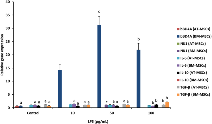
Gene expression of antibacterial and immunomodulatory factors in BM-MSCs and AT-MSCs exposed to different concentrations of LPS. Supplementation of 10, 50 and 100 µg/mL of LPS increased (P < 0.05) bBD4A in BM-MSCs compared to control. Although, IL6 was downregulated (P < 0.05) by 100 µg/mL of LPS (0.57-fold) in AT-MSCs, IL10 gene expression was increased (P < 0.05) in these cells using the same concentration of LPS. Treatment with 100 µg/mL of LPS reduced (P < 0.05) TGFβ gene expression in AT-MSCs; however, this treatment increased (P < 0.05) expression of this gene in BM-MSCs. Superscripts (a, b,c) indicate significant differences (P < 0.05) among concentrations of LPS and control for each gene.
Statistical analysis
Normality of data distribution was assessed using the Shapiro-Wilk test. Variables with non- normal distribution (clinical parameters, blood count, biochemical profile, EG and PMN) were analyzed using Kruskal-Wallis test to determine differences between days and treatments. Normally distributed variables, including erythrocyte indices, serum metabolites, and mRNA expression of immunomodulatory genes IL6, IL10, TNFɑ, TGFβ, and AMPs genes bBD4A and NK1 were analyzed by one-way ANOVA followed by Tukey’s post hoc test. Statistical significance was set at P < 0.05. Analyses were performed using Info Stat software (Córdoba, Argentina, 2008).
Results
Pathogen detection and antimicrobial resistance analysis in bovine endometritis samples
Characterization of uterine secretions revealed that of the 24 samples from cows with clinical endometritis, 19 (79.16%) were positive for E. Coli, whereas Providencia Stuertti (8.33%), S. Epidermidis (4.16%), S. Chromogenes (4.16%) and Streptococcus Spp (4.16%) were detected at lower frequencies (Table 2).
Antimicrobial resistance analysis revealed that E. Coli showed resistance to enrofloxacin (4.17%), sulfa + trimetoprim (4.17%) and cephalexin (20.83%). Resistance was also observed in P. Suartti to ampicillin, gentamicin, and tetracycline (8.33% each). Streptococcus spp. showed resistance to tetracycline (4.17%). S. Epidermis was resistant to tylosin (4.17%) and clindamycin (4.17%). S. Chromogenes was resistant to tylosin (4.17%) and clindamycin (4.17%).
LPS exposure modulated antimicrobial and Immunomodulatory gene expression in fetal MSCs
Stimulation of bovine fetal MSCs with E. Coli lipopolysaccharide (LPS; 10–100 µg mL−1) altered the expression of several antimicrobial and immune-related genes (Fig. 1). In AT-MSCs bBD4A transcripts were undetectable at all concentrations of LPS, whereas BM-MSCs exhibited a marked up-regulation (P < 0.05) reaching 14.3-, 31.3-, 21.9-fold at 10, 50 and 100 µg/mL, respectively compared to control. NK1 expression was detected only in BM-MSCs treated with 50 µg/mL LPS. IL6 expression was unchanged in AT-MSCs but downregulated (0.57-fold; P < 0.05) in BM-MSCs exposed to 100 µg/mL LPS. Conversely, IL10 mRNA increased (1.2-fold; P < 0.05) in AT-MSCs treated with 100 µg/mL LPS but was not detected in BM-MSCs. Treatment with 100 µg/mL LPS reduced TGFβ expression (0.98-fold; P < 0.05) yet enhanced it (2.1-fold) in BM-MSCs.
Conditioned media from fetal MSCs reduce E. Coli survival in vitro
The antimicrobial effect of conditioned media (CM) derived from AT-MSCs, BM-MSCs, and FBs was evaluated against E. Coli ATCC 25,922 (Fig. 2). After 1–3 h of inoculation, E. Coli counts expressed as percentage of initial (0 h) CFU/mL (100%) were lower (P < 0.05) in CM from AT-MSCs (75.9 ± 40.9; 71.6 ± 60.5; 26 ± 25.5% at 1, 2 and 3 h, respectively) compared with CM from BM-MSCs (143.3 ± 80.9; 201.2 ± 118.6; 301.8 ± 171.1%), FBs (171.4 ± 98.7; 218.3 ± 141.7; 440.5 ± 303.1%) and DMEM (152.1 ± 10.4; 579.3 ± 130.5; 1275.4 ± 112%). Concentrating CM 10-fold (CCM), reduced (P < 0.05; Fig. 2B) E. Coli counts in CCM from AT-MSCs (1 h: 80.4 ± 0.4; 2 h: 56.4 ± 3.1; 3 h: 32.9 ± 7.1%) and FBs (1 h: 125.8 ± 14.1; 2 h: 113 ± 2.4; 3 h: 100.8 ± 16.2) in comparison to DMEM (1 h: 152.1 ± 10.4; 2 h: 579.3 ± 130.5; 3 h: 1275.4 ± 112%).

Evaluation of the in vitro antibacterial potential of CM derived from bovine fetal BM-MSCs and AT-MSCs against E. Coli ATCC25922. (A) First the antibacterial effect of CM derived from BM-MSCs, AT-MSCs, and FB was evaluated against E. Coli strain ATCC25922. (B) CM were concentrated (10-fold, CCM) with the aim of increasing their antibacterial capacity. (C) The in vitro antibacterial effect of CM from AT-MSCs, BM-MSCs and FBs pre-exposed to E. Coli (ACM) were in vitro evaluated against E. Coli. (D) ACM were also concentrated (10-fold) (CACM) and used against E. Coli. (E) The in vitro antibacterial effect against E. Coli of CM from AT-MSCs, BM-MSCs and FB was compared to each CCM. This was performed with the aim of verifying whether the effect of media concentration produced an increase in their antibacterial potential. (F) Comparative survival analysis of E. Coli strain ATCC25922 in CM and ACM from bovine fetal BM-MSCs, AT-MSCs and FBs. Superscripts (a, b,c) indicate significant differences (P < 0.05) between treatments within incubation hours.
Pre-exposure of MSCs to E. Coli for 6 h before CM collection (ACM; Fig. 1C) significantly enhanced bacterial killing in AT-MSCs (1 h: 56.7 ± 36.7; 2 h: 36 ± 32; 3 h: 34.7 ± 32.7%), BM-MSCs (1 h: 81.3 ± 6.3; 2 h: 23.7 ± 3.7; 3 h: 15.5 ± 7.9%), and FB (1 h: 77.4 ± 10.1; 2 h: 15.5 ± 4.2; 3 h: 11.3 ± 3.8%) in comparison to DMEM medium (1 h: 152.1 ± 10.4; 2 h: 579.3 ± 130.5; 3 h: 1275.4 ± 112%). Concentrating ACM 10-fold (CACM; Fig. 1D) decreased (P < 0.05) E. Coli counts in BM-MSCs (90 ± 35; 40 ± 10; 30 ± 15%), AT-MSCs (90.9 ± 54.5; 57.9 ± 48.8; 39.4 ± 30.3%) and FBs (96.2 ± 16.2; 72.4 ± 42.4; 42.5 ± 14.5%), compared to DMEM (152.1 ± 10.4; 579.3 ± 130.5; 1275.4 ± 112%).
Comparison of CM to CCM showed (Fig. 1E) that concentration of CM did not increase the anti-microbial effect against E. Coli. Activated CM (ACM) decreased E. Coli counts (P < 0.05; Fig. 1F) in BM-MSCs and FB when compared to CM, however, in AT-MSCs no significant effects were detected.
Proteomic analysis identifies SPARC as an abundant component of the sMSC
Mass spectrometry analysis of lyophilized AT-MSC-derived sMSC identified an average of 421 proteins across three replicates (Fig. 3A) and their proportions were similar in the distribution curves plotted (Fig. 3B). The majority were involved in molecular binding and catalytic activity (Fig. 3C). The most abundant proteins or peptided included of secreted protein acidic and cysteine rich (SPARC) (4.76 µg; 11%), metallopeptidase inhibitor 1 (TIMP1) (3.38 µg; 8%), collagen alpha − 2 (COL1A2) (1.98 µg; 5%), albumin (ALB) (1.619 µg; 4%), endothelial plasminogen activator inhibitor-1 (SERPINE 1PAI1) (1.517 µg; 3%), 72 kDa Type IV collagenase (MMP2) (0.935 µg; 2%), Fibronectin (FN1) (0.667 µg; 2%) and of Serotransferrin (TF) (0.51 µg; 1%) (Fig. 3D).

Mass spectrometry proteomic analysis in bovine sMSC. (A) Comparative percentage distribution of the three replicates of sMSC analyzed by mass spectrometry. (B) Percentage distribution of proteins or peptides with the highest concentration in sMSC-derived secretome used in the present study. (C) Distribution of proteins in relation to biological processes (left) and molecular function (D) Venn diagram of the three secretome samples detected by mass spectrometry .lc-ms/ms technology.
Intrauterine administration of sMSC is safe in healthy dairy cows
Cows receiving intrauterine sMSC (42.5 µg per dose) showed no clinical abnormalities or adverse reactions during the 21-day observation period. Clinical variables, including HR, RR and RT remained within physiological ranges and did not differ between sMSC and placebo group (Table 3). Hematological profiles revealed transient variations within normal limits: leukocyte count expressed as cells x10^3/mm3 decrease (P < 0.05) between day 0 (11.2 ± 7.02) and day 7 (6.02 ± 0.87) and increased (P < 0.05) again from day 14 (9.86 ± 7.52) to day 21 (16.2 ± 8.01) (Table 4). Segmented neutrophil counts expressed as percentage increased (P < 0.05) from day 0 (60.25 ± 11.39) to day 7 (72 ± 6.74) to then decrease (P < 0.05) from day 14 (68.37 ± 19.89) to day 21 (49.62 ± 19.83). Lymphocyte counts expressed as percentage followed a similar pattern compared to leukocytes, decreasing (P < 0.05) from day 0 (32.87 ± 12.18) to day 7 (20.87 ± 6.01) and increasing (P < 0.05) from day 14 (24.75 ± 19.35) to 21 (45 ± 19.83). Eosinophils and monocytes expressed as percentage did not show significant differences.
Biochemical parameters expressed in mg/dL, including uremia, BUN, creatinine, and calcium showed significant (P < 0.05) differences among sampling days and most of them remained within reference values. Other biochemical parameters did not show significant differences. All values are summarized in Table 5.
sMSC treatment reduces uterine inflammation and promotes recovery in cows with clinical endometritis
In cows with grade 2 endometritis, both intrauterine sMSC and cefapirin (ATBiu) treatments improved uterine cytology and discharge scores relative to placebo. PMN count expressed in percentage of total cells in endometrial cytology samples did not show significant differences between ATBiu and sMSC groups on the experimental period (Fig. 4A). PMN counts were lower (P < 0.05) in sMSC than in placebo controls at all time points (day 7: 29.78 ± 34.08 vs. 100; day 14: 61.89 ± 43.01 vs. 97.39 ± 6.4; and day 21: 52.56 ± 32.12 vs. 98.94 ± 1.64). Similarly, ATBiu showed lower (P < 0.05) PMN counts than PCB (day 7: 58.17 ± 41.76; day 14 56.72 ± 47.78; and day 21: 41.72 ± 32.58).

Polymorphonuclear neutrophil count, rectal temperature and endometritis grade in dairy cows treated with two doses of sMSC separated by 14 days. Control groups of cows included respective two dose treatments with ATBiu and PCB. PMN counts (A), rectal temperature (B) and endometritis grade (C) were determined every 7 days during a 21-day experimental period in cows treated with sMSC, ATBiu or PCB on days 0 and 14. Different letters (a, b) indicate significant (p ˂0.05) differences between treatments.
Clinical variables were not significantly different among groups (Table 6). Regrading clinical recovery, EG expressed in grade (0 to 3) decreased (P˂0.05) following sMSC therapy compared with PCB (day 7: 0.67 ± 0.52 vs. 2 ± 0.63), (day 14: 1 ± 0.6 vs. 1.8 ± 0.9) and (day 21: 0.5 ± 0.84 vs. 2.33 ± 0.52). Similarly, cows treated with ATBiu decreased (P˂0.05) EG when compared to PCB at (day 7: 1.67 ± 0.82; day 14:1.5 ± 0.78; and day 21:0.83 ± 0.98).
Discussion
In the present study, E. Coli was the predominant bacterium (79.2%) isolated from uterine secretions of cows with endometritis and 20.8% of isolates were resistant to cephalexin, a first-generation cephalosporin commonly used in veterinary medicine. These findings are consistent with previous reports in Chile and other regions4,26, highlighting E. Coli as a major etiological and antimicrobial-resistant agent in bovine endometritis. Although cefapirin, is routinely used to treat this condition and has proven efficacy5, the growing prevalence of multidrug-resistance, threatens its long-term effectiveness6,27,28. This situation underscores the need for alternative, non-antibiotic therapeutic approaches for uterine infections in bovine.
We first evaluated the in vitro antibacterial activity of CM derived from bovine fetal MSCs against E. Coli ATCC25922. CM derived from AT-MSCs exhibited a marked antibacterial activity, reducing E. Coli survival by up to 98% of count after 3 h of incubation. This is in agreement with findings in equine, where CM from AT-MSCs reduced E. Coli growth by 95% and its effectiveness was greater than BM-MSCs29. Our data confirmed that antibacterial capacity varies according to MSCs tissue source. In BM-MSCs, LPS exposure induced gene expression of bBD4A, a peptide known to disrupt bacterial membranes and promote cell lysis30. Conversely, LPS down-regulated bBD4A expression in AT-MSCs, suggesting distinct activation pathways between tissues. NK1, another antimicrobial peptide, was also up-regulated in LPS-stimulated BM-MSCs but not AT-MSCs. These findings indicate that the antibacterial properties of MSCs arise from tissue-specific responses to bacteria stimuli, particularly though LPS-mediated activation.
E. Coli ATCC25922 is a standard reference strain extensively used in microbiology as a quality-control organism for antimicrobial susceptibility testing and as a model in studies of bovine health, including endometritis research31,32,33. Although this strain is not pathogenic for cattle, it provides a reproducible benchmark for comparing antimicrobial results across laboratories and for this purpose is recommended as quality control strain by the CLSI34. This ensures standardized assay conditions and facilitates interpretation of antibacterial effects independent of the variability inherent to field isolates. This approach allows meaningful comparison between treatments while maintaining methodological consistency with international antimicrobial testing guidelines.
It has been reported that MSCs recognize pathogens-associated molecules through Toll-like receptors (TLRs)35 that can trigger differential immunomodulatory responses36,37. In this study, LPS (100 µg/mL) treatment modulated cytokines in both MSC types, upregulating IL10 and downregulating IL6 and in AT-MSCs. LPS downregulated TGFβ expression in AT-MSCs while increased it in BM-MSCs, indicating that this response occurs in a tissue-dependent manner. These cytokines are central to the regulation of inflammation and macrophage activity. Similar effects have been reported in E. Coli-preactivated human BM-MSCs used to treat E. Coli-induced pneumonia in mice, where IL10 induction was associated with reduced inflammation and enhanced macrophage phagocytosis38. These results support the notion that the immunomodulatory actions of MSCs contribute to bacterial control but also modulate excessive inflammation and promote tissue recovery.
Proteomic analysis of the lyophilized MSC-derived secretome (sMSC) revealed 421 proteins of which 36% corresponded to 8 proteins and 64% to other proteins. A previous report of endometrial sMSC detected a total of 699 proteins38, indicating that protein expression is tissue dependent. However, in both studies, ~ 70% proteins match and are associated with extracellular matrix. Here, we detected Alipoprotein AI and Cystatin proteins that is reported to exert antimicrobial action against E. Coli39,40,41. We also found SPARC as the most abundant protein (11%) in sMSC, which plays a key role in extracellular matrix remodeling and immune regulation42, which suggest a potential contribution to both antibacterial and regenerative processes observed here. SPARC interacts with Toll-like receptor- and TGFβ mediated signaling pathways to inhibit NFkB (nuclear factor enhancer of activated B-cell Kappa light chains) and TGFβ, limiting inflammatory response42,43. This mechanism could help explain the reduction in endometrial inflammation following sMSC administration. Other abundant proteins, including TIMP1, MMP2, and fibronectin, are also associated with tissue repair and antimicrobial defense, reinforcing the therapeutic potential of the secretome.
The intrauterine administration of sMSC was well tolerated in healthy cows. Clinical, hematological, and biochemical parameters remained within physiological ranges, confirming the safety of local application. Transient changes in lymphocytes and leukocytes counts are consistent with normal physiological variations during initial stages of lactation. Importantly, endometrial cytology did not reveal local inflammation, further demonstrating no adverse effects. To our knowledge, this is the first report evaluating the safety of intrauterine use of lyophilized sMSC derived from AT-MSCs in cattle. A previous study in dairy buffaloes showed that intravenous (IV) and intracervical (IC) administration of AT-MSCs produced no systemic immune response44, supporting the safety of this approach.
In cows with clinical endometritis, the sMSC treatment improved both cytological and clinical outcomes. PMN counts, and EG decreased significantly after two intrauterine administrations, achieving an effect comparable to ATBiu treated animals. These results demonstrate that sMSC effectively reduces uterine inflammation and promotes clinical recovery without used of antibiotics. In contrast, in placebo treated animals, EG increased to grade 3, in which the presence of pus was detected in 100% of the secretion. Similar therapeutic effects have been reported for MSCs in experimental models of endometritis45 and other inflammatory conditions in dairy buffaloes44.
Taken together, our findings indicate that the bovine MSC-derived secretome exerts both antibacterial and immunomodulatory effects that alleviate uterine inflammation in endometritic cows. In this line, the presence of SPARC and other bioactive proteins likely mediates tissue repair and resolution of inflammation. Although, direct in vivo antibacterial activity of sMSCs, in terms of bacterial count in endometrial samples, was not evaluated here, its application significantly improved uterine health and clinical recovery. Future studies should address dose optimization, treatment intervals, and large-scale field validation. Overall, sMSC represents a promising, antibiotic-free therapeutic alternative for managing bovine endometritis and mitigating antimicrobial resistance in dairy production.
Data availability
The datasets used and/or analyzed during the current study are available from the corresponding author on reasonable request.
References
-
LeBlanc, S. J. Review: postpartum reproductive disease and fertility in dairy cows. Animal 17, 100781 (2023).
-
Galvão, K. N. Symposium review: The uterine microbiome associated with the development of uterine disease in dairy cows. J. Dairy Sci. 102(12), 11786–11797 (2019).
-
LeBlanc, S. J. et al. The effect of treatment of clinical endometritis on reproductive performance in dairy cows. J. Dairy. Sci. 85, 2237–2249 (2002).
-
Husnain, A. et al. Induced endometritis in early lactation compromises production and reproduction in dairy cows. J. Dairy. Sci. 106, 4198–4213 (2023).
-
Dubuc, J. et al. Short communication: efficacy of a second intrauterine cephapirin infusion for the treatment of purulent vaginal discharge and endometritis in postpartum dairy cows. J. Dairy. Sci. 104, 3559–3563 (2021).
-
Wei, X. et al. Prevalence of multidrug-resistant CTX-M extended spectrum beta-lactamase-producing Escherichia coli from different bovine faeces in China. Front. Vet. Sci. 9, 738904 (2022).
-
Zhang, K. et al. Prevalence and molecular characterization of extended-spectrum β-lactamase-producing Escherichia coli isolates from dairy cattle with endometritis in Gansu province, China. BMC Vet. Res. 20, 19 (2024).
-
Sensebe, L., Krampera, M., Schrezenmeier, H., Bourin, P. & Giordano, R. Mesenchymal stem cells for clinical application. Vox Sang. 98, 93–107 (2010).
-
Bianco, P., Robey, P. G. & Simmons, P. J. Mesenchymal stem cells: revisiting history, concepts, and assays. Cell. Stem Cell. 2, 313–319 (2008).
-
De Ugarte, D. A. et al. Differential expression of stem cell mobilization-associated molecules on multi-lineage cells from adipose tissue and bone marrow. Immunol. Lett. 89, 267–270 (2003).
-
Visozo, L. E., Martínez-González, M. A. & Pérez-Castrillo, S. Regenerative potential of mesenchymal stem cells: mechanisms and applications. Vet. Res. Commun. 41, 237–245 (2017).
-
Maguire, G. Stem cell therapy without the cells. Commun. Integr. Biol. 6, e26631 (2013).
-
Konala, V. B. et al. The current landscape of the mesenchymal stromal cell secretome: a new paradigm for cell-free regeneration. Cytotherapy 18, 13–24 (2016).
-
Rogulska, O. et al. Storage conditions affect the composition of the lyophilized secretome of multipotent mesenchymal stromal cells. Sci. Rep. 14, 10243 (2024).
-
Shipkova, M. & Wieland, E. Surface markers of lymphocyte activation and markers of cell proliferation. Clin. Chim. Acta. 413, 1338–1349 (2012).
-
Rhatomy, S., Pawitan, J. A., Kurniawati, T., Fiolin, J. & Dilogo, I. H. Allogeneic umbilical cord mesenchymal stem cell conditioned medium (secretome) for treating posterior cruciate ligament rupture: a prospective single-arm study. Eur. J. Orthop. Surg. Traumatol. 33, 669–675 (2023).
-
Kichenbrand, C., Velot, E., Menu, P. & Moby, V. Dental pulp stem cell-derived conditioned medium: an attractive alternative for regenerative therapy. Tissue Eng. Part. B Rev. 25, 78–88 (2019).
-
Sheldon, I. M., Lewis, G. S., LeBlanc, S. & Gilbert, R. O. Defining postpartum uterine disease in cattle. Theriogenology 65, 1516–1530 (2006).
-
Huaman, O. et al. Immunomodulatory and Immunogenic properties of mesenchymal stem cells derived from bovine fetal bone marrow and adipose tissue. Res. Vet. Sci. 124, 212–222 (2019).
-
Kong, A. et al. MSFragger: ultrafast and comprehensive peptide identification in mass spectrometry–based proteomics. Nat. Methods. 14, 513–520 (2017).
-
O’Driscoll, K. et al. Differences in leukocyte profile, gene expression, and metabolite status of dairy cows with or without sole ulcers. J. Dairy Sci. 98(3), 1685–1695 (2015).
-
Reece, W. O., Erickson, H. H., Goff, J. P. & Uemura, E. E. Duke’s Physiology of Domestic Animals 13th edn (Wiley Blackwell, 2015).
-
Bari, E. et al. Pilot production of mesenchymal stem/stromal freeze-dried secretome for cell-free regenerative nanomedicine: a validated GMP-compliant process. Cells 7, 190 (2018).
-
Kasimanickam, R. et al. A comparison of the cytobrush and uterine lavage techniques to evaluate endometrial cytology in clinically normal postpartum dairy cows. Can. Vet. J. 46, 255–259 (2005).
-
Hidalgo-Ríos, S. et al. Peripheral inflammatory indexes neutrophil/lymphocyte ratio (NLR) and red cell distribution width (RDW) as prognostic biomarkers in advanced solitary fibrous tumour (SFT) treated with pazopanib. Cancers 14, 4186 (2022).
-
Borie, C. et al. Etiology of bovine metritis in dairy herds of the five and metropolitan regions (Chile) and bacterial resistance to different antimicrobial drugs. Av. En Ciencias Veterinarias. 19, 23–30 (2004).
-
Ma, Z., Ginn, A., Kang, M., Galvão, K. N. & Jeong, K. C. Genomic and virulence characterization of intrauterine pathogenic Escherichia coli with multi-drug resistance isolated from cow Uteri with metritis. Front. Microbiol. 9, 3137 (2018).
-
De Boer, M., Heuer, C., Hussein, H. & McDougall, S. Minimum inhibitory concentrations of selected antimicrobials against Escherichia coli and Trueperella pyogenes of bovine uterine origin. J. Dairy. Sci. 98, 4427–4438 (2015).
-
Cortés-Araya, Y. et al. Comparison of antibacterial and immunological properties of mesenchymal stem/stromal cells from equine bone marrow, endometrium, and adipose tissue. Stem Cells Dev. 27, 1518–1525 (2018).
-
Cahuascanco, B. et al. Bovine fetal mesenchymal stem cells exert antiproliferative effect against mastitis causing pathogen Staphylococcus aureus. Vet. Res. 50, 25 (2019).
-
Sivakumar, L. et al. Isolation and identification of bacteriophage against Escherichia coli ATCC 25922 and their biofilm Inhibition studies. Sci.Rep. 24(1), 26964 (2025).
-
Effect of estradiol et al. After bacterial infection on the Wnt/β-catenin pathway in bovine endometrium epithelial cells and organoids. Theriogenology 15 219, 75–85 (2024).
-
Expression pattern of et al. Cathelicidins in dairy cows during endometritis and role of bovine endometrial epithelial cells in production of cathelicidins. Front. Vet. Sci. Sep. 20, 8:675669 (2021).
-
Tomazi, T. et al. Antimicrobial susceptibility patterns of Escherichia coli phylogenetic groups isolated from bovine clinical mastitis. J. Dairy. Sci. ;101(10), 9406–9418 (2018).
-
Najar, M., Krayem, M., Meuleman, N., Bron, D. & Lagneaux, L. Mesenchymal stromal cells and Toll-like receptor priming: a critical review. Immune Netw. 17, 89–102 (2017).
-
Maiti, A. & Jiranek, W. A. Inhibition of methicillin-resistant Staphylococcus aureus-induced cytokines mRNA production in human bone marrow derived mesenchymal stem cells by 1,25-dihydroxyvitamin D3. BMC Cell. Biol. 15, 11 (2014).
-
Devaney, J. et al. Human mesenchymal stromal cells decrease the severity of acute lung injury induced by E. coli in the rat. Thorax 70, 625–635 (2015).
-
De Moraes, C. N. et al. Shotgun proteomic analysis of the secretome of bovine endometrial mesenchymal progenitor/stem cells challenged or not with bacterial lipopolysaccharide. Vet. Immunol. Immunopathol. 187, 42–47. (2017).
-
Beck, W. H. et al. Apolipoprotein A-I binding to anionic vesicles and lipopolysaccharides: role for lysine residues in antimicrobial properties. Biochim. Biophys. Acta. 1828, 1503–1510 (2013).
-
Wang, L. et al. Antimicrobial activity and molecular mechanism of the CRES protein. PLoS One. 7, e48368 (2012).
-
Ravenscroft, H. et al. Novel antibacterial properties of the human dental pulp multipotent mesenchymal stromal cell secretome. Am. J. Pathol. 192, 956–969 (2022).
-
Jiang, S. et al. SPARC: A potential target for functional nanomaterials and drugs. Front. Mol. Biosci. 10, 1235428 (2023).
-
Guo, Q. et al. NF-κB in biology and targeted therapy: new insights and translational implications. Sig Transduct. Target. Ther. 9, 53 (2024).
-
Bhaskar, V. et al. Allogenic adipose derived mesenchymal stem cells are effective than antibiotics in treating endometritis. Sci. Rep. 13(1), 11280 (2023).
-
Li, J. et al. Therapeutic effect of human umbilical cord mesenchymal stem cells and their conditioned medium on LPS-induced endometritis in mice. Tissue Cell. 88, 102346 (2024).
Funding
This study was supported by grants 1191114 and 1231145 from the National Research and Development Agency of Chile (ANID) from the Ministry of Education, Government of Chile. Funding was also provided by Academic Insertion Program (PIA) from the Pontifical Catholic University of Chile.
Ethics declarations
Competing interests
The authors declare no competing interests.
Ethics approval and consent to participate
All the procedures for the safety and efficacy trials were approved by the Institutional Animal Care and Use Committee (CICUA) of the University of Chile (Certificate Nº: 23728-VET-UCH).
Additional information
Publisher’s note
Springer Nature remains neutral with regard to jurisdictional claims in published maps and institutional affiliations.
Rights and permissions
Open Access This article is licensed under a Creative Commons Attribution-NonCommercial-NoDerivatives 4.0 International License, which permits any non-commercial use, sharing, distribution and reproduction in any medium or format, as long as you give appropriate credit to the original author(s) and the source, provide a link to the Creative Commons licence, and indicate if you modified the licensed material. You do not have permission under this licence to share adapted material derived from this article or parts of it. The images or other third party material in this article are included in the article’s Creative Commons licence, unless indicated otherwise in a credit line to the material. If material is not included in the article’s Creative Commons licence and your intended use is not permitted by statutory regulation or exceeds the permitted use, you will need to obtain permission directly from the copyright holder. To view a copy of this licence, visit http://creativecommons.org/licenses/by-nc-nd/4.0/.
About this article

Cite this article
Díaz, C., Frankenberg, D., Retamal, P. et al. Evaluation of an experimental therapy based on bovine mesenchymal stem cell (MSC) secretome for the treatment of endometritis in dairy cows. Sci Rep 15, 43626 (2025). https://doi.org/10.1038/s41598-025-27499-4
-
Received:
-
Accepted:
-
Published:
-
Version of record:
-
DOI: https://doi.org/10.1038/s41598-025-27499-4