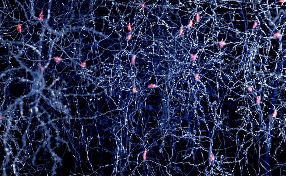
At the core of neuronal communication lie action potentials, brief, rapid electrical impulses that travel down the axon to transmit information for brain function. Abnormalities in neuron firing are associated with brain disease, including Parkinson’s, Alzheimer’s, epilepsy, and schizophrenia. Historically, voltage imaging instrumentation has struggled with low sensitivity, thereby challenging the ability to effectively track neuron firing in cell populations.
In a new study published in Cell titled, “Imaging high-frequency voltage dynamics in multiple neuron classes of behaving mammals,” researchers from Stanford University have now developed ultra-sensitive optical instruments that can detect high-frequency dynamics of fluorescent genetically encoded voltage indicators to reveal neuronal brain activity in mice. The study leverages fiber photometry, a neuroimaging technique that uses optical fibers to deliver excitation light and collect emitted fluorescence, allowing researchers to measure population-level neural activity in vivo.
“We’re getting a very broad view of waves propagating across the brain,” said Mark Schnitzer, PhD, corresponding author of the study and professor of biology and applied physics at Stanford. “We can look at multiple brain areas at once and see the brain waves sweeping across the cortex, the brain’s outermost layer of nerve tissue, with cell-type specificity.”
The study builds upon a decade of work on optical techniques termed transmembrane electrical measurements performed optically (TEMPO), which was first reported in a 2016 Cell paper. Schnitzer, as well as Michael Lin, PhD, professor of neurobiology and bioengineering at Stanford, were among the original TEMPO team. Lin is also co-author on the current study.
The researchers demonstrated the use of two new complementary TEMPO instruments. First, a fiber optic sensor that can track the electrical activity in freely moving and head-fixed behaving mice with ten times more sensitivity than existing technologies. Additionally, the sensor allows hour-long recordings and monitors two neuron classes per fiber-optic probe. Second, an optical mesoscope that can provide an 8 mm-wide brain image and show neural activity across the mouse neocortex, the layer of the brain responsible for high-level functions such as perception and cognition.
According to the authors, TEMPO revealed distinctive brain wave phenomena and can empower investigations of how waves influence neural computation. In awake mice, TEMPO revealed new sensory-evoked neural interactions and traveling gamma and 3–7 Hz waves in the visual cortex and bidirectional propagation for both hippocampal theta and beta waves. By reporting rhythms up to ~100 Hz, TEMPO can show contributions of different neuron types to the brain’s high-frequency waves and their roles in health and disease.
“It seems the brain has an internal clock that synchronizes neural activity, but these traveling waves may also actively reorganize neural circuits across large distances, beyond just local connections,” said co-lead author Radosław Chrapkiewicz, PhD, director of engineering in the Schnitzer lab. “This could play an important role in further bio-inspired AI models.”
The authors stated that the instruments and data analytics reported in the study enable a wide range of studies in high-frequency voltage dynamics in identified neuron classes of behaving animals.
“There are a lot of very important applications in the field of neuroscience for understanding pathology and different dynamics in the brain,” said Simon Haziza, PhD, research scientist at Stanford and lead author of the study. “We are just scratching the surface.”
The post New Voltage Sensing Technology Images Brain Activity to Understand Disease appeared first on GEN – Genetic Engineering and Biotechnology News.