
Introduction
Nanotechnology has been known as the most popular research area lately1. Nanoparticles of noble metals are of interest due to their biocompatibility. They are effectively used in areas such as medicine, food, agriculture, pharmacy, and water treatment2. Physical and chemical methods have been employed to obtain metal nanoparticles. A significant amount of energy is spent, and a large amount of toxic chemicals are produced for nanomaterials obtained by traditional methods. Therefore, there is a widespread trend worldwide to obtain environmentally friendly, non-toxic, and affordable nanoparticles that adhere to the principles of “green chemistry.”
Different natural materials can be used to synthesize nanoparticles biologically. Plants are essential because they contain bioactive compounds that act as reducing agents in nanoparticle fabrication3. Natural products show effective biological action because of their presence of bioactive compounds. The use of natural products in food and medicine is as old as human history4,5,6. Due to the side effects of chemical products, there is a growing tendency to use natural products instead of chemicals in food and medicinal products7. Additionally, the natural product-mediated synthesis of nanoparticles could be an effective material for use in nutrition and pharmaceuticals. In this context, Centaurea saligna-mediated nanoparticles could be a crucial material for biotechnology and food applications.
The Centaurea genus, which belongs to the Asteraceae family, comprises 700 species distributed across Asia, Africa, the Americas, and Europe. Centaurea species grow widely in Turkey8. The Centaurea species exhibits significant biological effects, including anticancer, anti-ulcerogenic, anti-inflammatory, and antioxidant properties. The research on this plant yielded the separation of fascinating compound classes, including flavonoids, terpenes, steroids, phenolics, and alkaloids. Centaurea species are used as folk remedies for various diseases such as urethritis, abscesses, hemorrhoids, peptic ulcers, and colds9. The usage of Centaurea saligna for the synthesis of silver nanoparticles is significant for biological activity. Since this plant includes significant bioactive compounds that act as reducing, capping, and stabilizing agents.
Reactive oxygen species (ROS) are molecules that damage the human body10. These molecules are by-products of cellular metabolism, particularly during aerobic respiration. They include non-radical molecules such as hydrogen peroxide, free radicals such as superoxide, and radicals11. Due to their unpaired electrons, ROS efficiently react with proteins and DNA. High levels of ROS can result in oxidative stress, which damages cellular components and contributes to aging, as well as diseases like cancer, neurodegenerative disorders, and cardiovascular diseases12. Antioxidants neutralize ROS by donating electrons to them. Both endogenous and dietary antioxidants play a key role in oxidative damage13.
It is essential to develop new antibacterial drugs against drug resistance. Inorganic materials in the form of silver nanoparticles can be alternative drugs to control pathogenic microbial infections. Silver nanoparticles have been determined to have antibacterial effects14,15. Silver nanoparticles affect different parts of bacteria and inhibit their growth. Nanoparticles also cause membrane damage, leakage of cell components, and inhibition of crucial proteins, thus stopping microbial growth. AgNPs are responsible for killing and inhibiting microorganisms16.
Herein, phytochemical analysis of Centaurea saligna, including quantitative analysis of phenolics with antioxidant and antimicrobial activity, was carried out. To our knowledge, this is the first study on this subject.
Results and discussion
Synthesis and UV–Vis analysis
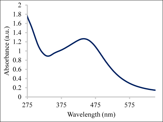
AgNPs@cs were synthesized from the leaves of Centaurea saligna using green approaches, which are cost-effective, efficient, scalable, and environmentally friendly. The bioactive compounds found in the leaves of C. saligna act as reducing agents. Because the Centaurea species contains significant biologically active compounds, silver nanoparticles synthesized from this plant are expected to exhibit high biological activity. UV–Vis analysis revealed a maximum absorption at 441 nm, indicating the formation of silver nanoparticles (Fig. 1). The absorption at 400–450 nm confirms the synthesis of silver nanoparticles. The signal observed at this wavelength is well-documented for a variety of metal nanoparticles with sizes ranging from 2 to 100 nm due to surface plasmon resonance17.

UV–Vis spectrum of AgNPs@cs. The maximum absorption was observed at 441 nm.
Since scutellarin was the primary compound in C. saligna, the mechanism was shown with this molecule (Fig. 2).

Proposed reaction mechanism for the formation of AgNPs@cs.
Fourier transform infrared spectroscopy (FTIR) analysis
The responsible molecules for reducing agents were determined by FTIR analysis. The signal at 3261 cm−1 and 2921 cm−1 may be due to the hydroxyl stretching and C-H stretching of the alkane, respectively. The absorption peaks at 1594 cm−1 and 1381 cm−1 could be attributed to the N–H bending of amine and the OH bending, respectively. The stretching of the ether peak and C-F stretching signal were observed at 1243 cm−1 and 1009 cm−1, respectively (Fig. 3). The FTIR values obtained in this study are consistent with those in previous studies. Silver nanoparticles were synthesized using the walnut leaf18, and the OH bending signal was observed at 3258 cm−1. Silver nanoparticles were produced using Tagetes erecta leaves19, and the OH signal was observed at 3305 cm−1, CH stretching signals of alkanes occurred at 2973 cm−1 and 2885 cm−1. The interaction of silver ions with flavonoids, terpenoids, and phenolics created the colloidal suspension. Therefore, the reduction of silver ions by comparable natural components was ascertained using an FTIR20.

FTIR spectrum of AgNPs@cs.
X-ray diffraction (XRD) measurement
XRD measurement presented the particle size of nanoparticles as well as crystal structures. X’Pert HighScore Plus software was used to calculate particle size. The signal (2θ) observed at 38.12°, 44.26°, 64.53°, 77.47°, and 81.51° degrees corresponded to the crystal planes [1 1 1, 2 0 0, 2 2 0, 3 1 1, and 2 2 2] (ICDD, no:96-500-0219), verifying the structure as face-centered cubic21 (Fig. 4). The dimension of the particle was calculated as 18.49 nm using the Scherrer Equation (Eq. (1)).
$${text{D}} = , 0.{9 }lambda , / , beta {text{ cos }}theta$$
(1)

XRD spectrum and full width at half maximum values of AgNPs@cs. Average particle size was calculated as 18.49 nm.
Transmission electron microscopy (TEM) analysis
TEM analysis determined the morphology and size of green synthesized nanoparticles (Fig. 5). The particle size was 16.2 nm. Due to the different physical and chemical properties of nanoparticles, their sizes and shapes are significant. The TEM image revealed the homogeneously distributed monodisperse AgNPs@cs with a spherical shape. The monodispersity could be due to the capping of bioactive compounds.

TEM images and particle size distribution of AgNPs@cs.
The intense signals observed at 3.3 keV in the EDX spectrum confirmed the formation of AgNPs@cs (Fig. 6). This characteristic signal at 3.0–3.3 keV is attributed to the surface plasmon resonance of silver nanoparticles22. Elemental analysis exhibited remarkable yield (79.13%).

EDX spectrum and elemental analysis of nanostructure. The silver nanoparticles were synthesized in high yield (79.13%).
Zeta potential analysis
The surface charge of particles in suspension is determined by zeta potential measurement, which provides insight the stability since zeta potential is an indicator of electrostatic interactions between particles. A high zeta potential (positive or negative) generally reveals good dispersion and stability as particles repel each other and resist aggregation. A low zeta potential indicates that particles are more likely to aggregate, leading to instability. The values of zeta potential between the 30 mV and 60 mV show the good stability of nanoparticles23. The zeta potential of AgNPs@cs was calculated as − 20.3 mV, indicating moderate stability (Fig. 7).

Zeta potential of AgNPs@cs. The zeta potential (−20.3 mV) reveals the stability.
Quantitative analysis of natural compounds
Quantitative analysis is essential for the synthesis of AgNPs, as it indicates natural compounds that reduce, stabilize, and cap the nanoparticles. The analysis of bioactive compounds in Centaurea saligna water extract was determined by LC–MS/MS and scutellarin (8.67 mg/g extract), shikimic acid (3.97 mg/g extract), chrysin (2.67 mg/g extract), and chlorogenic acid (0.55 mg/g extract) were found as major compounds by LC–MS/MS (Table 1). Quantification of phenolics found in Robinia pseudoacacia leaves and flowers was executed. Syringic acid (24.78 µg/g extract) was found as a major compound in leaves and rutin (199.74 µg/g extract) was found in flowers24. In another study, chlorogenic acid (250.171 µg/g extract) was found as a major product of Silybum marianum flowers25. Phytochemical analysis of phenolic compounds in Hypericum heterophyllum flowers resulted in the determination of chlorogenic acid as a major compound26. Syringa vulgaris was reported to contain hesperidin as an major compound27. The activity may be due to the corresponding bioactive compounds. These studies are essential in terms of the isolation of bioactive compounds from plants, the determination of their structures, and revealing their biological activities.
Antioxidant activity
The antioxidant activity of the extract and AgNPs@cs was evaluated using the DPPH, ABTS, and FRAP assays. In DPPH free radical scavenging activity, nanoparticles displayed significant activity with the value of 9.27 ± 0.14 (IC50, µg/mL), and the activity of the extract was determined as 10.47 ± 0.31 (IC50, µg/mL). However, the effect of standard BHT was calculated as 12.45 ± 0.41 (IC50, µg/mL). In the ABTS radical cation scavenging effect, the same trend was observed. AgNPs@cs displayed a better effect (7.43 ± 0.15, IC50, µg/mL) than that of the extract (9.44 ± 0.35, IC50, µg/mL) and standard BHT (8.13 ± 0.09, IC50, µg/mL). In FRAP activity, nanoparticles displayed higher activity (5.4 ± 0.35, µmol TE/mg extract) than that of the extract (4.78 ± 0.15, µmol TE/mg extract) but lower activity than standard BHT (6.29 ± 0.13, µmol TE/mg extract) (Fig. 8). In Fig. 8, the different letters (a, b, c) reveal the statistical differences between each group. It was reported that the silver nanoparticles were synthesized using the plants. The walnut leaves were used for the synthesis of silver nanoparticles, which showed high activity against the L929, MCF-7, and H1299 cell lines28. Another study reported that Lactuca anatolica root aqueous extract was used for the synthesis of AgNPs that displayed good antioxidant, antibacterial, and enzyme inhibition activities29.

Antioxidant activity of the extract and nanoparticles. The different letters (a, b, c) indicate the statistical differences in the sample activity. One-way ANOVA followed by Tukey’s multiple comparison test was used to compare the activities of each sample. A level of probability of < 0.05 indicates statistical significance.
Antibacterial activity
The antibacterial effect of nanoparticles and extract was tested using gram-positive and gram-negative bacteria. The activity was measured using the minimum inhibitory concentration assay. AgNPs@cs displayed better activity than the extract. Various concentrations of samples were incubated with the bacteria in an aqueous LB broth. The extract effect against the B. subtilis, S. aureus, E. coli, P. aeruginosa were found to be 21, 42, 84, and 84 μg/mL, respectively. However, the AgNPs@cs activity was calculated as 21, 21, 42, and 42 μg/mL, respectively. The standard Amoxicillin activity was found to be 10.5, 21, 21, and 21 against B. subtilis, S. aureus, E. coli, and P. aeruginosa, respectively (Table 2). The nanoparticles displayed the same activity as the standard against S. aureus. It is well known that silver metal exhibits perfect antibacterial activity. The bioactive compounds are expected to enhance the activity.
Conclusion
The synthesis of AgNPs@cs using the Centaurea saligna leaves represents an eco-friendly, scalable, cost-effective method. This study showed that plant-derived bioactive chemicals can successfully act as stabilizing and reducing agents, resulting in stable and valuable nanoparticles. The produced nanoparticles had high antibacterial and antioxidant activity, demonstrating their potential for use in biomedical applications such as infection prevention, medication administration, and wound healing. Quantitative analysis of phenolic compounds in C. saligna was investigated. The major compounds, scutellarin, shikimic acid, and chrysin may be responsible for bioactivities. Moreover, these compounds acted as reducing, stabilizing, and capping agents. In the future study, in vivo toxicity, biocompatibility, and long-term stability may be assessed.
Methods
Plant materials
Centaurea saligna (K.Koch) Wagenitz was collected from Bingöl, South of Ortakoy, at an altitude of 1600–1700 m. Dr. Lütfi Behcet, Bingol University, identified the plant, and a voucher specimen was kept at the Bingol University herbarium (BIN6121).
Synthesis and characterization of silver nanoparticles
The dry leaves of Centaurea saligna (5.0 g) were heated with 150 mL of deionized water at 40 °C for 2 h30. The solution was filtered, and then it was treated with AgNO3 (1.0 mM, 50 mL) for 2 h at 55 °C. The reaction mixture was subjected to centrifugation for 15 min at 5000 rpm and then dried by lyophilization. The synthesized AgNPs@cs were characterized by the analytical procedures. UV–Vis (Hitachi U-2900) spectrophotometer demonstrated the maximum absorption of nanoparticles. Transmission electron microscopy (TEM) (Empyrean, Malvern Panalytical diffractometer) measurement presented the morphology of nanostructures. Fourier transform infrared spectroscopy (Jasco FT/IR 4700) presented the functional groups of molecules. The crystal structure with particle size was defined by X-ray diffraction (Empyrean, Malvern Panalytical diffractometer). The elemental analysis was carried out by an EDAX detector and Energy Dispersive X-ray (EDX). The stability of nanostructures was evaluated using the Zetasizer Nano ZSP (Malvern) instrument30.
Antioxidant activity
AgNPs@cs and extract were subjected to the DPPH• experiment. The stock solution of extract and silver nanoparticles (1.0 mg/mL) was prepared. The treatment of samples with the DPPH solution yielded a decrease in absorbance, which determined the activity. After preparing the ABTS radical cation, the samples reacted with the corresponding ABTS radical cation. The extract and AgNPs@cs were subjected to the FRAP assay. The phosphate buffer and potassium ferric cyanide were mixed with samples. Absorbance measurement presented the activity of extract and nanostructure31.
Evaluation of antibacterial activity
The antibacterial activity of extract and nanoparticles was evaluated using the minimum inhibition concentration method. The gram-positive and gram-negative bacteria in the Luria broth medium were used. The extract and nanoparticles were dissolved in deionized water. pH adjustment was carried out using NaOH. The colonies were measured after the slides were cultured for eighteen hours at 37 °C. The slides exposed to microorganisms were placed in an autoclaved Luria broth medium (1%) and solidified with agar (20 mg/mL). The test samples were dissolved in deionized water. The colonies were measured after the slides were cultured for eighteen hours at 37 °C. Deionized water was used for serial dilution, and the concentration was diluted from 168 to 10.50 μg/mL. The 96-well plate was used for the experiment. Bacterial concentrations in the Luria broth medium (5.0 mL) ranged from 105 to 106 CFU/mL (cfu: colony forming units). A positive control was the Luria broth medium. The negative control was inoculated broth. LB agar plates were used to plate the material (100 μL)32.
Quantitative analysis of phenolic compounds
Quantitative analysis of phenolic compounds in Centaurea saligna leaves was ascertained by LC–ESI–MS/MS analysis. An Agilent Technologies 1260 Infinity II system, coupled with a 6460 Triple Quadrupole mass spectrometer, was employed for the study. The extract (50 mg) was dissolved in methanol/hexane (1/1) into the Eppendorf. Afterward, the mixture was centrifuged for 5 min. A sample (100 µl) was taken from the methanol phase of the solution, and this sample was diluted to 900 µl by adding an equal volume of deionized water (450 µl) and methanol (450 µl). After filtration (0.45 µm), the sample was pipetted into a vial. The reverse phase column was employed. The mobile phase included the A: formic acid (0.1%) and ammonium formate dissolved in water (5.0 mM), and B: formic acid (0.1%) dissolved in acetonitrile. The program was set as 12% for 0 min, 23% for 8 min, 70% for 9–15 min, and 100% for 16–30 min and was applied in the B mobile phase26.
Statistical analysis
One-way ANOVA in GraphPad Prism (8.0.1) was used for statistical analysis. Tukey’s multiple comparison test was used to compare the activities of each sample. The findings were presented as mean values ± standard deviations. The different letters (a, b, c) indicate the statistical differences between each group (P < 0.05).
Data availability
All data generated during this study are included in the manuscript.
References
-
Martínez, G. et al. Environmental impact of nanoparticles’ application as an emerging technology: A review. Materials 14, 166. https://doi.org/10.3390/ma14010166 (2021).
-
Kumari, S., Tehri, N., Gahlaut, A. & Hooda, V. Actinomycetes mediated synthesis, characterization, and applications of metallic nanoparticles. Inorg. Nano-Met. Chem. 51, 1386–1395. https://doi.org/10.1080/24701556.2020.1835978 (2020).
-
Gecer, E. N. Green synthesis of silver nanoparticles from Salvia aethiopis L. and their antioxidant activity. J. Inorg. Organomet. Polym. Mater. 31, 4402–4409. https://doi.org/10.1007/s10904-021-02057-3 (2021).
-
Topçu, G. et al. Diterpenes from the berries of Juniperus excelsa. Phytochemistry 50, 1195–1199. https://doi.org/10.1016/S0031-9422(98)00675-X (1999).
-
Elmastas, M., Ozturk, L., Gokce, I., Erenler, R. & Aboul-Enein, H. Y. Determination of antioxidant activity of marshmallow flower (Althaea officinalis L.). Anal. Lett. 37, 1859–1869. https://doi.org/10.1081/AL-120039431 (2004).
-
Demirtas, I., Erenler, R., Elmastas, M. & Goktasoglu, A. Studies on the antioxidant potential of flavones of Allium vineale isolated from its water-soluble fraction. Food Chem. 136, 34–40. https://doi.org/10.1016/j.foodchem.2012.07.086 (2013).
-
Sahin Yaglioglu, A. et al. Antiproliferative activity of pentadeca-(8E, 13Z) dien-11-yn-2-one and (E)-1,8-pentadecadiene from Echinacea pallida (Nutt.) Nutt. roots. Med. Chem. Res. 22, 2946–2953. https://doi.org/10.1007/s00044-012-0297-2 (2013).
-
Bouafia, M., Benarfa, A., Gourine, N. & Yousfi, M. Seasonal variation of fatty acid composition, tocopherol content and antioxidant activity of lipid extracts from Centaurea sp. Food Biosci. 37, 100728. https://doi.org/10.1016/j.fbio.2020.100728 (2020).
-
Twaij, H. A., Kery, A. & Al-Khazraji, N. K. Some pharmacological, toxicological and phytochemical investigations on Centaurea phyllocephala. J. Ethnopharmacol. 9, 299–314. https://doi.org/10.1016/0378-8741(83)90037-5 (1983).
-
Elmastaş, M., Telci, İ, Akşit, H. & Erenler, R. Comparison of total phenolic contents and antioxidant capacities in mint genotypes used as spices/Baharat olarak kullanılan nane genotiplerinin toplam fenolik içerikleri ve antioksidan kapasitelerinin karşılaştırılması. Turk. J. Biochem. 40, 456–462. https://doi.org/10.1515/tjb-2015-0034 (2015).
-
Erenler, R. et al. Chemical constituents, quantitative analysis and antioxidant activities of Echinacea purpurea (L.) Moench and Echinacea pallida (Nutt.) Nutt. J. Food Biochem. 39, 622–630. https://doi.org/10.1111/jfbc.12168 (2015).
-
Erenler, R. et al. Isolation and identification of chemical constituents from Origanum majorana and investigation of antiproliferative and antioxidant activities. J. Sci. Food Agr. 96, 822–836. https://doi.org/10.1002/jsfa.7155 (2016).
-
Erenler, R. et al. Chemical constituents isolated from Origanum solymicum with antioxidant activities. Eurasia Proc. Sci. Tech. Eng. Math. 1, 139–145 (2017).
-
Dag, B. Green synthesis, characterization, and antioxidant activity of silver nanoparticles using Stachys annua L. subsp Annua var. annua.. Particul. Sci. Technol. 40, 512–520. https://doi.org/10.1080/02726351.2021.1966689 (2022).
-
Erenler, R. & Dag, B. Biosynthesis of silver nanoparticles using Origanum majorana L. and evaluation of their antioxidant activity. Inorg. Nano-Met. Chem. 52, 485–492. https://doi.org/10.1080/24701556.2021.1952263 (2022).
-
Karan, T., Erenler, R., Gonulalan, Z. & Kolemen, U. Biogenic synthesis of silver nanoparticles using Sambucus nigra leaves: Elucidation, antimicrobial, antioxidant activities and quantification of phenolics. Chem. Pap. 78, 473–481. https://doi.org/10.1007/s11696-023-03103-9 (2024).
-
Kumar, H. et al. Antioxidant functionalized nanoparticles: A combat against oxidative stress. Nanomaterials 10, e1334. https://doi.org/10.3390/nano10071334 (2020).
-
Seçme, A., Bozer, B. M., Kocaman, A. Y., Erenler, R. & Calimli, M. H. Synthesis, characterization, and anticancer properties of Ag nanoparticles derived from walnut leaves tested on cells of L929, MCF-7 and H1299. J. Drug Deliv. Technol. 94, 105478. https://doi.org/10.1016/j.jddst.2024.105478 (2024).
-
Erenler, R., Gecer, E. N., Genc, N. & Yanar, D. Antioxidant activity of silver nanoparticles synthesized from Tagetes erecta L. leaves. Int. J. Chem. Technol. 5, 141–146. https://doi.org/10.32571/ijct.1005275 (2021).
-
Sahin Yaglioglu, A., Erenler, R., Gecer, E. N. & Genc, N. Biosynthesis of silver nanoparticles using Astragalus flavesces leaf: Identification, antioxidant activity, and catalytic degradation of methylene blue. J. Inorg. Organomet. Polym. Mater. 32, 3700–3707. https://doi.org/10.1007/s10904-022-02362-5 (2022).
-
Erenler, R. et al. Biosynthesis, characterisation, and antioxidant activity of silver nanoparticles using Schinus molle L. Trends Sci. 20, 6105–6105. https://doi.org/10.48048/tis.2023.6105 (2023).
-
Kaviya, S., Santhanalakshmi, J., Viswanathan, B., Muthumary, J. & Srinivasan, K. Biosynthesis of silver nanoparticles using Citrus sinensis peel extract and its antibacterial activity. Spectrochim. Acta A Mol. Biomol. Spectrosc. 79, 594–598. https://doi.org/10.1016/j.saa.2011.03.040 (2011).
-
Hedberg, J. et al. Interactions between surfactants and silver nanoparticles of varying charge. J. Coll. Interface Sci. 369, 193–201. https://doi.org/10.1016/j.jcis.2011.12.004 (2012).
-
Başar, Y., Hosaflıoğlu, İ & Erenler, R. Phytochemical analysis of Robinia pseudoacacia flowers and leaf: Quantitative analysis of natural compounds and molecular docking application. Turk. J. Biodiv. 7, 1–10. https://doi.org/10.38059/biodiversity.1446241 (2024).
-
Başar, Y. & Erenler, R. Phytochemical analysis of Silybum marianum flowers: Quantitative analysis of natural compounds and molecular docking application. Turk. J. Biodiv. 7, 20–31. https://doi.org/10.38059/biodiversity.1450643 (2024).
-
Erenler, R., Yaman, C., Demirtas, I. & Alma, M. H. Phytochemical investigation of Hypericum heterophyllum flowers: LC-ESI-MS/MS analysis, total phenolic and flavonoid contents, antioxidant activity. Nat. Prod. J. 13, e120123212672. https://doi.org/10.2174/2210315513666230112165545 (2023).
-
Erenler, R., Karan, T. & Hosaflioglu, İ. Phytochemical analysis of Syringa vulgaris: Quantitative analysis of natural compounds by LC-ESI-MS/MS. Turk. J. Biodiv. 6, 75–78. https://doi.org/10.38059/biodiversity.1312872 (2023).
-
Seçme, A., Bozer, B. M., Kocaman, A. Y., Erenler, R. & Calimli, M. H. Synthesis, characterization, and anticancer properties of Ag nanoparticles derived from walnut leaves tested on cells of L929, MCF-7 and H1299. J. Drug Deliv. Technol. https://doi.org/10.1016/j.jddst.2024.105478 (2024).
-
Nayel, N. et al. Characterization and comparative investigation of in vitro bioactivities for Lactuca anatolica root aqueous extract and their green-chemical synthesized nanoparticles; molecular docking studies. BioNanoScience https://doi.org/10.1007/s12668-024-01353-9 (2024).
-
Erenler, R., Gecer, E. N., Hosaflioglu, I. & Behcet, L. Green synthesis of silver nanoparticles using Stachys spectabilis: Identification, catalytic degradation, and antioxidant activity. Biochem. Bioph. Res. Commun. 659, 91–95. https://doi.org/10.1016/j.bbrc.2023.04.015 (2023).
-
Erenler, R. & Gecer, E. N. Synthesis of silver nanoparticles using Sideritis montana L. leaf extract: Characterization, catalytic degradation of methylene blue and antioxidant activity. J. Nano. Res. 75, 17–28 (2022).
-
Karan, T., Gonulalan, Z., Erenler, R., Kolemen, U. & Eminagaoglu, O. Green synthesis of silver nanoparticles using Sambucus ebulus leaves extract: Characterization, quantitative analysis of bioactive molecules, antioxidant and antibacterial activities. J. Mol. Struct. 1296, 136836. https://doi.org/10.1016/j.molstruc.2023.136836 (2024).
Acknowledgements
The author thanks Dr. Lütfi Behcet for identifying the plant.
Ethics declarations
Competing interests
The authors declare that they have no competing interest.
Additional information
Publisher’s note
Springer Nature remains neutral with regard to jurisdictional claims in published maps and institutional affiliations.
Rights and permissions
Open Access This article is licensed under a Creative Commons Attribution-NonCommercial-NoDerivatives 4.0 International License, which permits any non-commercial use, sharing, distribution and reproduction in any medium or format, as long as you give appropriate credit to the original author(s) and the source, provide a link to the Creative Commons licence, and indicate if you modified the licensed material. You do not have permission under this licence to share adapted material derived from this article or parts of it. The images or other third party material in this article are included in the article’s Creative Commons licence, unless indicated otherwise in a credit line to the material. If material is not included in the article’s Creative Commons licence and your intended use is not permitted by statutory regulation or exceeds the permitted use, you will need to obtain permission directly from the copyright holder. To view a copy of this licence, visit http://creativecommons.org/licenses/by-nc-nd/4.0/.
About this article

Cite this article
Gecer, E.N. Phytochemistry of Centaurea saligna: silver nanoparticle synthesis, quantification of natural compounds, antioxidant and antibacterial activity. Sci Rep 15, 26307 (2025). https://doi.org/10.1038/s41598-025-11970-3
-
Received:
-
Accepted:
-
Published:
-
DOI: https://doi.org/10.1038/s41598-025-11970-3