Introduction
With the increasing environmental problems, there has been a focus on green chemistry. As a result, the synthesis of nanomaterials through clean environmental methods has also increased significantly1,2. Green chemistry seeks to minimize the use of hazardous substances that impact human health and the environment. Designing safer products minimizes potential risks associated with nano-applications3. Green chemistry has revolutionized nanotechnology by introducing environmentally friendly, cost-effective, and sustainable methods in nanoparticle synthesis4. Studies have utilized bacteria, mushrooms, plant extracts, and enzymes to produce silver nanoparticles (AgNPs)5. Bioproduced silver nanoparticles have significant physical and chemical properties that find wide applications in medicine6,7. Plant extract-based synthesis of nanoparticles is a clean, cheap, and quick method that produces non-toxic and diverse nanoparticles8. Plants can help transform metal ions into nanoparticles using their metabolites. These metabolites can act as reducing and capping agents, either primary (monosaccharides, proteins, enzymes, and lipids) or secondary (polyphenolics, flavonoids, alkaloids, and terpenes)9.
AgNPs exhibit versatile applications in electronics, optics, biosensing, catalysis, plasmonics, antimicrobial agents, and cancer therapy10. AgNPs have various effects, including anti-cancer, anti-inflammatory, antioxidant, and wound-healing properties, as well as antihelminthic and anti-larval effects11. AgNPs effectively combat multidrug-resistant microorganisms by disrupting cell function, penetrating membranes, and preventing microbial growth and biofilm formation12,13.
Nanoparticle properties depend on size, shape, and synthesis method14.
The alarming rise of multidrug-resistant bacterial strains is a direct result of excessive use of antibiotics. This misuse has also paved the way for biofilms to form, rendering antibiotics ineffective and posing a grave threat to public health15. Biofilms are formed when planktonic bacteria attach to a surface, create a microcolony, and eventually develop into a community enclosed in an extracellular matrix. This provides high protection and allows them to thrive in various environments16,17. Biofilms have high resistance to antimicrobial agents and the host immune system, leading to uncontrollable infections18.
Biofilms cause 60% of human diseases and are challenging to eradicate19,20. Microorganisms like Staphylococcus aureus, Staphylococcus epidermidis, Pseudomonas aeruginosa, and Escherichia coli form biofilms21.
Meyer et al. used brine shrimp (Artemia salina) as a simple zoological organism to test lethality22. Measuring cytotoxicity is easy and cheap with the brine shrimp lethality assay23.
Prunus mahaleb L., known as Mahaleb, Mahlab cherry, and St. Lucy cherry, is a species of Rosaceae family, subfamily Prunoidae24,25. P. mahaleb is a deciduous tree that grows up to 5 m. It is found in central Europe, the Mediterranean, southwestern Asia, and northwestern Africa26. Cherry plants are traditionally used to treat diarrhea, high blood pressure, kidney stones, nausea, and swelling of the stomach, intestines, and kidneys. They are also known for their skin benefits27. It has been used in folk remedies to treat sensory organs, the heart, asthma, and liver pain24. The study focuses on evaluating the cytotoxic and anti-biofilm capabilities of silver nanoparticles synthesized using extracts from the stems, fruit pericarps, and leaves of P. mahaleb.
Material and methods
Extraction and fraction of plant
Prunus mahaleb L. plant was collected from Marivan, Iran, in August 2017, with the necessary permissions obtained from the relevant authorities. A voucher specimen (UKH 2706) was deposited in the herbarium of the University of Kashan to ensure accessibility for future studies. Dr. Toluei verified the plant identification. The plant materials, including leaves, stems, and fruit pericarps, were washed, dried, and ground into powder. The methanolic extract samples were obtained using soxhlet extractor and then evaporated to obtain solid masses (rotary evaporator, Heidolf, Rotary Evaporator, model-RE 801, Germany). The methanol extract (3 g)was dissolved in 300 ml of distilled water and then divided into n-hexane, ethyl acetate, and water fractions. The fractions were concentrated using a vacuum evaporator and dried in a vacuum oven. The aqueous fraction was kept for further process.
Bioproduction of AgNPs
AgNPs were produced by mixing aqueous extracts of P. mahaleb stem, leaf, and fruit pericarp with silver nitrate solutions at concentrations of 1 mM in water, maintained at pH 7. The mixture was then incubated for 9, 3, and 28 days (for stem, leaf, and fruit pericarp extracts, respectively) at 60 °C without light. The yellow solutions turned brown, were centrifuged at 12,000 rpm for 20 min, washed with distilled water, and then dried in an oven at 60 °C for 24 h.
Characterization of AgNPs
AgNPs were detected using a UV–visible spectrophotometer (350-800 nm) and identified via X-ray diffraction with a Philips-X ‘pertpro diffractometer (2θ range: 10°−80°). The samples were analyzed using a Nicolet Magna-550 spectrometer at room temperature after being diffused in KBr pellets for Fourier transform infrared (FT-IR) spectroscopy. Samples were analyzed using a Philips EM208 LEO-1455VP scanning electron microscope at 200 kV to observe morphology and particle sizes. Compound elements were determined accurately using EDX with 20 kV acceleration.
Brine shrimp lethality assay (BSLA)
The cytotoxicity property of the AgNPs was determined by BSLA following Meyer et al.22. The brine shrimp lethality bioassay determines the cytotoxic ability of various bioactive compounds28. The study used brine shrimp (Artemia salina) as a screening tool. The shrimp eggs were hatched in artificial seawater with 3.8% NaCl for 48 h at 30 °C, with proper lighting and oxygenation. AgNPs were mixed with 3.2% dimethyl sulfoxide (DMSO) and seawater to create concentrations from 0.1 µg/ml to 300 µg/ml.
Ten mature shrimp were utilized in the experimental and control tubes, with Vincristine sulfate (VS) and DMSO as the positive and negative controls, respectively29. After 24 h, the number of surviving Nauplii was assessed, and the percentage of lethality was calculated. Use the formula 1 for accurate mortality percentage calculation.
$$Percentage , of , the , dead , larvae , after , 24 h, = ,left[ {left( {m{-}M} right) , / , S} right], times ,100$$
(1)
Formula: m = average dead larvae in sample, M = average dead larvae in blank control, S = average live larvae in blank control.
Biofilm formation assay
Antibiotic-resistant clinical strains were obtained from urine and lung specimens of patients in Qom, Iran. Table 1 lists the characteristics of each strain.
Antibiotic susceptibility profiling of the clinical isolates was performed using the disk diffusion method (Kirby-Bauer) by the Clinical and Laboratory Standards Institute (CLSI) guidelines (2023). Bacterial suspensions were adjusted to a turbidity equivalent to a 0.5 McFarland standard and uniformly spread on Mueller–Hinton agar plates using sterile swabs. Commercial antibiotic disks were placed on the agar surface, and the plates were incubated at 37 °C for 24 h. Inhibition zone diameters were measured using a caliper, and isolates were categorized as Resistant, Intermediate, or Susceptible based on CLSI interpretive criteria.
The microtiter plate method was used to determine the formation of biofilm. 0.2 ml of the bacterial suspension (0.5 Macfarland) diluted (1:100), and TSB was added to the tests and control wells, respectively. The plate was incubated at 37 °C for 24 h. Then, the wells were washed with sterilized phosphate-buffered saline (PBS) to remove loosely adhered cells. Next, the samples were fixed with methanol, stained with crystal violet, washed, and had the optical density measured at 570 nm using an ELISA microplate reader. Biofilm formation was calculated using Stepanovic ‘s method30.
Effect of AgNPs on biofilm inhibition
AgNPs were evaluated at a 2 μg/mL concentration for their ability to combat biofilm formation by clinical strains. A mixture of 0.1 ml of AgNPs solution and 0.1 ml of bacterial suspension was added to each well. The plate was then incubated at 37 °C for 24 h, and biofilm formation was assessed using an ELISA reader set at 570 nm after staining. Calculate the percentage of biofilm inhibition using formula 2.
$$% , biofilm , inhibition , = frac{(C – B) – (T – B)}{{(C – B)}} , times ,100$$
(2)
In this formula, C is OD570 of positive control, B is OD570 of negative control, and T is OD570 of cells with AgNPs31.
Results and discussion
Characterization of nanocomposites
In this study, silver nanoparticles (AgNPs) were successfully synthesized using aqueous extracts derived from three distinct parts of the Prunus mahaleb plant (leaves, stems, and fruit pericarps). The synthesis was visually confirmed by a noticeable color change from yellow to brown, attributed to the surface plasmon resonance of silver nanoparticles, and further validated by UV–Vis spectrophotometry.
The synthesis approach employed here offers several distinct advantages over conventional green synthesis methods. By utilizing extracts from three plant parts simultaneously, a diverse array of bioactive compounds was introduced into the reaction, enhancing the efficiency, uniformity, and functionality of the resulting nanoparticles. Moreover, precise control over reaction conditions—including constant pH, stable temperature, and specific incubation times—ensured the production of nanoparticles with consistent size, morphology, and superior stability. In contrast, traditional green synthesis methods often rely on a single plant part with minimal control over reaction parameters, resulting in nanoparticles that are less uniform and less stable.
Additionally, the inherent antioxidant and antimicrobial properties of P. mahaleb not only facilitated the reduction of silver ions but also significantly enhanced the biological properties and stability of the synthesized nanoparticles. These characteristics establish the present method as an optimized, eco-friendly, and efficient alternative to traditional green synthesis.
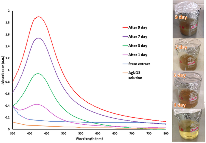
The formation of AgNPs was confirmed using UV–Vis spectrophotometry. As shown in Table S1, silver nitrate solution without plant extract exhibited no significant absorption in the 350–800 nm range. In contrast, the synthesized silver nanocomposites displayed characteristic absorption peaks, directly correlating with nanoparticle formation. Figures 1, 2, and 3 illustrate the absorption spectra and observable color changes of the synthesized nanoparticles derived from the leaf, stem, and fruit pericarp extracts.

Absorption spectra and color change of Ag/AgO synthesis from stem extract over time.

Absorption spectra and color change of Ag/Ag2O nanocomposited synthesis from leaf extract over time.

Absorption spectra color change of Ag/AgCl synthesis from fruit pericarp extract over time.
The Ag/Ag2O nanoparticles had the highest absorption rate and the lowest wavelength among other extracts. Peak intensities for AgNPs gradually increased as time passed, highlighting the importance of synthesis time in achieving optimal characteristics. The nanocomposites synthesized from plant extracts showed increasing peak intensities over time: 3 days for leaf extract, 9 days for stem extract, and 12 days for fruit pericarp extract. Adsorption intensity measures nanoparticle presence in a solution. Higher intensity means more nanoparticles. Our study contributes to nanoparticle synthesis research and will benefit industries that are developing new materials. Ahmad et al. found that as time and reaction progress, the concentration of nanoparticles increases, resulting in more color change in the reaction solution. Their conclusion establishes a direct proportionality between the intensity of the color change and the concentration of the nanoparticles32.
A study utilized Teucrium polium L. extract to create an Ag/AgCl nanocomposite with compelling antibacterial and cytotoxic properties33. Moreover, Ag/AgCl nanocomposites were produced by Teucrium polium and used as sensors for detecting Hg +34. In another study, copper nanoparticles were created using P. mahaleb L as antibacterial, cytotoxic, and anti-mutagenic agents35.
XRD analysis
X-ray diffraction patterns of nanocomposites synthesized from each plant part extract are shown in Fig. 4. These patterns provide valuable insight into unique structural characteristics.

XRD pattern of AgNPs synthesized from stem (a), leave (b), and fruit pericarp (c) extracts.
The XRD patterns display different peaks at specific 2θ values for nanocomposites synthesized from stem, leaf, and fruit pericarp extract. For stem extract, there are five peaks at 33.28°, 38.20°, 44.39°, 64.53°, and 77.50°. On the other hand, Ag/Ag2O nanocomposites have seven peaks at 27.96°, 32.36°, 38.23°, 44.36°, 46.24°, 64.62°, and 77.49°. Similarly, Ag/AgCl nanocomposites synthesized from fruit pericarp extract also show seven peaks at 27.85°, 32.25°, 38.12°, 44.32°, 46.26°, 64.58°, and 77.42°. We used the Scherrer equation to calculate the average size of the crystallite particles.
The nanocomposites extracted from stems, leaves, and fruit pericarps have average sizes of 23.31 nm, 24.46 nm, and 30.49 nm, respectively, calculated using the Scherrer Eq36..
SEM analysis
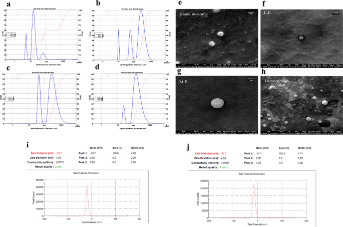
Figure 5 shows SEM images of nanocomposites from different plant parts with their size and morphology. Figure S1 compares the abundance distribution charts of nanocomposites synthesized from each plant part extract using Digimizer software. Table 2 displays the average and maximum abundance distribution of nanocomposite size synthesized from each plant part extract.

SEM images of AgNPs synthesized from stem (a), leave (b), and fruit pericarp (c) extracts.
The Ag/AgO nanocomposites exhibited the smallest size, as observed in the study. The particle count was 100, and all synthesized nanoparticles were spherical. The spherical shape of nanoparticles affects the contact surface and ion release, making them ideal for various uses. This shape has a higher surface-to-volume ratio than others and aligns with XRD analysis results.
EDX analysis
Figure 6 displays the energy-dispersive X-ray spectrometry analysis results of plant-extract-synthesized nanocomposites.

EDX spectra of AgNPs synthesized from stem (a), leave (b), and fruit pericarp (c) extracts.
EDX spectra show silver in AgNPs from stem, leaf, and fruit pericarp (9.01%, 42.34%, and 18.25% w/w). Carbon, oxygen, and nitrogen signals were also detected. The FT-IR analysis provides strong evidence that the surface of metallic nanoparticles has been enriched by extracellular organic materials that originated from the plant extract. The chlorine element in the nanocomposite synthesized from fruit pericarp extract was confirmed by XRD analysis.
FT-IR analysis
The FTIR spectra of the plant extracts (stem, leaf, and fruit) and the synthesized nanomaterials (Ag/AgO, Ag/Ag₂O, and Ag/AgCl nanocomposites) are presented in Figs. 7A, B, and C. In the FTIR spectrum of the stem extract (Fig. 7A-a), a broad absorption band at 3430.93 cm⁻1 is observed, corresponding to the O–H stretching vibrations of hydroxyl groups, which are typically found in polyphenolic compounds. This peak shifts to 3422.01 cm⁻1 in the spectrum of silver nanoparticles (Fig. 7A-b), indicating the involvement of hydroxyl groups in the reduction and stabilization of silver nanoparticles. The C-H stretching vibrations at 2920.76 cm⁻1 in the stem extract remain almost unchanged in the silver nanoparticles (2921.13 cm⁻1), suggesting minimal interaction of aliphatic chains. Notably, the C = C stretching vibrations at 1599.51 cm⁻1 (A) shift to 1628.00 cm⁻1 in the nanoparticles spectrum, highlighting the role of aromatic compounds in nanoparticle stabilization. Peaks at 1384.13 cm⁻1 (A) and 1381.49 cm⁻1 (B) are attributed to C-O or C-N stretching, with slight shifts in the nanoparticles, suggesting potential involvement. The characteristic peak at 471.87 cm⁻1 in the silver nanoparticles spectrum corresponds to Ag–O bonding, confirming the formation of silver nanoparticles.

FT-IR spectroscopy results: (A) stem extract (a) and Ag/AgO nanocomposites synthesized from stem extract (b); (B) leaf extract (a) and Ag/Ag₂O nanocomposites synthesized from leaf extract (b); (C) fruit pericarp extract (a) and Ag/AgCl nanocomposites synthesized from fruit pericarp extract (b).
For the leaf extract (Fig. 7B-a), the broad peak at 3425.00 cm⁻1 corresponds to O–H stretching, which shifts to 3433.44 cm⁻1 in the Ag/Ag₂O nanocomposites (Fig. 7B-b), indicating the involvement of hydroxyl groups in nanoparticle formation. The C-H stretching vibrations at 2924.48 cm⁻1 in the leaf extract are absent in the Ag/Ag₂O spectrum, suggesting the modification of aliphatic compounds during synthesis. The aromatic C = C stretching vibration at 1610.62 cm⁻1 in the leaf extract shifts to 1602.68 cm⁻1 in the nanocomposites, highlighting interactions with the Ag/Ag₂O surface.
In the case of the fruit extract (Fig. 7 C-a), the broad peak at 3410.34 cm⁻1 corresponds to O–H stretching, which shifts to 3429.54 cm⁻1 in the Ag/AgCl nanocomposites (Fig. 7C-b), indicating the participation of hydroxyl groups in nanoparticle synthesis. The C-H vibrations at 2926.64 cm⁻1 shift to 2921.82 cm⁻1. The aromatic C = C stretching at 1630.07 cm⁻1 shifts to 1634.46 cm⁻1, confirming the role of aromatic compounds in nanoparticle formation. Peaks at 1384.87 cm⁻1 (extract) and 1382.92 cm⁻1 (nanomaterial) correspond to C-O or C-N stretching. The characteristic peak at 595.03 cm⁻1 in the Ag/AgCl nanocomposites is associated with Ag–Cl bonding, confirming the successful formation of Ag/AgCl nanocomposites.
These spectral differences collectively demonstrate that the bioactive compounds in the plant extracts (stem, leaf, and fruit) directly participate in the reduction of silver ions and the stabilization of the synthesized silver-based nanomaterials.
Phenolic compounds play a dual role in the green synthesis of metal nanoparticles by acting as both reducing and capping agents. The presence of functional groups such as hydroxyl and carboxyl allows them to effectively reduce metal ions (e.g., Au3⁺, Ag⁺) while simultaneously stabilizing the formed nanoparticles through surface adsorption. This results in well-dispersed nanoparticles with high colloidal stability, as indicated by strongly negative zeta potential values and the absence of significant aggregation. In addition, phenolic compounds are well known for their intrinsic bioactivities, including antioxidant, antimicrobial, anti-inflammatory, and anticancer properties. When conjugated onto the nanoparticle surface, they enhance the biological functionality of the nanomaterials through synergistic effects. Consequently, the use of phenolic-rich plant extracts offers a sustainable and effective strategy not only for nanoparticle synthesis but also for improving their performance in biomedical and environmental applications37,38,39.
In the fruit pericarp extract spectroscopy, the peak at 595.03 indicates C–Cl bonding, consistent with XRD analysis results of its nanoparticles. One of the main factors in reducing silver ions and synthesizing nanoparticles is the presence of phenolic compounds in the plant. Stirbescu et al. used P. mahaleb L. extract to synthesize silver nanoparticles and found it a perfect bio-reducing agent. However, they used a different plant source and hydroalcoholic extract than our study40.
There are reports of other Prunus species in this regard, such as in 2017, when Saravanakumar et al. synthesized silver nanoparticles from the aqueous extract of Prunus japonica leaves. Their results showed that the silver nanoparticles were spherical with an average size of 24 nm41.
In continuation of the nanomaterial characterization, the data presented in Table 2 demonstrate that the physicochemical and biological properties of silver-based nanocomposites are significantly influenced by the specific plant part used during green synthesis. Among the samples studied, the Ag/AgCl nanocomposite synthesized from fruit pericarp exhibited superior properties, including a relatively larger average particle size (61.66 nm), a dominant size distribution in the 50–60 nm range, and the highest optical absorbance at 426.5 nm (2.363). This formulation also showed the strongest biofilm inhibition activity and the highest bioactivity, with an LC50 exceeding 300 µg/ml.
In contrast, nanocomposites synthesized from leaf (Ag/Ag₂O) and stem (Ag/AgO) extracts had smaller particle sizes (45.44 nm and 43.55 nm, respectively), lower absorbance values, and maximum absorbance at shorter wavelengths (414 nm and 426 nm, respectively). These samples demonstrated only moderate biofilm inhibition and lower bioactivity, with LC50 values around 28 ± 0.4 µg/ml.
The observed variations can be attributed to differences in the phytochemical composition of each plant part, which may affect the reducing and stabilizing capacity during nanoparticle formation, as well as the crystalline phases formed. These findings highlight the importance of selecting suitable plant materials for green synthesis to optimize the functional performance of silver-based nanocomposites. Such strategic selection can significantly enhance their applicability in biomedical and antimicrobial domains.
Although SBET data were not measured in the present study, previous literature indicates that the specific surface area of green-synthesized silver nanoparticles (AgNPs) can vary considerably depending on the phytochemical composition of the plant extract used during synthesis. For example, Hazrat Ali et al. (2023) reported a specific surface area of 28.2 m2/g for AgNPs synthesized using Moringa oleifera leaf extract42. Similarly, Sathishkumar et al. (2016) synthesized AgNPs utilizing Coriandrum sativum (parsley) leaf extract, with Brunauer–Emmett–Teller (BET) analysis revealing a porous structure and a surface area of 33.72 m2/g43. In another study, BET analysis of AgNPs synthesized using Brassica oleracea var. botrytis (cauliflower) extract demonstrated a specific surface area of 19.22 m2/g44. These findings collectively underscore the pivotal role of plant-derived phytochemicals in influencing nanoparticle morphology and surface characteristics.
BSLT study
One of the reliable methods for evaluating the toxicity of nanoparticles is the Brine Shrimp Lethality Assay (BSLA). This simple, safe, cost-effective approach does not require feeding the test organism throughout the experimental period and only requires a small amount of the test substance45. The acute toxicity of silver nanoparticles (AgNPs) in Artemia salina is primarily attributed to the interaction of silver ions (Ag⁺) with chitin, the main structural component of the organism ‘s cuticle46. Previous studies have shown no statistically significant difference (P > 0.05) between BSLA and the MTT assay in evaluating nanoparticle cytotoxicity. Therefore, BSLA can be considered a rapid and economical alternative for preliminary nanoparticle toxicity screening47.
In this study, silver nanoparticles synthesized using leaf, stem, and fruit peel extracts of Prunus mahaleb were investigated. The results indicated that AgNPs derived from the leaf and stem extracts exhibited LC₅₀ values of 28 ± 0.40 and 28 ± 0.42 µg/ml, respectively (Table 3). In contrast, AgNPs synthesized from fruit peel had an LC₅₀ greater than 300 µg/ml, suggesting significantly lower toxicity. For further evaluation, these results were compared with those of other biosynthesized nanoparticles.
Fluorescent AgNPs synthesized using Teucrium polium extract exhibited an LC₅₀ of 2.4 µg/ml33; Fe₃O₄ nanoparticles synthesized by Bacillus zhangzhouensis had an LC₅₀ of 0.5 µg/ml48; and Ag/AgCl nanocomposites produced by Bacillus paralicheniformis showed an LC₅₀ of 1 µg/ml49. Likewise, biologically synthesized AgNPs from Marinospirillum alkaliphilum also demonstrated high toxicity, with an LC₅₀ of 1 µg/ml50. In comparison, biosynthesized copper nanoparticles produced using Prunus mahaleb in a separate study exhibited an LC₅₀ of 3.6 µg/mL, indicating higher toxicity than the silver nanoparticles synthesized in the present study.
Furthermore, similar studies have examined the anticancer activity of silver nanoparticles using both BSLA and MTT assays. For instance, Zia et al. (2018) reported that AgNPs synthesized from Bergenia ciliata rhizomes showed anticancer activity with an LD₅₀ of 116.9 mg/ml51. Kumar et al. (2012) found that AgNPs from the alga Sargassum ilicifolium had an LD₅₀ of 10 nM52. Swamy et al. (2015) observed the highest cytotoxicity of AgNPs synthesized from Momordica cymbalaria fruit extract at a concentration of 100 µg/ml using the MTT assay53. Nazari and Kashi (2021) also reported that AgNPs synthesized by Marinospirillum alkaliphilum exhibited potent antibacterial and anticancer properties, particularly against multidrug-resistant bacteria50.
Collectively, these findings suggest that silver nanoparticles synthesized using Prunus mahaleb extracts possess lower toxicity compared to many other biologically synthesized nanoparticles, indicating their strong potential for safe applications in biomedical and environmental fields.
Biofilm inhibition
The antibiotic resistance profiles of the isolated clinical strains are summarized in Fig. 8. Cephalosporins such as Cefixime (62.12%), whereas greater susceptibility was noted for aminoglycosides like Amikacin (45.45%) and Gentamicin (48.48%). Isolates were classified as Resistant, Intermediate, or Susceptible based on CLSI guidelines (2023). A high rate of resistance was observed against the third-generation cephalosporin cefixime (62.12%), whereas greater susceptibility was detected for aminoglycosides, including amikacin (45.45%) and gentamicin (48.48%). Intermediate resistance was generally low across most antibiotics tested. These profiles are particularly relevant in the context of biofilm inhibition, as many of the tested strains exhibit multidrug-resistant phenotypes.

Antibiotic resistance profiles of clinical bacterial isolates.
The eighteen clinical isolates were further categorized into three groups: strong, moderate, and weak biofilm producers based on their biofilm-forming capacity. All biosynthesized Ag/AgCl nanocomposites, regardless of plant part origin, demonstrated antibiofilm activity against these strains. As shown in Fig. 9, biofilm inhibition varied depending on the plant extract used for nanocomposite synthesis.

The comparison chart of biofilm inhibition percentage of AgNPs. The 95% confidence intervals for the measurements are illustrated by the error bars shown in the chart.
Among the tested nanocomposites, those synthesized using fruit pericarp extract exhibited the highest overall biofilm inhibition. Notably, the nanocomposites derived from stem and leaf extracts also showed substantial inhibitory effects across the strain panel. Table 4 summarizes the maximum and minimum biofilm inhibition percentages recorded for each nanocomposite preparation, highlighting variations in efficacy based on the plant part used in synthesis.
Singh et al. found that AgNPs made from Cannabis sativa extract inhibit biofilm formation in gram-negative bacteria. The nanoparticles were particularly effective against P. aeruginosa and E. coli. However, they did not inhibit biofilm formation in S. epidermidis12.
Singh et al. found that AgNPs from Borago officinalis extract inhibit biofilm formation in S. aureus and P. aeruginosa at 10 μg/ml54.
Kordzanganeh et al. discovered that biosynthesized Ag/AgCl nanocomposites by Bacillus paralicheniformis strain Tmas-01 exhibit an anti-biofilm effect on bacterial strains isolated from patients49. In 2021, an Ag/AgCl nanocomposite of Staphylococcus pasteuri was created, exhibiting an antimicrobial effect against antibiotic-resistant strains isolated from patients55.
In other studies, metal oxide nanoparticles such as ZnO, NiO, and Fe₂O₃ were green synthesized using Permelia perlata extract56,57,58, while MgO and Cr₂O₃ nanoparticles were prepared using Taxus wallichiana leaf extract59,60. These nanoparticles exhibited significant photocatalytic, antibacterial, and antioxidant activities, along with effective degradation of acridine orange dye. These results underscore the potential of natural extracts for the eco-friendly synthesis of bioactive nanoparticles with diverse environmental and biomedical applications.
Conclusion
This study successfully demonstrated that aqueous extracts derived from various anatomical parts of Prunus mahaleb L. can effectively serve as both reducing and stabilizing agents in the green synthesis of silver-based nanocomposites. The simultaneous use of stem, leaf, and fruit pericarp extracts not only enhanced the synthesis efficiency and nanoparticle stability but also led to the formation of nanocomposites with distinct structural and biofunctional characteristics. The Ag/AgO, Ag/Ag₂O, and Ag/AgCl nanocomposites, synthesized respectively from stem, leaf, and fruit pericarp extracts, exhibited notable differences in particle size, chemical composition, crystalline structure, optical absorption, and biological activities. Among these, the Ag/AgO nanocomposite showed the smallest particle size and the highest cytotoxicity. In contrast, the Ag/AgCl nanocomposite displayed the most pronounced anti-biofilm activity against the antibiotic-resistant strain E. coli (4828). These findings highlight the critical role of plant part selection in determining the physicochemical and biological properties of the resulting nanomaterials. Furthermore, the spherical morphology of the nanoparticles contributed to their increased surface-area-to-volume ratio, thereby enhancing their antimicrobial and anticancer efficacy. The green synthesis approach introduced in this work is not only environmentally sustainable but also holds significant potential for developing multifunctional nanoparticles applicable in biomedicine, cancer therapy, and the control of antibiotic-resistant infections. Finally, future studies are recommended to explore the underlying biochemical mechanisms of phytoreduction in greater detail, assess the in vivo efficacy and safety of the synthesized nanocomposites, and investigate the development of nanoparticle-based pharmaceutical formulations for clinical applications.
Data availability
The data analyzed in this study is obtainable from the corresponding author upon reasonable request.
References
-
Sintubin, L. et al. Lactic acid bacteria as reducing and capping agent for the fast and efficient production of silver nanoparticles. Appl. Microbiol. Biotechnol.84(4), 741–749 (2009).
-
Mukherjee, P. et al. Green synthesis of highly stabilized nanocrystalline silver particles by a non-pathogenic and agriculturally important fungus T asperellum. Nanotechnology19(7), 075103 (2008).
-
Ahmad, N.; Sharma, S. Green synthesis of silver nanoparticles using extracts of Ananas comosus. 2012.
-
Lateef, A. et al. Cocoa pod husk extract-mediated biosynthesis of silver nanoparticles: its antimicrobial, antioxidant and larvicidal activities. J. Nanostruct. Chem. 6(2), 159–169 (2016).
-
alias Antonysamy, M. J., Santhanam, A., Thangaiah, S., Narayanan, J.,. Green synthesis of silver nanoparticles using Cyathea nilgirensis Holttum and their cytotoxic and phytotoxic potentials. Particulate Sci. Technol. 36(5), 578–582 (2018).
-
Zhang, G. et al. Green chemistry-type one-step synthesis of silver nanostructures based on MoV–MoVI mixed-valence polyoxometalates. Chem. Mater.19(24), 5821–5823 (2007).
-
Kattumuri, V. et al. Gum arabic as a phytochemical construct for the stabilization of gold nanoparticles: In vivo pharmacokinetics and X-ray-contrast-imaging studies. Small3(2), 333–341 (2007).
-
Mohanpuria, P., Rana, N. K. & Yadav, S. K. Biosynthesis of nanoparticles: Technological concepts and future applications. J. Nanopart. Res.10(3), 507–517 (2008).
-
Patil Shriniwas, P. Antioxidant, antibacterial and cytotoxic potential of silver nanoparticles synthesized using terpenes rich extract of Lantana camara L. leaves. Biochem. Biophys. Rep. 10, 76–81 (2017).
-
Hamedi, S., Shojaosadati, S. A. & Mohammadi, A. Evaluation of the catalytic, antibacterial and anti-biofilm activities of the Convolvulus arvensis extract functionalized silver nanoparticles. J. Photochem. Photobiol., B167, 36–44 (2017).
-
Heydari, R. Biological applications of biosynthesized silver nanoparticles through the utilization of plant extracts. Herb. Med. J. 2(2), 87–95 (2017).
-
Singh, P. et al. Green synthesis of gold and silver nanoparticles from Cannabis sativa (industrial hemp) and their capacity for biofilm inhibition. Int. J. Nanomed. 13, 3571 (2018).
-
Dhas, T. S., Kumar, V. G., Karthick, V., Angel, K. J. & Govindaraju, K. Facile synthesis of silver chloride nanoparticles using marine alga and its antibacterial efficacy. Spectrochim. Acta Part A Mol. Biomol. Spectrosc. 120, 416–420 (2014).
-
Syafiuddin, A., Hadibarata, T., Salim, M. R., Kueh, A. B. H. & Sari, A. A. A purely green synthesis of silver nanoparticles using Carica papaya, Manihot esculenta, and Morinda citrifolia: Synthesis and antibacterial evaluations. Bioprocess Biosyst. Eng.40(9), 1349–1361 (2017).
-
Galvez, A. M., Ramos, K. M., Teja, A. J. & Baculi, R. bacterial exopolysaccharide-mediated synthesis of silver nanoparticles and their application on bacterial biofilms. J. Microbiol. Biotechnol. Food Sci. 2019, 970–978 (2019).
-
O ‘Toole, G., Kaplan, H. B. & Kolter, R. Biofilm formation as microbial development. Annu. Rev. Microbiol.54(1), 49–79 (2000).
-
Huigens, R. W. et al. Inhibition of Pseudomonas aeruginosa biofilm formation with bromoageliferin analogues. J. Am. Chem. Soc.129(22), 6966–6967 (2007).
-
Brooun, A., Liu, S. & Lewis, K. A dose-response study of antibiotic resistance in Pseudomonas aeruginosa biofilms. Antimicrob. Agents Chemother.44(3), 640–646 (2000).
-
Drenkard, E. Antimicrobial resistance of Pseudomonas aeruginosa biofilms. Microbes Infect. 5(13), 1213–1219 (2003).
-
Spoering, A. L. & Lewis, K. Biofilms and planktonic cells of Pseudomonas aeruginosa have similar resistance to killing by antimicrobials. J. Bacteriol.183(23), 6746–6751 (2001).
-
Donlan, R. M. & Costerton, J. W. Biofilms: Survival mechanisms of clinically relevant microorganisms. Clin. Microbiol. Rev.15(2), 167–193 (2002).
-
Meyer, B. et al. Brine shrimp: A convenient general bioassay for active plant constituents. Planta Med. 45(05), 31–34 (1982).
-
Umaru, I., Badruddin, F. & Umaru, H. Cytotoxicity brine shrimp activity of leptadenia hastata (Per) decne leaves, stem-bark and roots extract. Int. J. Biochem. Physiol. 3(2), 01–09 (2018).
-
Aydin, C. & Konak, M. PH—Postharvest technology: Some physical properties of Turkish Mahaleb. Biosyst. Eng.82(2), 231–234 (2002).
-
Mastelić, J., Jerković, I. & Mesić, M. Volatile constituents from flowers, leaves, bark and wood of Prunus mahaleb L. Flavour Fragr. J.21(2), 306–313 (2006).
-
Kollmann, J. & Pflugshaupt, K. Population structure of a fleshy-fruited species at its range edge–the case of Prunus mahaleb L in northern Switzerland. Bot. Helv.115(1), 49–61 (2005).
-
Mariod, A. A., Ibrahim, R. M., Ismail, M. & Ismail, N. Antioxidant activities of phenolic rich fractions (PRFs) obtained from black mahlab (Monechma ciliatum) and white mahlab (Prunus mahaleb) seedcakes. Food Chem.118(1), 120–127 (2010).
-
Manivasagan, P. et al. Antimicrobial and cytotoxic activities of an actinobacteria (Streptomyces sp. PM-32) isolated from an offshore sediments of the Bay of Bengal in Tamilnadu. Adv. Biol. Res. 3(5–6), 231–236 (2009).
-
Apu, A. et al. Antimicrobial activity and brine shrimp lethality bioassay of the leaves extract of dillenia indica linn. J. Young Pharm. 2(1), 50–53 (2010).
-
Stepanović, S., Vuković, D., Dakić, I., Savić, B. & Švabić-Vlahović, M. A modified microtiter-plate test for quantification of staphylococcal biofilm formation. J. Microbiol. Methods 40(2), 175–179 (2000).
-
Nikolić, M., Vasić, S., Đurđević, J., Stefanović, O. & Čomić, L. Antibacterial and anti-biofilm activity of ginger (Zingiber officinale (Roscoe)) ethanolic extract. Krag. J. Sci.36, 129–136 (2014).
-
Ahmad, N. et al. Rapid synthesis of silver nanoparticles using dried medicinal plant of basil. Colloids Surf. B, Biointerfaces81(1), 81–86 (2010).
-
Yazdani, M., Kashi, F. J. & Hosseini, E. S. An environmentally safe approach for the facile synthesis of anti-mutagenic fluorescent quantum dots: Property investigation and the development of novel antimicrobial applications. Arab. J. Chem. 16(7), 104811 (2023).
-
Rakhshan, N., Mansournia, M. & Kashi, F. J. Plant extract-strategy using Teucrium Polium stems to green synthesize Ag/AgCl bionanocomposite imprinted on Fe3O4/kaolinite and potentials in catalytic and chemosensor applications. Arab. J. Chem. 15(4), 103719 (2022).
-
Dashtizadeh, Z., Kashi, F. J. & Ashrafi, M. Phytosynthesis of copper nanoparticles using Prunus mahaleb L and its biological activity. Mater. Today Commun. 27, 102456 (2021).
-
Klug, H. P.; Alexander, L. E. X-ray diffraction procedures: for polycrystalline and amorphous materials; 1974.
-
Huang, X., Devi, S., Bordiga, M., Brennan, C. S. & Xu, B. Phenolic compounds mediated biosynthesis of gold nanoparticles and evaluation of their bioactivities: A review. Int. J. Food Sci. Technol.58(4), 1673–1694 (2023).
-
Alegria, E. C. et al. Effect of phenolic compounds on the synthesis of gold nanoparticles and its catalytic activity in the reduction of nitro compounds. Nanomaterials8(5), 320 (2018).
-
Abdel-Aty, A. M., Barakat, A. Z., Bassuiny, R. I. & Mohamed, S. A. Statistical optimization, characterization, antioxidant and antibacterial properties of silver nanoparticle biosynthesized by saw palmetto seed phenolic extract. Sci. Rep.13(1), 15605. (2023).
-
Stirbescu, N.-M.; Ion, R.-M.; Suica-Bunghez, I.-R.; Radulescu, C. Synthesis of silver nanoparticles using hydroalcoholic extract of Prunus mahaleb (mahaleb cherry) and Malus purpurea (blutapfel). 18th International Balkan Workshop on Applied Physics and Materials Science, At Constanta, Romania 2018.
-
Saravanakumar, A. et al. Low-cost and eco-friendly green synthesis of silver nanoparticles using Prunus japonica (Rosaceae) leaf extract and their antibacterial, antioxidant properties. Artif. Cells, Nanomed., Biotechnol. 45(6), 1165–1171 (2017).
-
Ali, M. H. et al. Analysis of crystallographic structures and properties of silver nanoparticles synthesized using PKL extract and nanoscale characterization techniques. ACS Omega8(31), 28133–28142 (2023).
-
Sathishkumar, P. et al. Anti-acne, anti-dandruff and anti-breast cancer efficacy of green synthesised silver nanoparticles using Coriandrum sativum leaf extract. J. Photochem. Photobiol., B 163, 69–76 (2016).
-
Kadam, J., Dhawal, P., Barve, S. & Kakodkar, S. Green synthesis of silver nanoparticles using cauliflower waste and their multifaceted applications in photocatalytic degradation of methylene blue dye and Hg 2+ biosensing. SN Appl. Sci. 2, 1–16 (2020).
-
Rajakumar, G. et al. Evaluation of anti-cholinesterase, antibacterial and cytotoxic activities of green synthesized silver nanoparticles using from Millettia pinnata flower extract. Microb. Pathog.103, 123–128 (2017).
-
de Paiva Pinheiro, S. K. et al. Assessing toxicity mechanism of silver nanoparticles by using brine shrimp (Artemia salina) as model. Chemosphere347, 140673 (2024).
-
Rajabi, S., Ramazani, A., Hamidi, M. & Naji, T. Artemia salina as a model organism in toxicity assessment of nanoparticles. DARU J. Pharm. Sci.23, 1–6 (2015).
-
Eshghi, S. & Kashi, F. J. Bacterial synthesis of magnetic Fe3O4 nanoparticles: Decolorization acid red 88 using FeNPs/Ca-Alg beads. Arab. J. Chem. 15(9), 104032 (2022).
-
Kordzangeneh, H. & Jookar Kashi, F. A new Bacillus paralicheniformis sp. Tmas-01 as bioreactor for synthesis of Ag/AgCl composite–different effects of biological and Rodamin B dye decolorization, anticancer, genotoxic activity. Arch. Microbiol.204(12), 706 (2022).
-
Nazari, N. & Kashi, F. J. A novel microbial synthesis of silver nanoparticles: Its bioactivity, Ag/Ca-Alg beads as an effective catalyst for decolorization Disperse Blue 183 from textile industry effluent. Sep. Purif. Technol. 259, 118117 (2021).
-
Zia, G. et al. In vitro studies on cytotoxic, DNA protecting, antibiofilm and antibacterial effects of biogenic silver nanoparticles prepared with Bergenia ciliata rhizome extract. Curr. Pharm. Biotechnol. 19(1), 68–78 (2018).
-
Kumar, P. et al. Antibacterial activity and in-vitro cytotoxicity assay against brine shrimp using silver nanoparticles synthesized from Sargassum ilicifolium. Dig. J. Nanomater. Biostruct. 7(4), 1447–1455 (2012).
-
Swamy, M. K., Akhtar, M. S., Mohanty, S. K. & Sinniah, U. R. Synthesis and characterization of silver nanoparticles using fruit extract of Momordica cymbalaria and assessment of their in vitro antimicrobial, antioxidant and cytotoxicity activities. Spectrochim. Acta Part A Mol. Biomol. Spectrosc.151, 939–944 (2015).
-
Singh, H., Du, J. & Yi, T.-H. Green and rapid synthesis of silver nanoparticles using Borago officinalis leaf extract: Anticancer and antibacterial activities. Artif. Cells Nanomed. Biotechnol.45(7), 1310–1316 (2017).
-
Fakher, S. N. & Kashi, F. J. Microbial synthesized Ag/AgCl nanoparticles using Staphylococcus pasteuri sp. nov., ZAR1: antimutagenicity, antimicrobial agent. J. Inorg. Organomet. Polym. Mater. 31, 1688–1703 (2021).
-
Ahmad, W., Ahmed, S., Kumar, S. & Joshi, H. C. Facile one step microwave-assisted bioextract-mediated green synthesis of ZnO NPs and subsequent investigation of their antibacterial and photocatalytic activity. Chem. Pap.78(15), 8309–8320 (2024).
-
Ahmad, W., Joshi, H., Ahmed, S., Kumar, S. & Wilson, I. Parmelia perlata mediated microwave-assisted one-pot green synthesis of NiO nanoparticles a noble approach: Antibacterial and photocatalytic activity evaluation. Chem. Phys. Lett.853, 141524 (2024).
-
Ahmad, W.; Ahmed, S.; Kumar, S. Facile one step microwave assisted biofabrication of Fe2O3 NPs: potential application as solar light-driven photocatalyst in the photodegradation of acridine orange. International Journal of Environmental Analytical Chemistry. 1–16 2024.
-
Ahmad, W. & Kumar, S. T axus wallichiana leaf extract-mediated microwave-assisted one-pot biosynthesis of MgO NPs for biomedical and photocatalytic applications. Emerg. Mater. 7(3), 1081–1090 (2024).
-
Ahmad, W., Joshi, A., Kumar, S., Rana, R. & Arora, A. Bio-extract-mediated microwave-assisted synthesis of Cr2O3 nanoparticles: Characterization, antibacterial, antioxidant, and photocatalytic activity evaluation. MRS Adv. 9(12), 970–978 (2024).
Acknowledgements
We are grateful to University of Kashan for supporting this work.
Funding
This research received no specific grant from public, commercial, or not-for-profit funding agencies.
Ethics declarations
Competing interests
The authors declare no competing interests.
Ethics approval
All procedures are fully compliant with applicable institutional, national, and international regulations and standards.
Additional information
Publisher’s note
Springer Nature remains neutral with regard to jurisdictional claims in published maps and institutional affiliations.
Supplementary Information
Rights and permissions
Open Access This article is licensed under a Creative Commons Attribution-NonCommercial-NoDerivatives 4.0 International License, which permits any non-commercial use, sharing, distribution and reproduction in any medium or format, as long as you give appropriate credit to the original author(s) and the source, provide a link to the Creative Commons licence, and indicate if you modified the licensed material. You do not have permission under this licence to share adapted material derived from this article or parts of it. The images or other third party material in this article are included in the article’s Creative Commons licence, unless indicated otherwise in a credit line to the material. If material is not included in the article’s Creative Commons licence and your intended use is not permitted by statutory regulation or exceeds the permitted use, you will need to obtain permission directly from the copyright holder. To view a copy of this licence, visit http://creativecommons.org/licenses/by-nc-nd/4.0/.
About this article

Cite this article
Dashtizadeh, Z., Jookar Kashi, F. Comparison of anti-biofilm and cytotoxic activity of Ag/AgO, Ag/Ag2O, and Ag/AgCl nanocomposites synthesized using stem, leaf, and fruit pericarp of Prunus mahaleb L. Sci Rep 15, 26450 (2025). https://doi.org/10.1038/s41598-025-11756-7
-
Received:
-
Accepted:
-
Published:
-
DOI: https://doi.org/10.1038/s41598-025-11756-7