- Research Briefing
- Published:
Subjects
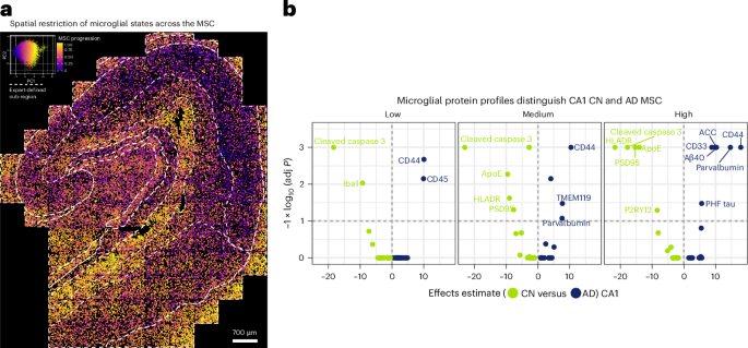
Single-cell spatial proteomics paired with chromatin accessibility mapping identifies a diverse range of human microglial immune states. In Alzheimer’s disease, hippocampal microglia tend to lose antigen-presenting function and become disengaged from inhibitory synapses. Our dual-modality approach illustrates the importance of integrating local tissue architecture and cell identity to fully understand disease-associated immune remodeling.
This is a preview of subscription content, access via your institution
Access options
Access Nature and 54 other Nature Portfolio journals
Get Nature+, our best-value online-access subscription
$32.99 / 30 days
cancel any time
Subscribe to this journal
Receive 12 print issues and online access
$209.00 per year
only $17.42 per issue
Buy this article
- Purchase on SpringerLink
- Instant access to full article PDF
Prices may be subject to local taxes which are calculated during checkout

References
-
Prinz, M., Jung, S. & Priller, J. Microglia biology: one century of evolving concepts. Cell 179, 292–311 (2019). A comprehensive review charting the historical and conceptual development of microglial research.
-
Keren-Shaul, H. et al. A unique microglia type associated with restricting development of Alzheimer’s disease. Cell 169, 1276–1290.e17 (2017). Seminal primary study that first defined disease-associated microglia and highlighted subtype heterogeneity in Alzheimer’s disease.
-
Vijayaragavan, K. et al. Single-cell spatial proteomic imaging for human neuropathology. Acta Neuropathol. Commun. 10, 158 (2022). Peer-reviewed protocol and dataset describing the MIBI workflow for high-plex in situ proteomics of human brain tissue.
-
Mrdjen, D. et al. High-dimensional single-cell mapping of central nervous system immune cells reveals distinct myeloid subsets in health, aging and disease. Immunity 48, 380–395.e6 (2018). This paper introduced mass cytometry-based single-cell proteomics to profile immune diversity in the central nervous system, inspiring the continuum approach.
-
Corces, M. R. et al. Single-cell epigenomic analyses implicate causal variants at inherited risk loci for Alzheimer’s and Parkinson’s diseases. Nat. Genet. 52, 1158–1168 (2020). This paper provides the single-nucleus ATAC-seq dataset and analytical framework we used to construct the epigenetic continuum.
Additional information
Publisher’s note Springer Nature remains neutral with regard to jurisdictional claims in published maps and institutional affiliations.
This is a summary of: Mrdjen, D. et al. Spatial proteomics reveals Alzheimer’s disease-specific human microglial states. Nat. Immunol. https://doi.org/10.1038/s41590-025-02203-w (2025).
Rights and permissions
About this article

Cite this article
Spatial map of microglial diversity beyond proteopathy. Nat Immunol (2025). https://doi.org/10.1038/s41590-025-02205-8
-
Published:
-
DOI: https://doi.org/10.1038/s41590-025-02205-8