Introduction
Protein aggregation is often observed in various biological processes, characterized by the accumulation of improperly processed or misfolded proteins within cells1,2. This aberrant occurrence is frequently found in neurodegenerative disorders, for example in polyglutamine (polyQ) diseases, which arise from the unstable expansion of CAG trinucleotide repeats in the specific gene sequences3. Previous research indicated that protein aggregation could serve a protective role by isolating misfolded or damaged proteins, thereby potentially maintaining cellular functions4,5. During this process, polyQ-expanded proteins can effectively sequester cellular essential factors6 including transcription factors7,8,9 ubiquitin adaptor proteins10,11 and molecular chaperones12,13,14 into aggregates or inclusions. Such sequestration may reduce the solubility and availability of the sequestered proteins, potentially compromising their biological functions and altering cellular fates6,15.
The polyQ-tract protein is prone to aggregation, and its expansion exacerbates this tendency, but it may exhibit relative inertness within cells16. By leveraging these biochemical traits, we proposed a therapeutic strategy to design and construct a series of polyQ fusion proteins by combining a polyQ tract with a peptide sequence that specifically interacts with the target protein. These designer polyQ fusions effectively sequester target proteins into aggregates, diminishing their cellular availability and influencing their functionality17. In this study, we have applied a designer polyQ fusion protein to modulate nuclear factor kappa-B (NF-κB) signaling pathway by sequestering the transcription factor P65 into aggregates.
The NF-κB signaling is an important cellular pathway that regulates immune responses, inflammation, cell survival and apoptosis18. Regulation of the NF-κB pathway primarily occurs through the action of inhibitor of kappa-B (IκB) proteins, which then recruit NF-κB in the cytoplasm and prevent its activation19,20. Upon stimulation by various signals, such as TNF-α, IL-1β and reactive oxygen species (ROS), the IκB kinase (IKK) complex phosphorylates IκB, leading to its degradation and the release of NF-κB19,20,21,22. Subsequently, the P65 (RelA) subunit of NF-κB translocates into the nucleus, where it regulates the expression of target genes involved in inflammatory responses, immune functions, and cell survival18,23,24. Excessive activation of the NF-κB signaling has been implicated in various diseases, such as inflammation24,25, autoimmune disorder26,27 and cancer18,28. Therefore, maintaining the precise control of this pathway is crucial for regulating cellular responses and averting pathological outcomes29.
Our previous study demonstrated that the designer polyQ fusion proteins, polyQ-IRF and polyQ-PMI, can specifically bind to and effectively sequester USP7/HDM2 into aggregates, leading to depleting the availabilities of USP7 and/or HDM2 in A549 cells and enhancing the P53 functionality in the USP7-HDM2-P53 axis17. This polyQ-fusion strategy is feasible to modulate the P53 stability and functionality, furnishing a therapeutic potential for cancers. Based on the polyQ-fusion technology, we have engineered a polyQ fusion protein to modulate the NF-κB signaling pathway by directly sequestering cellular P65 into aggregates or inclusions. This study provides a novel therapeutic approach for treating inflammation and autoimmune diseases, which are closely associated with excessive P65 activation.
Results
Design and construction of the PolyQ fusion protein targeting P65 directly
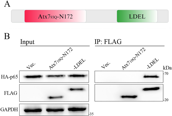
It was reported that the deubiquitinating enzyme USP7 can directly bind to the P65 subunit of NF-κB via its LDEL motif (sequence: LDKALDELMDGDIIVFQK) within the UbL2 domain and deubiquitinate the P65 protein30. We thus designed a polyQ fusion protein by combining the N-terminal fragment of ataxin-7 (Atx7)12,17 with the LDEL peptide sequence, referred to as Atx793Q-N172-LDEL (Fig. 1A). This fusion protein was designed and constructed aiming to modulate the NF-κB signaling pathway by sequestering P65 directly.

Design of the polyQ-LDEL fusion and examination of its interaction with P65. (A) Schematic diagram of a designer polyQ fusion protein, Atx793Q-N172-LDEL. Atx793Q-N172, the N-terminal 172-residue fragment of Atx793Q. LDEL, a homologous peptide sequence, LDKALDELMDGDIIVFQK, from the UbL2 domain of USP7. (B) Examining the interaction between Atx710Q-N172-LDEL and P65 by co-IP assay. HEK 293T cells were co-transfected with FLAG-tagged Atx710Q-N172 or Atx710Q-N172-LDEL and HA-P65. At 48 h post-transfection, the cell lysates were subjected to co-IP assay using anti-FLAG agarose beads. Vec., vector; Atx710Q-N172, the N-terminal 172-residue fragment of Atx710Q; LDEL, Atx710Q-N172-LDEL, a polyQ fusion combined Atx710Q-N172 with LDEL.
We firstly characterized the interaction between polyQ-LDEL and P65 by using the Atx710Q-N172 template but without aggregation propensity12. HEK 293T cells were co-transfected with FLAG-tagged Atx710Q-N172 or Atx710Q-N172-LDEL and HA-tagged P65, and the cell lysates were subjected to co-IP experiment. The data showed that Atx710Q-N172-LDEL could precipitate P65 remarkably compared to Atx710Q-N172 (Fig. 1B), indicating that Atx710Q-N172-LDEL specifically interacts with P65 via its fused LDEL peptide sequence. As an inference, our designed polyQ fusion protein Atx793Q-N172-LDEL can also interacts with cellular P65 in its aggregated form.
PolyQ-LDEL fusion sequesters P65 into aggregates or inclusions
We performed S/P fractionation to characterize whether the polyQ-LDEL fusion could sequester endogenous P65 protein. The results showed that over-expression of Atx793Q-N172-LDEL in HEK 293T cells significantly reduced the protein level of endogenous P65 in the supernatant fraction, while remarkably increased its accumulation in the pellet (Fig. 2A). We also assessed sequestration of exogenous P65 by this polyQ-LDEL fusion and observed a notable increase of the P65 protein sequestered into the pellet fraction (Fig. S1). We then used immunofluorescence imaging to visualize the sequestration and co-localization in HEK 293T cells. While Atx793Q-N172 formed inclusion bodies in the cytoplasm, it was not competent to sequester endogenous P65 into the inclusions. Conversely, the cytoplasmic inclusions formed by Atx793Q-N172-LDEL exhibited clear co-localization with the endogenous P65 protein (Fig. 2B). These results indicate that the polyQ-LDEL fusion can effectively sequester either endogenous or exogenous P65 into cytoplasmic inclusions in cells and substantially diminish the P65 solubility. This specific sequestration may thereby reduce the solubility and availability of the target protein, potentially interfering with its biological function in the cellular milieu.

The polyQ-LDEL fusion sequesters endogenous P65 into aggregates. (A) S/P fractionation for characterizing the distribution of endogenous P65 in supernatant and pellet affected by the polyQ-LDEL fusion. HEK 293T cells were transfected with FLAG-tagged Atx793Q-N172 or Atx793Q-N172-LDEL, followed by culturing for 48 h. The P65 levels in the supernatant and pellet fractions were then detected and analyzed. Sup., supernatant; Pel., pellet. Vec., vector; Atx793Q-N172, the N-terminal 172-residue fragment of Atx793Q; LDEL, Atx793Q-N172-LDEL, a polyQ fusion combined Atx793Q-N172 with LDEL. Data are shown as Mean ± SD (n = 3). *, p < 0.05; **, p < 0.01; N.S., no significance. (B) Immunofluorescence imaging for characterizing the co-localization of endogenous P65 with the inclusions formed by the polyQ-LDEL fusion. HEK 293T cells were transfected with FLAG-tagged Atx793Q-N172 or Atx793Q-N172-LDEL, and after cultured for 48 h, the cells were fixed and immunostained with anti-FLAG (green) and anti-P65 (red) antibodies. Nuclei were stained with Hoechst (blue). Scale bar = 10 μm. Bottom: co-localization analysis of the fluorescence signals for the distance represented by white lines.
PolyQ-LDEL fusion affects nuclear translocation of P65 in cells
To verify whether the sequestration of P65 into cytoplasmic aggregates or inclusions affects its nuclear translocation, we over-expressed FLAG-tagged Atx793Q-N172 or Atx793Q-N172-LDEL in HEK 293T cells, followed by treating with the TNF-α protein to induce P65 translocation into the nucleus. Immunofluorescence imaging revealed that both the free and LDEL-fused species could form cytoplasmic inclusions in HEK 293T cells. Over-expression of Atx793Q-N172-LDEL effectively sequestered endogenous P65 into cytoplasmic inclusions, preventing its nuclear translocation even upon TNF-α stimulation (Fig. 3A), but the free Atx793Q-N172 form did not affect the nuclear translocation of P65.

The polyQ-LDEL fusion affects nuclear translocation of P65 in cells. (A) Immunofluorescence imaging for assessing the nuclear translocation of P65 affected by the polyQ-LDEL fusion. HEK 293T cells were transfected with FLAG-tagged Atx793Q-N172 or Atx793Q-N172-LDEL. After cultured for 48 h, the cells were treated with TNF-α (10 ng/mL) for 40 min. Subsequently, the cells were fixed and immunostained with anti-FLAG (green) and anti-P65 (red) antibodies. Nuclei were stained with Hoechst (blue). TNF-α, a cytokine for activating NF-κB signaling. Vec., vector; Atx793Q-N172, the N-terminal 172-residue fragment of Atx793Q; LDEL, Atx793Q-N172-LDEL. Scale bar = 10 μm. (B) Nuclear/cytoplasmic fractionation for assessing the nuclear translocation of P65 affected by the polyQ-LDEL fusion. HEK 293T cells were transfected with FLAG-tagged Atx793Q-N172 or Atx793Q-N172-LDEL. After cultured for 48 h, the cells were treated with TNF-α (10 ng/mL) for 40 min. Subsequently, the cells were collected and subjected to nuclear/cytoplasmic fractionation and detection of the P65 levels in the cytoplasm and nucleus. H3, histone 3. Data are presented as Mean ± SD (n = 3). *, p < 0.05; N.S., no significance.
Nuclear/cytoplasmic fractionation exhibited a significant reduction in the nuclear P65 content upon TNF-α treatment, after over-expression of Atx793Q-N172-LDEL compared to the free Atx793Q-N172 group (Fig. 3B). These results demonstrate that sequestration of endogenous P65 by the polyQ-LDEL fusion significantly affects the translocation of P65 into the nucleus.
PolyQ-LDEL fusion modulates NF-κB signaling by sequestering P65 into aggregates
The transcriptional factor P65 enters the nucleus in response to external stimuli, such as TNF-α, then P65 binds to the NF-κB response elements for regulating inflammation and immune responses within cells18,31. We next performed a dual-luciferase reporter assay by using an NF-κB-FL reporter plasmid (Fig. 4A) to investigate the effects of P65 sequestration by polyQ-LDEL on the NF-κB pathway32. The results showed that transfection of NF-κB-FL significantly increased the luciferase activity (FLuc/RLuc) (Fig. 4B), whether Atx793Q-N172 or its LDEL-fused form is present or not, suggesting that the reporter system is functional and suitable for NF-κB signaling assay in HEK 293T cells. We then treated the transfected cells with TNF-α to activate NF-κB signaling. As usual, transfection of NF-κB-FL remarkably increased the luciferase activity (Fig. 4C, column 2). However, co-transfection of Atx793Q-N172-LDEL significantly reduced the luciferase activity (Fig. 4C, column 4), whereas that of Atx793Q-N172 had little effect only. As a contrast, Atx710Q-N172 is not an aggregation-prone template, its fusion with the LDEL sequence specifically interacts with P65 (Fig. 1B). We also over-expressed the Atx710Q-N172-fused forms in HEK293T cells and observed that neither Atx710Q-N172 nor Atx710Q-N172-LDEL affected the luciferase activity (Fig. S2), indicating that the simple LDEL-P65 binding without aggregation and sequestration does not influence NF-κB signaling activity. Together, these findings suggest that polyQ-LDEL can suppress NF-κB signaling possibly by sequestering P65 into aggregates and impeding its nuclear translocation.

The polyQ-LDEL fusion modulates the NF-κB signaling activity. (A) Schematic diagram of the NF-κB-FL reporter. The NF-κB response element harbors a DNA sequence of tandem GGGAATTTCC, which drives the expression of the Firefly luciferase gene through the mini-promoter on the plasmid. minP, mini-promoter. (B, C) Effects of the polyQ-LDEL fusion on the luciferase activities. The pGL3-NF-κB-FL plasmid as well as the pGL3 vector harboring a Renilla luciferase gene was co-transfected with FLAG-tagged Atx793Q-N172 or Atx793Q-N172-LDEL into HEK 293T cells. After cultured for 48 h, the cells were treated without (B) or with (C) TNF-α (10 ng/mL) for 40 min, and then the cell lysates were subjected to luciferase activity assay (FLuc/RLuc). The empty vector was set as a control. Insert: immunoblots showing the expression levels of Atx793Q-N172 and its LDEL-fused form. Vec., vector; Atx793Q-N172, the N-terminal 172-residue fragment of Atx793Q; LDEL, Atx793Q-N172-LDEL. Data are presented as Mean ± SD (n = 3). *, p < 0.05; ***, p < 0.001; ****, p < 0.001; N.S., no significance.
PolyQ-LDEL fusion down-regulates expression of the P65-regulated genes
When cells are stimulated by TNF-α, it may bind to TNF-α receptors on the cell surface and activate the NF-κB pathway18. Once NF-κB signaling is activated, P65/P50 may translocate into nucleus and binds to the DNA elements, which facilitates the transcription of a series of inflammation-related genes, including TNF-α, IL-6, IL-1β, and COX-233. Thus, we examined the expression of two NF-κB downstream genes, TNF-α and IL-6, which might be influenced by polyQ-LDEL. The results showed that, without TNF-α treatment, over-expression of either the free or LDEL-fused form did not affect the expression levels of TNF-α (Fig. 5A) and IL-6 (Fig. 5B) in HEK 293T cells. However, upon TNF-α treatment, the protein levels of TNF-α (Fig. 5A, column 4) and IL-6 (Fig. 5B, column 4) increased remarkably. Over-expression of Atx793Q-N172-LDEL significantly reduced the protein levels of TNF-α (Fig. 5A, column 6) and IL-6 (Fig. 5B, column 6), whereas that of Atx793Q-N172 exhibited little effect on their expression. These findings suggest that the polyQ-LDEL fusion down-regulates the expression of inflammatory factors in response to TNF-α activation.

Effects of the polyQ-LDEL fusion on the downstream gene expression. TNF-α expression. (B) IL-6 expression. HEK 293T cells were transfected with FLAG-tagged Atx793Q-N172 or Atx793Q-N172-LDEL. After cultured for 30 h, the cells were treated with TNFα (10 ng/mL) for 40 min. The cell lysates were then subjected to immunoblotting with an anti-TNF-α or anti-IL6 antibody to assess the protein level of TNF-α (A) or IL-6 (B). Vec., vector; Atx793Q-N172, the N-terminal 172-residue fragment of Atx793Q; LDEL, Atx793Q-N172-LDEL. Data are presented as Mean ± SD (n = 3). *, p < 0.05; **, p < 0.01; N.S., no significance.
Discussion
P65 (RelA) is a critical subunit of the NF-κB transcription factor family34 which regulates the expression of numerous cytokines associated with inflammation and immunity18. Dysregulation of the NF-κB pathway has been implicated in the pathologies of inflammation and autoimmune disorders35. Thus, the NF-κB pathway, particularly involving P65, becomes a significant target for therapeutic intervention.
Our previous studies established a polyQ fusion strategy to modulate the biological processes and thereby enable therapeutic application17. A polyQ fusion protein may be constructed by combination of a polyQ tract and a peptide sequence that specifically binds to the target protein. The LDEL motif within the UBL2 domain of USP7 mediates the interaction between USP7 and P6530. Based on the interacting motif, we have engineered a polyQ fusion protein that can specifically sequester P65 into inclusions, affect its nuclear translocation, reduce the P65 abundancy in the nucleus, and thereby prevent activation of the NF-κB signaling. Thus, this polyQ fusion technology may provide an alternative approach for modulating the NF-κB signaling pathway, highlighting its potential for therapeutic application in inflammation and autoimmune disorders caused by the excessive P65 activity.
However, it should be mentioned that polyQ-expanded protein aggregation has been shown to induce impairments of some cellular pathways, such as ubiquitin-proteasome pathway (UPS), at least in cell overexpression systems36,37. As known, UPS dysfunction can alter expression of the protein components including IκB and IKKβ38,39 both of which play key roles in NF-κB signaling. Thus, while applying the polyQ fusion technology in future studies, it is important to verify the expression and degradation of these protein components to rule out any potential UPS-related effects on the NF-κB signaling.
In some aspects, the sequestration effect of polyQ fusion is similar to that of the small-molecule inhibitors and interference RNAs40,41. Some small-molecule inhibitors, such as parthenolide and BAY 11-7082, can inhibit the phosphorylation of IκBα and the subsequent nuclear translocation of P6542,43; while silencing P65 by siRNA or shRNA can directly reduce its expression and thereby inhibit NF-κB signaling44,45. However, compared to the off-target effects of small-molecule inhibitors and siRNAs, this approach by sequestering P65 into inclusions may offer a higher specificity for directly targeting the transcription factor. Further research will focus on the sequestration effects of polyQ fusions in animal models and examine the feasibility of this technology in therapeutic application to inflammation and autoimmune diseases.
In summary, a polyQ fusion protein has been developed to modulate the NF-κB signaling pathway by sequestering the transcription factor P65 into aggregates. This sequestration effect impedes the nuclear entry process of P65 and consequently suppresses NF-κB signaling, leading to down-regulation of the downstream genes, especially TNF-α and IL-6. This technical approach is potentially applicable to therapeutic intervention of inflammation and autoimmune diseases.
Materials and methods
Plasmids, antibodies, and reagents
The DNA sequences encoding Atx710Q-N172 and Atx793Q-N172 were cloned in the FLAG-pcDNA3.1 vector12. The cDNAs encoding Atx710Q-N172-LDEL and Atx793Q-N172-LDEL were inserted into the FLAG-pcDNA3.1 vector via EcoR I/Xba I sites. The DNA sequences for P65 were cloned into the HA-pcDNA3.1 vector using BamH I/Xba I sites. All the DNA sequences were verified by DNA sequencing (Table S1), and the PCR primers used in this study were listed in nucleotide sequences (Table S2).
The anti-FLAG antibody was purchased from Sigma, and the anti-P65 was purchased from Santa Cruz. Anti-TNF-α, anti-IL-6, and anti-GAPDH antibodies were purchased from Proteintech, anti-H3 was from Cell Signaling Technology, and anti-HA was from Sigma-Aldrich Corporation. All secondary antibodies were purchased from Jackson ImmunoResearch Laboratories (Table S3). Recombinant human soluble TNF-α protein was purchased from Beyotime Corporation.
Cell culture and transfection
HEK 293T cells, sourced from the Cell Bank of the Chinese Academy of Sciences (Shanghai), were cultured in Dulbecco’s Modified Eagle’s medium (HyClone) supplemented with 10% fetal bovine serum (Gibco) and penicillin-streptomycin. Cells were maintained at 37 °C in a humidified atmosphere containing 5% CO2. Plasmid transfections were carried out using PolyJet reagent (SignaGen) according to the manufacturer’s instructions.
Co-immunoprecipitation
Co-immunoprecipitation (co-IP) was performed as previously described46. Approximately 48 h post-transfection, the HEK 293T cells were harvested and lysed with a RIPA buffer (50 mM Tris-HCl, pH 7.5, 150 mM NaCl, 1 mM EDTA, 1% NP-40, and protease inhibitor cocktail (Roche)). After cell lysis, the lysates were centrifuged at 13,000 rpm for 20 min at 4 °C. Following centrifugation, the supernatants were collected and incubated with anti-FLAG agarose beads (Abmart) overnight at 4 °C. The beads were then washed three times with the RIPA buffer before boiling in 40 µL of 2x loading buffer (4% SDS). The protein eluates from the beads were analyzed by immunoblotting.
Supernatant/pellet fractionation
The supernatant/pellet (S/P) fractionation experiment was performed according to the previously established protocol47. Approximately 48 h post-transfection, the HEK 293T cells were harvested and lysed in 90 µL of the RIPA buffer on ice for 30 min. The lysates were centrifuged at 13,000 rpm for 15 min at 4 °C. Subsequently, 80 µL of the supernatant was mixed with 20 µL of 4x loading buffer (8% SDS). The pellet was thoroughly washed thrice with the RIPA buffer at 4 °C and re-suspended in 40 µL of 4x loading buffer (8% SDS). Equal volumes of both supernatant and pellet fractions were analyzed by SDS-PAGE and Western blotting.
Nuclear and cytoplasmic protein extraction and Western blotting
The nuclear and cytoplasmic proteins were obtained separately from HEK 293 cells by using the Nuclear and Cytoplasmic Protein Extraction Kit (Beyotime Biotech., Shanghai, P0028) following the manufacturer’s instructions. For protein analysis, the cell lysates were subjected to SDS-PAGE and then transferred onto PVDF membranes (Millipore). Before antibody incubation, the blots were sectioned as required and blocked with 5% skim milk in a PBS buffer (10 mM Na2HPO4, 2 mM KH2PO4,137 mM NaCl, 2.7mM KCl, ) containing 0.1% Tween-20 for 1 h at room temperature. Specific protein detection was accomplished using the appropriate primary and secondary antibodies, followed by visualization using an ECL detection kit (Thermo Fisher Scientific). Quantitative analysis of the protein bands was carried out by using the integral grayscale values recorded with Sage Capture V3.2.2 software (Sage Science, http://www.sagecreation.com.cn/en/).
Dual-luciferase reporter assay
The pGL3-NF-κB-FL reporter plasmid was purchased from Bioscien (Shanghai). The dual-luciferase reporter assay was conducted using a Luciferase Reporter Assay kit (YEASEN, Shanghai) following the manufacturer’s instructions48. Briefly, HEK 293T cells were co-transfected with pGL3-NF-κB-FL and FLAG-tagged Atx793Q-N172 or Atx793Q-N172-LDEL, as well as the pGL3 vector harboring a Renilla luciferase gene for recording control. To verify the transactivation function of endogenous P65, the recombinant human soluble protein TNF-α (Beyotime, Shanghai) was included in the cell culture (10 ng/ml) to activate the NF-κB signaling pathway. After treated for 40 min, the cells were collected and the luciferase activity was measured. The relative luciferase activity was calculated as the ratio of Firefly luciferase over Renilla luciferase activities (FLuc/RLuc).
Immunofluorescence imaging
HEK 293T cells cultured on glass coverslips were processed approximately 48 h post-transfection or for the specified culture duration. Initially, the cells were washed with a PBS buffer (10 mM Na2HPO4, 1.8 mM KH2PO4, 140 mM NaCl, 2.7 mM KCl, pH 7.3) and fixed with 4% paraformaldehyde for 15 min. After three washes with the PBS buffer, the cells were permeabilized with 0.1% Triton X-100 and then blocked in a solution containing 5% bovine serum albumin (BSA) in the PBS buffer for 1 h at room temperature. Subsequently, the specific primary antibodies were applied, and the cells were incubated overnight at 4 °C. Following primary antibody incubation, the cells were washed with the PBS buffer and incubated with either fluorescein isothiocyanate (FITC)-conjugated or tetramethyl rhodamine isothiocyanate (TRITC)-conjugated secondary antibodies (Jackson ImmunoResearch Laboratories). The nuclei were stained with Hoechst 33,342 (Thermo Fisher Scientific). Imaging was performed using a Leica TCS SP8 WLL confocal microscope (Leica Microsystems), and the images were captured using LAS X V3.5 software (Leica, https://www.leica-microsystems.com/) and presented without modification. For co-localization analysis, the fluorescence intensities from the respective channels were quantified using ImageJ V1.53 software (NIH, https://imagej.net/) and visualized as plots generated with GraphPad Prism V8.0.2 (GraphPad Software, https://www.graphpad.com/).
Quantification and statistical analysis
Quantitative analysis of the Western blots was conducted using ImageJ V1.53 software (NIH, https://imagej.net/). The integral grayscale values of the indicated protein bands were normalized against those of the respective controls. Data were derived from three independent experiments and are presented as Mean ± SD. Statistical analyses were conducted using GraphPad Prism 8.0.2 (GraphPad software, https://www.graphpad.com/). One-way ANOVA algorithm was used to compare differences among three or more experimental groups, followed by Tukey’s multiple comparisons test to assess significance between group pairs. Statistical significance was defined as p < 0.05, where the p-values were indicated in graphs as ∗ (p < 0.05), ∗∗ (p < 0.01), ∗∗∗ (p < 0.001), **** (p < 0.0001) or N.S. (no significance).
Data availability
The datasets generated and/or analyzed during the current study are available in the GenBank repository (https://www.ncbi.nlm.nih.gov/genbank/) with the accession numbers PV413212 (Atx710Q-N172-LDEL) and PV413213 (Atx793Q-N172-LDEL).
Abbreviations
- Atx7:
-
Ataxin-7
- co-IP:
-
Co-immunoprecipitation
- HDM2:
-
Human double minute 2 homolog
- IκB:
-
Inhibitor of kappa-B
- IKK:
-
IκB kinase
- LDEL:
-
A P65-binding motif from the UbL2 domain of USP7
- NF-κB:
-
Nuclear factor kappa-B
- PMI:
-
P53/MDM2 inhibitor
- polyQ:
-
Polyglutamine
- ROS:
-
Reactive oxygen species
- S/P:
-
Supernatant/pellet
- UPS:
-
Ubiquitin-proteasome pathway
- USP7:
-
Ubiquitin-specific protease 7
- vIRF1:
-
Viral interferon regulatory factor 1
References
-
Housmans, J. A. J., Wu, G., Schymkowitz, J. & Rousseau, F. A guide to studying protein aggregation. FEBS J. 290, 554–583. https://doi.org/10.1111/febs.16312 (2023).
-
Chiti, F. & Dobson, C. M. Protein misfolding amyloid formation, and human disease: a summary of progress over the last decade. Annu. Rev. Biochem. 86, 27–68. https://doi.org/10.1146/annurev-biochem-061516-045115 (2017).
-
Lieberman, A. P., Shakkottai, V. G. & Albin, R. L. Polyglutamine repeats in neurodegenerative diseases. Annu. Rev. Pathol. 14, 1–27. https://doi.org/10.1146/annurev-pathmechdis-012418-012857 (2019).
-
Arrasate, M., Mitra, S., Schweitzer, E. S., Segal, M. R. & Finkbeiner, S. Inclusion body formation reduces levels of mutant Huntingtin and the risk of neuronal death. Nature 431, 805–810. https://doi.org/10.1038/nature02998 (2004).
-
Mahul-Mellier, A. L. et al. The process of lewy body formation, rather than simply alpha-synuclein fibrillization, is one of the major drivers of neurodegeneration. Proc. Natl. Acad. Sci. U S A. 117, 4971–4982. https://doi.org/10.1073/pnas.1913904117 (2020).
-
Yang, H. & Hu, H. Y. Sequestration of cellular interacting partners by protein aggregates: implication in a loss-of-function pathology. FEBS J. 283, 3705–3717. https://doi.org/10.1111/febs.13722 (2016).
-
Jobe, F. et al. Respiratory syncytial virus sequesters NF-κB subunit p65 to cytoplasmic inclusion bodies to inhibit innate immune signaling. J. Virol. 94, 563. https://doi.org/10.1128/jvi.01380-20 (2020).
-
Cortes, C. J. et al. Polyglutamine-expanded androgen receptor interferes with TFEB to elicit autophagy defects in SBMA. Nat. Neurosci. 17, 1180–1189. https://doi.org/10.1038/nn.3787 (2014).
-
Yang, J. et al. A prion-like domain of TFEB mediates the co-aggregation of TFEB and mHTT. Autophagy 19, 544–550. https://doi.org/10.1080/15548627.2022.2083857 (2023).
-
Yang, H. et al. PolyQ-expanded Huntingtin and ataxin-3 sequester ubiquitin adaptors hHR23B and UBQLN2 into aggregates via conjugated ubiquitin. FASEB J. 32, 2923–2933. https://doi.org/10.1096/fj.201700801RR (2018).
-
Farrawell, N. E. et al. Ubiquitin Homeostasis Is Disrupted in TDP-43 and FUS Cell Models of ALS. iScience 23, 101700, (2020). https://doi.org/10.1016/j.isci.2020.101700
-
Yue, H. W., Hong, J. Y., Zhang, S. X., Jiang, L. L. & Hu, H. Y. PolyQ-expanded proteins impair cellular proteostasis of ataxin-3 through sequestering the co-chaperone HSJ1 into aggregates. Sci. Rep. 11, 7815. https://doi.org/10.1038/s41598-021-87382-w (2021).
-
Park, S. H. et al. PolyQ proteins interfere with nuclear degradation of cytosolic proteins by sequestering the Sis1p chaperone. Cell 154, 134–145. https://doi.org/10.1016/j.cell.2013.06.003 (2013).
-
Yu, A. et al. Tau protein aggregates inhibit the protein-folding and vesicular trafficking arms of the cellular proteostasis network. J. Biol. Chem. 294, 7917–7930. https://doi.org/10.1074/jbc.RA119.007527 (2019).
-
Hu, H. Y. & Liu, Y. J. Sequestration of cellular native factors by biomolecular assemblies: physiological or pathological? Biochim. Biophys. Acta Mol. Cell. Res. 1869, 119360. https://doi.org/10.1016/j.bbamcr.2022.119360 (2022).
-
Orr, H. T. & Zoghbi, H. Y. Trinucleotide repeat disorders. Annu. Rev. Neurosci. 30, 575–621. https://doi.org/10.1146/annurev.neuro.29.051605.113042 (2007).
-
Zhang, X. L. et al. Designer PolyQ fusion proteins sequester USP7/HDM2 for modulating P53 functionality. iScience 28, 112025. https://doi.org/10.1016/j.isci.2025.112025 (2025).
-
Hoesel, B. & Schmid, J. A. The complexity of NF-κB signaling in inflammation and cancer. Mol. Cancer. 12, 86. https://doi.org/10.1186/1476-4598-12-86 (2013).
-
Ghosh, S. & Baltimore, D. Activation in vitro of NF-kappa B by phosphorylation of its inhibitor I kappa B. Nature 344, 678–682. https://doi.org/10.1038/344678a0 (1990).
-
Napetschnig, J. & Wu, H. Molecular basis of NF-kappaB signaling. Annu. Rev. Biophys. 42, 443–468. https://doi.org/10.1146/annurev-biophys-083012-130338 (2013).
-
Guo, Q. et al. NF-kappaB in biology and targeted therapy: new insights and translational implications. Signal. Transduct. Target. Ther. 9, 523. https://doi.org/10.1038/s41392-024-01757-9 (2024).
-
Jacobs, M. D. & Harrison, S. C. Structure of an IkappaBalpha/NF-kappaB complex. Cell 95, 749–758. https://doi.org/10.1016/s0092-8674(00)81698-0 (1998).
-
Vallabhapurapu, S. & Karin, M. Regulation and function of NF-kappaB transcription factors in the immune system. Annu. Rev. Immunol. 27, 693–733. https://doi.org/10.1146/annurev.immunol.021908.132641 (2009).
-
Liu, T., Zhang, L., Joo, D. & Sun, S. C. NF-kappaB signaling in inflammation. Signal. Transduct. Target. Ther. 2, 17023. https://doi.org/10.1038/sigtrans.2017.23 (2017).
-
Zheng, X. et al. FAM76B regulates NF-κB-mediated inflammatory pathway by influencing the translocation of hnRNPA2B1. eLife 12, e85659. https://doi.org/10.7554/eLife.85659 (2023).
-
Chen, G. et al. The NF-kappaB transcription factor c-Rel is required for Th17 effector cell development in experimental autoimmune encephalomyelitis. J. Immunol. 187, 4483–4491. https://doi.org/10.4049/jimmunol.1101757 (2011).
-
Campbell, I. K., Gerondakis, S., O’Donnell, K. & Wicks, I. P. Distinct roles for the NF-kappaB1 (p50) and c-Rel transcription factors in inflammatory arthritis. J. Clin. Invest. 105, 1799–1806. https://doi.org/10.1172/JCI8298 (2000).
-
Caruana, B. T. & Byrne, F. L. The NF-kappaB signalling pathway regulates GLUT6 expression in endometrial cancer. Cell. Signal. 73, 109688. https://doi.org/10.1016/j.cellsig.2020.109688 (2020).
-
Yu, H., Lin, L., Zhang, Z., Zhang, H. & Hu, H. Targeting NF-kappaB pathway for the therapy of diseases: mechanism and clinical study. Signal. Transduct. Target. Ther. 5, 209. https://doi.org/10.1038/s41392-020-00312-6 (2020).
-
Mitxitorena, I. et al. The deubiquitinase USP7 uses a distinct ubiquitin-like domain to deubiquitinate NF-kB subunits. J. Biol. Chem. 295, 11754–11763. https://doi.org/10.1074/jbc.RA120.014113 (2020).
-
Kempe, S., Kestler, H., Lasar, A. & Wirth, T. NF-kappaB controls the global pro-inflammatory response in endothelial cells: evidence for the regulation of a pro-atherogenic program. Nucleic Acids Res. 33, 5308–5319. https://doi.org/10.1093/nar/gki836 (2005).
-
Pessara, U. & Koch, N. Tumor necrosis factor alpha regulates expression of the major histocompatibility complex class II-associated invariant chain by binding of an NF-kappa B-like factor to a promoter element. Mol. Cell. Biol. 10, 4146–4154. https://doi.org/10.1128/mcb.10.8.4146-4154.1990 (1990).
-
Wang, S., Liu, Z., Wang, L. & Zhang, X. NF-kappaB signaling pathway, inflammation and colorectal cancer. Cell. Mol. Immunol. 6, 327–334. https://doi.org/10.1038/cmi.2009.43 (2009).
-
Ashburner, B. P., Westerheide, S. D. & Baldwin, A. S. Jr The p65 (RelA) subunit of NF-kappaB interacts with the histone deacetylase (HDAC) corepressors HDAC1 and HDAC2 to negatively regulate gene expression. Mol. Cell. Biol. 21, 7065–7077. https://doi.org/10.1128/MCB.21.20.7065-7077.2001 (2001).
-
Giridharan, S. & Srinivasan, M. Mechanisms of NF-kappaB p65 and strategies for therapeutic manipulation. J. Inflamm. Res. 11, 407–419. https://doi.org/10.2147/JIR.S140188 (2018).
-
Bence, N. F., Sampat, R. M. & Kopito, R. R. Impairment of the ubiquitin-proteasome system by protein aggregation. Sci. (New York N Y). 292, 1552–1555. https://doi.org/10.1126/science.292.5521.1552 (2001).
-
Jana, N. R., Zemskov, E. A., Wang, G. & Nukina, N. Altered proteasomal function due to the expression of polyglutamine-expanded truncated N-terminal Huntingtin induces apoptosis by caspase activation through mitochondrial cytochrome c release. Hum. Mol. Genet. 10, 1049–1059. https://doi.org/10.1093/hmg/10.10.1049 (2001).
-
Scherer, D. C., Brockman, J. A., Chen, Z., Maniatis, T. & Ballard, D. W. Signal-induced degradation of I kappa B alpha requires site-specific ubiquitination. Proc. Natl. Acad. Sci. U S A. 92, 11259–11263. https://doi.org/10.1073/pnas.92.24.11259 (1995).
-
Carter, R. S., Pennington, K. N., Arrate, P., Oltz, E. M. & Ballard, D. W. Site-specific monoubiquitination of IkappaB kinase IKKbeta regulates its phosphorylation and persistent activation. J. Biol. Chem. 280, 43272–43279. https://doi.org/10.1074/jbc.M508656200 (2005).
-
Wang, J. Y. et al. PolyQ-expanded ataxin-2 aggregation impairs cellular processing-body homeostasis via sequestering the RNA helicase DDX6. J. Biol. Chem. 300, 107413. https://doi.org/10.1016/j.jbc.2024.107413 (2024).
-
Liu, Y. J., Wang, J. Y., Zhang, X. L., Jiang, L. L. & Hu, H. Y. Ataxin-2 sequesters raptor into aggregates and impairs cellular mTORC1 signaling. FEBS J. 291, 1795–1812. https://doi.org/10.1111/febs.17081 (2024).
-
Hehner, S. P., Hofmann, T. G., Droge, W. & Schmitz, M. L. The antiinflammatory sesquiterpene lactone parthenolide inhibits NF-kappa B by targeting the I kappa B kinase complex. J. Immunol. 163, 5617–5623 (1999).
-
Lee, J., Rhee, M. H., Kim, E. & Cho, J. Y. BAY 11-7082 is a broad-spectrum inhibitor with anti-inflammatory activity against multiple targets. Mediators Inflamm. 2012, 416036. https://doi.org/10.1155/2012/416036 (2012).
-
Guo, J., Fu, Y. C. & Becerra, C. R. Dissecting role of regulatory factors in NF-kappaB pathway with SiRNA. Acta Pharmacol. Sin. 26, 780–788. https://doi.org/10.1111/j.1745-7254.2005.00140.x (2005).
-
Yang, Q. et al. AAV-based ShRNA Silencing of NF-kappaB ameliorates muscle pathologies in Mdx mice. Gene Ther. 19, 1196–1204. https://doi.org/10.1038/gt.2011.207 (2012).
-
Liu, Y. J., Wang, J. Y., Zhang, X. L., Jiang, L. L. & Hu, H. Y. Ataxin-2 sequesters raptor into aggregates and impairs cellular mTORC1 signaling. FEBS J. https://doi.org/10.1111/febs.17081 (2024).
-
Jiang, L. L., Guan, W. L., Wang, J. Y., Zhang, S. X. & Hu, H. Y. RNA-assisted sequestration of RNA-binding proteins by cytoplasmic inclusions of the C-terminal 35-kDa fragment of TDP-43. J. Cell. Sci. 135, jcs259380. https://doi.org/10.1242/jcs.259380 (2022).
-
Solberg, N. & Krauss, S. Luciferase assay to study the activity of a cloned promoter DNA fragment. Methods Mol. Biol. 977, 65–78. https://doi.org/10.1007/978-1-62703-284-1_6 (2013).
Acknowledgements
We would like to thank the Core Facility for Molecular Biology and the Core Facility for Cell Biology at CEMCS, CAS, for providing technical support in biochemical analysis and microscopic imaging.
Ethics declarations
Competing interests
The authors declare no competing interests.
Additional information
Publisher’s note
Springer Nature remains neutral with regard to jurisdictional claims in published maps and institutional affiliations.
Supplementary Information
Rights and permissions
Open Access This article is licensed under a Creative Commons Attribution-NonCommercial-NoDerivatives 4.0 International License, which permits any non-commercial use, sharing, distribution and reproduction in any medium or format, as long as you give appropriate credit to the original author(s) and the source, provide a link to the Creative Commons licence, and indicate if you modified the licensed material. You do not have permission under this licence to share adapted material derived from this article or parts of it. The images or other third party material in this article are included in the article’s Creative Commons licence, unless indicated otherwise in a credit line to the material. If material is not included in the article’s Creative Commons licence and your intended use is not permitted by statutory regulation or exceeds the permitted use, you will need to obtain permission directly from the copyright holder. To view a copy of this licence, visit http://creativecommons.org/licenses/by-nc-nd/4.0/.
About this article

Cite this article
Zhang, XL., Jiang, LL., Duan, HT. et al. A designer polyQ fusion protein modulates NF-κB signaling by sequestering P65/RelA into aggregates. Sci Rep 15, 27351 (2025). https://doi.org/10.1038/s41598-025-13237-3
-
Received:
-
Accepted:
-
Published:
-
DOI: https://doi.org/10.1038/s41598-025-13237-3