
Data availability
All data associated with this study are available within manuscript files. The RNA-seq data generated in this study have been deposited into the Gene Expression Omnibus database with accession number GSE298239. Raw data and processed data that support the findings in this study are available in a public repository (https://doi.org/10.5281/zenodo.15870084).
Code availability
Conventional RNA-seq analysis pipeline with the R package ‘edgeR’ was performed in this study. The R package ‘tidyverse’ was used to analyze metabolomics data, and ‘ggplot2’ was used to visualize analyzed data. The R codes used for this study have been deposited in a public repository (https://doi.org/10.5281/zenodo.15870084).
References
-
Bianconi, E. et al. An estimation of the number of cells in the human body. Ann. Hum. Biol. 40, 463–471 (2013).
-
Cheung, A. L. Isolation and culture of human umbilical vein endothelial cells (HUVEC). Curr. Protoc. Microbiol. https://doi.org/10.1002/9780471729259.mca04bs4 (2007).
-
Herold, J. & Kalucka, J. A rapid adaptable method for isolation of endothelial cells from human adipose tissue. Methods Mol. Biol. 2441, 235–250 (2022).
-
Medina, R. J. et al. Endothelial progenitors: a consensus statement on nomenclature. Stem Cells Transl. Med. 6, 1316–1320 (2017).
-
Pellegata, A. F., Tedeschi, A. M. & De Coppi, P. Whole organ tissue vascularization: engineering the tree to develop the fruits. Front. Bioeng. Biotechnol. 6, 56 (2018).
-
Palikuqi, B. et al. Adaptable haemodynamic endothelial cells for organogenesis and tumorigenesis. Nature 585, 426–432 (2020).
-
Wakabayashi, T. et al. CD157 marks tissue-resident endothelial stem cells with homeostatic and regenerative properties. Cell Stem Cell 22, 384–397 (2018).
-
Lin, Y. et al. ABCG2-expressing clonal repopulating endothelial cells serve to form and maintain blood vessels. Circulation 150, 451–465 (2024).
-
Andrade, J. et al. Control of endothelial quiescence by FOXO-regulated metabolites. Nat. Cell Biol. 23, 413–423 (2021).
-
Ricard, N., Bailly, S., Guignabert, C. & Simons, M. The quiescent endothelium: signalling pathways regulating organ-specific endothelial normalcy. Nat. Rev. Cardiol. 18, 565–580 (2021).
-
Boitano, A. E. et al. Aryl hydrocarbon receptor antagonists promote the expansion of human hematopoietic stem cells. Science 329, 1345–1348 (2010).
-
Lin, Y., Yoder, M. C. & Yoshimoto, M. Lymphoid progenitor emergence in the murine embryo and yolk sac precedes stem cell detection. Stem Cells Dev. 23, 1168–1177 (2014).
-
Wiggins, B. G. et al. Endothelial sensing of AHR ligands regulates intestinal homeostasis. Nature 621, 821–829 (2023).
-
Major, J. et al. Endothelial AHR activity prevents lung barrier disruption in viral infection. Nature 621, 813–820 (2023).
-
Murray, I. A., Patterson, A. D. & Perdew, G. H. Aryl hydrocarbon receptor ligands in cancer: friend and foe. Nat. Rev. Cancer 14, 801–814 (2014).
-
Stockinger, B., Di Meglio, P., Gialitakis, M. & Duarte, J. H. The aryl hydrocarbon receptor: multitasking in the immune system. Annu. Rev. Immunol. 32, 403–432 (2014).
-
Yamada, T. et al. Constitutive aryl hydrocarbon receptor signaling constrains type I interferon-mediated antiviral innate defense. Nat. Immunol. 17, 687–694 (2016).
-
Dabir, P., Marinic, T. E., Krukovets, I. & Stenina, O. I. Aryl hydrocarbon receptor is activated by glucose and regulates the thrombospondin-1 gene promoter in endothelial cells. Circ. Res. 102, 1558–1565 (2008).
-
Fares, I. et al. Cord blood expansion. Pyrimidoindole derivatives are agonists of human hematopoietic stem cell self-renewal. Science 345, 1509–1512 (2014).
-
Zhao, B., Degroot, D. E., Hayashi, A., He, G. & Denison, M. S. CH223191 is a ligand-selective antagonist of the Ah (Dioxin) receptor. Toxicol. Sci. 117, 393–403 (2010).
-
Santostefano, M. et al. alpha-Naphthoflavone-induced CYP1A1 gene expression and cytosolic aryl hydrocarbon receptor transformation. Mol. Pharmacol. 43, 200–206 (1993).
-
Singh, K. P., Wyman, A., Casado, F. L., Garrett, R. W. & Gasiewicz, T. A. Treatment of mice with the Ah receptor agonist and human carcinogen dioxin results in altered numbers and function of hematopoietic stem cells. Carcinogenesis 30, 11–19 (2009).
-
Seok, S.-H. et al. Structural hierarchy controlling dimerization and target DNA recognition in the AHR transcriptional complex. Proc. Natl Acad. Sci. USA 114, 5431–5436 (2017).
-
Gutiérrez-Vázquez, C. & Quintana, F. J. Regulation of the immune response by the aryl hydrocarbon receptor. Immunity 48, 19–33 (2018).
-
Puga, A., Ma, C. & Marlowe, J. L. The aryl hydrocarbon receptor cross-talks with multiple signal transduction pathways. Biochem. Pharmacol. 77, 713–722 (2009).
-
Großkopf, H., Walter, K., Karkossa, I., von Bergen, M. & Schubert, K. Non-genomic AhR-signaling modulates the immune response in endotoxin-activated macrophages after activation by the environmental stressor BaP. Front. Immunol. 12, 620270 (2021).
-
Dasgupta, J. et al. Reactive oxygen species control senescence-associated matrix metalloproteinase-1 through c-Jun-N-terminal kinase. J. Cell. Physiol. 225, 52–62 (2010).
-
Raha, D. et al. The cancer stem cell marker aldehyde dehydrogenase is required to maintain a drug-tolerant tumor cell subpopulation. Cancer Res. 74, 3579–3590 (2014).
-
Dan Dunn, J., Alvarez, L. A., Zhang, X. & Soldati, T. Reactive oxygen species and mitochondria: a nexus of cellular homeostasis. Redox Biol. 6, 472–485 (2015).
-
Murray Stewart, T., Dunston, T. T., Woster, P. M. & Casero, R. A. Polyamine catabolism and oxidative damage. J. Biol. Chem. 293, 18736–18745 (2018).
-
Kuo, B. S., Korner, G., Dryjski, M. & Bjornsson, T. D. Role of polyamines in the stimulation of synthesis and secretion of plasminogen activator from bovine aortic endothelial cells. J. Cell. Physiol. 137, 192–198 (1988).
-
Morrison, R. F. & Seidel, E. R. Vascular endothelial cell proliferation: regulation of cellular polyamines. Cardiovasc. Res. 29, 841–847 (1995).
-
Perez-Leal, O. & Merali, S. Regulation of polyamine metabolism by translational control. Amino Acids 42, 611–617 (2012).
-
Asher, G., Bercovich, Z., Tsvetkov, P., Shaul, Y. & Kahana, C. 20S proteasomal degradation of ornithine decarboxylase is regulated by NQO1. Mol. Cell 17, 645–655 (2005).
-
Jaffe, E. A., Nachman, R. L., Becker, C. G. & Minick, C. R. Culture of human endothelial cells derived from umbilical veins. Identification by morphologic and immunologic criteria.J. Clin. Invest. 52, 2745–2756 (1973).
-
Rothhammer, V. & Quintana, F. J. The aryl hydrocarbon receptor: an environmental sensor integrating immune responses in health and disease. Nat. Rev. Immunol. 19, 184–197 (2019).
-
Nguyen, L. P. & Bradfield, C. A. The search for endogenous activators of the aryl hydrocarbon receptor. Chem. Res. Toxicol. 21, 102–116 (2008).
-
Barroso, A., Mahler, J. V., Fonseca-Castro, P. H. & Quintana, F. J. The aryl hydrocarbon receptor and the gut–brain axis. Cell. Mol. Immunol. 18, 259–268 (2021).
-
Sondermann, N. C. et al. Functions of the aryl hydrocarbon receptor (AHR) beyond the canonical AHR/ARNT signaling pathway. Biochem. Pharmacol. 208, 115371 (2023).
-
Pocar, P., Fischer, B., Klonisch, T. & Hombach-Klonisch, S. Molecular interactions of the aryl hydrocarbon receptor and its biological and toxicological relevance for reproduction. Reproduction 129, 379–389 (2005).
-
Ohtake, F. et al. Dioxin receptor is a ligand-dependent E3 ubiquitin ligase. Nature 446, 562–566 (2007).
-
Luecke-Johansson, S. et al. A molecular mechanism to switch the aryl hydrocarbon receptor from a transcription factor to an E3 ubiquitin ligase. Mol. Cell. Biol. 37, e00630-16 (2017).
-
Gomez-Salinero, J. M., Redmond, D. & Rafii, S. Microenvironmental determinants of endothelial cell heterogeneity. Nat. Rev. Mol. Cell Biol. 26, 476–495 (2025).
-
Baudin, B., Bruneel, A., Bosselut, N. & Vaubourdolle, M. A protocol for isolation and culture of human umbilical vein endothelial cells. Nat. Protoc. 2, 481–485 (2007).
-
Rustam, S. et al. A unique cellular organization of human distal airways and its disarray in chronic obstructive pulmonary disease. Am. J. Respir. Crit. Care Med. 207, 1171–1182 (2023).
-
Dobin, A. et al. STAR: ultrafast universal RNA-seq aligner. Bioinformatics 29, 15–21 (2013).
-
Liao, Y., Smyth, G. K. & Shi, W. featureCounts: an efficient general purpose program for assigning sequence reads to genomic features. Bioinformatics 30, 923–930 (2014).
-
Chen, Y., Chen, L., Lun, A. T. L., Baldoni, P. L. & Smyth, G. K. edgeR v4: powerful differential analysis of sequencing data with expanded functionality and improved support for small counts and larger datasets. Nucleic Acids Res. 53, gkaf018 (2025).
-
Nicholas, D. et al. Advances in the quantification of mitochondrial function in primary human immune cells through extracellular flux analysis. PLoS ONE 12, e0170975 (2017).
-
Chen, E. Y. et al. Enrichr: interactive and collaborative HTML5 gene list enrichment analysis tool. BMC Bioinformatics 14, 128 (2013).
Acknowledgements
The authors thank the members of the Weill Cornell Medicine Genomics Resources Core Facility, the Meyer Cancer Center Proteomics & Metabolomics Core, the Flow Cytometry Core Facility and the Yale Mass Spectrometry & Proteomics Resource for their outstanding technical support. Y.L. was supported by the NYSTEM Training Program in Stem Cell Biology and Regenerative Medicine, an American Heart Association Postdoctoral Fellowship (836459) and an American Heart Association Career Development Award (25CDA1444258). S. Rafii was supported by the Hartman Institute for Therapeutic Organ Regeneration; the Ansary Stem Cell Institute at Weill Cornell Medicine; the Division of Regenerative Medicine; National Institutes of Health grant R35 HL150809 and R01 grants DK136327, DK133332, DK136327 and DK137806; the Selma and Lawrence Ruben Daedalus Fund for Innovation; and the Juvenile Diabetes Research Foundation (JDRF) (renamed as Breakthrough T1D) (JDRF2-SRA-2022-1105-S-B and JDRF 230620-01).
Ethics declarations
Competing interests
. The authors declare no competing interests.
Peer review
Peer review information
Nature Cardiovascular Research thanks Terence Ryan and the other, anonymous, reviewer(s) for their contribution to the peer review of this work.
Additional information
Publisher’s note Springer Nature remains neutral with regard to jurisdictional claims in published maps and institutional affiliations.
Extended data
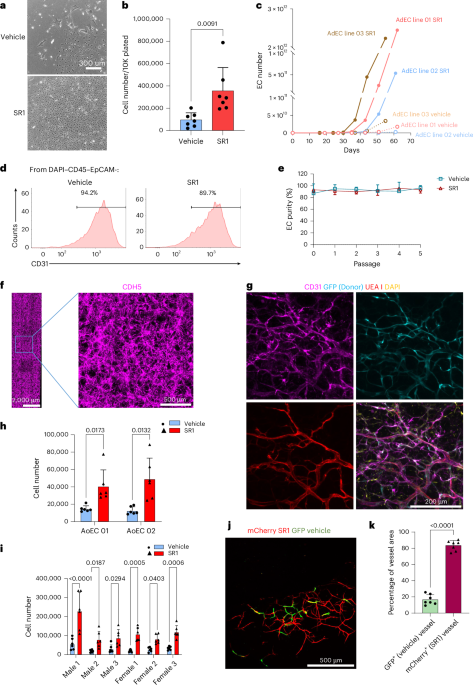
Extended Data Fig. 1 Canonical AHR inhibitor promotes adult human primary EC expansion.
a, b. Representative picture of EC colonies derived from human pancreatic acinar ECs (ASECs, a) and islet specific ECs (ISECs, b) after 13 days of culture with SR1. c. Serial expansion of HUVEC with vehicle or SR1. For each group, 2,000 HUVECs were plated at day 0. Every 8 days, cells were collected, counted, and 2,000 cells were re-plated. Y-axis shows the theoretic total number of ECs derived from each passage. (n = 7 independent experiments). d. Flow cytometry panels showing the expression of CD31 (PECAM1) and CDH5 by HUVEC after four 8-day expansions in vehicle or SR1. EC fraction indicates CD31+CDH5+ population. Data represents 4 experiments. e. Quantitation of the percentage of CD31+CDH5+ ECs after HUVECs were expanded in vehicle, or SR1 for 4 serial 8-day passages. p values, paired t test. (n = 4 experiments). f. Representative picture of donor derived vessels (GFP/mCherry+) from transplantation experiments using 1-1 mixed mCherry HUVECs after 2-week expansion with vehicle, and GFP HUVECs after 2-week expansion with SR1. g. Percentage of GFP+ and mCherry+ vessel areas from transplantations with ECs derived from 1-1 mixed mCherry HUVECs after 2-week expansion with vehicle, and GFP HUVECs after 2-week expansion with SR1 (n = 6 independent experiments). h. Percentage of GFP+ and mCherry+ vessel areas from transplantations with ECs derived from 1-1 mixed GFP HUVECs after 2-week expansion with vehicle, and mCherry HUVECs after 2-week expansion with SR1 (n = 7 independent experiments). i. Schematics of the in vivo vessel forming assay after HUVECs were expanded in vehicle or SR1. 10,000 mCherry expressing HUVECs were expanded with media containing 0.5 µM SR1, and 10,000 GFP expressing HUVECs were expanded with media containing vehicle (1: 10,000 DMSO) for 14 days. The GFP+ ECs and mCherry+ ECs derived from both cultures were mixed and transplanted into immunodeficient recipient mice. After 4 weeks, the gels were retrieved and the ratio of GFP+ and mCherry+ vessels were measured. j. Number of GFP+ (expanded in vehicle) and mCherry+ (expanded in SR1) HUVECs before transplantation as described in Fig. 1j (after 14 day-expansion. n = 7 independent experiments). k. Representative picture of GFP+ and mCherry+ vessels from transplantations with ECs derived from 10,000 GFP expressing HUVECs expanded in SR1 and 10,000 mCherry expressing HUVECs expanded in vehicle for 14 days. l. Quantitation of Extended Data Figure 1k. (n = 6 independent experiments).
Extended Data Fig. 2 Canonical AHR inhibitor promotes EC proliferation.
a. Cell retention of HUVECs after plating in vehicle, UM171, or SR1. 50,000 HUVECs were plated, and cells retained on the culture plates were counted after 14 h. Data represent mean ± s. d. p values, one-way ANOVA. (n = 6 independent experiments). b. Representative flow cytometry panels showing apoptosis analysis on HUVECs culture 72 h in vehicle, UM171, or SR1. c. Quantitation for the apoptosis assay (shown in Extended Data Fig. 2b). Y-axis indicates the percentage of Annexin V+ cells. Data represent mean ± s. d. p values, one-way ANOVA. (n = 5 independent experiments). d. Cell expansion of HUVECs in with 8 days vehicle, 8 days SR1, and 1 day SR1 (at day1) treatments. For each condition, 2,000 ECs were plated and cultured for 8 days. Data represent mean ± s. d. p values, RM one-way ANOVA with the Geisser-Greenhouse correction, matched values are both stacks and spread across a row. (n = 10 independent experiments). e. HUVECs expansion in primary culture with vehicle and SR1 (2,000 ECs for 8 days, left panel, n = 8 independent experiments), and in secondary culture with vehicle and SR1 (2,000 ECs from primary culture, for 8 days in vehicle or SR1, right panel, RM one-way ANOVA, with the Geisser-Greenhouse correction, matched values are both stacks and spread across a row, n = 8 independent experiments). f. Expansion of HUVECs treated with vehicle or SR1 under normal culture condition or with 10 ng/ml TNF and 10 ng/ml IL1β. For each condition, 20,000 cells were plated and cultured for 8 days. Data represent mean ± s. d. p values, two-way ANOVA. (n = 4 independent experiments). g. Expansion of 3 human primary adipose pericyte lines with treatment of vehicle, UM171, or SR1. For each condition, 20,000 cells were plated and cultured for 8 days. Data represent mean ± s. d. p values, two-way ANOVA with the Geisser-Greenhouse correction, matched values are both stacks and spread across a row. (n = 4-5 independent experiments). h. Expansion of 3 human primary airway basal cell lines with treatment of vehicle, UM171, or SR1. For each condition, 30,000 cells were plated and cultured for 8 days. Data represent mean ± s. d. p values, two-way ANOVA with the Geisser-Greenhouse correction, matched values are both stacks and spread across a row. (n = 3 independent experiments).
Extended Data Fig. 3 SR1 promotes EC proliferation through a non-canonical AHR pathway.
a. PCA plot of bulk RNAseq analysis of 6 HUVEC lines with vehicle or SR1 treatment for 12 or48 h. b. Cell expansion of HUVECs with treatment of vehicle, SR1, or GM6001 (MMP inhibitor). For each condition, 2,000 ECs were plated and cultured for 8 days. Data represent mean ± s. d. p values, two-way ANOVA with the Geisser-Greenhouse correction, matched values are both stacks and spread across a row. (n = 7 independent experiments). c. Volcano plot showing differentially expressed genes between SR1 and vehicle treated human adipose ECs in early passages (line AE30, p1, p2; line AE31, p1, p2; line AE32, p0, p1, p2; line AE38, p0, p1) and late passages (line AE30, p3, p4, p5; line AE31, p3, p4, p5; line AE32, p3, p4, p5). d. Expression of CYP1A1, CYP1B1, and AHR in HUVECs treated with vehicle, CH223191, or ANF by RT-qPCR. Data represent mean ± s. d. p values, two-way ANOVA. (n = 4 independent experiments). e. Western blot showing the knockdown efficiency of AHR by 3 shRNAs. NT, non-target. f. Western blot showing protein expression of the canonical AHR pathway downstream gene CYP1A1 in HUVECs with wild type (Vector) and mutant AHR (R40D and I154D) in response to FICZ.
Extended Data Fig. 4 SR1 diminishes intracellular ROS and regulates metabolic pathways via altering AHR–protein interactions.
a. Western blot showing AHR protein level in HUVECs treated with vehicle, SR1, UM171, CH223191 or ANF. b. Quantitation of AHR protein expression in HUVECs treated with vehicle, UM171, or SR1. Data represent mean ± s. d. p values, one-way ANOVA. (n = 4 experiments). c. Quantitation of AHR protein expression in HUVECs treated with vehicle, CH223191, or ANF. Data represent mean ± s. d. p values, one-way ANOVA. (n = 4 experiments). d. Silver stain of AHR binding proteins after vehicle or SR1 treatment resolved on SDS-PAGE. e. List of AHR binding proteins enriched in vehicle or SR1 treated HUVECs. f. STRING analysis for AHR binding proteins in HUVECs treated with vehicle or SR1. g. GO Molecular Function (GOMF) pathway analysis for up-regulated AHR binding proteins in HUVECs treated with SR1. Pathways with FDR < 0.05 are shown. h. GO Molecular Function (GOMF) pathway analysis for down-regulated AHR binding proteins in HUVECs treated with SR1. Pathways with FDR < 0.05 are shown. i. Western blot showing the level of LDHA and LDHB in HUVECs treated with vehicle, or SR1. j. Representative flow panel showing the level of superoxide in vehicle or SR1 treated HUVECs. k. Quantitation of superoxide level in vehicle or SR1 treated HUVECs. Data represent mean ± s. d. p values, pairwise t-test. (n = 9 independent experiments).
Extended Data Fig. 5 SR1 modulates non-canonical AHR pathway to promote EC expansion through a polyamine-dependent mechanism.
a. Metabolic analysis using Seahorse XF Mito Fuel Flex Test kit, without palmitate acid added. Etomoxir: blocks LCFA oxidation; UK5099: blocks glucose and/or pyruvate metabolism; BPTES: blocks glutamine oxidation (n represents replicates. For Vehicle, SR1, n = 10; UK5099 + Vehicle, Etomoxir + Vehicle, BPTES + SR1, n = 11; UK5099 + SR1, Etomoxir + SR1, BPTES + Vehicle, n = 9). b. Basal respiration rates from the assay described in Extended Data Fig. 5a. Data represent mean ± s. d. p values, one way ANOVA. (n represents replicates. For Vehicle, SR1, n = 10; UK5099 + Vehicle, Etomoxir + Vehicle, BPTES + SR1, n = 11; UK5099 + SR1, Etomoxir + SR1, BPTES + Vehicle, n = 9). c. Maximal respiration rates from the assay described in Extended Data Fig. 5a. Data represent mean ± s. d. p values, one way ANOVA. (n represents replicates. For Vehicle, SR1, n = 10; UK5099 + Vehicle, Etomoxir + Vehicle, BPTES + SR1, n = 11; UK5099 + SR1, Etomoxir + SR1, BPTES + Vehicle, n = 9). d. Spare respiration rates from the assay described in Extended Data Fig. 5a. Data represent mean ± s. d. p values, one way ANOVA. (n represents replicates. For Vehicle, SR1, n = 10; UK5099 + Vehicle, Etomoxir + Vehicle, BPTES + SR1, n = 11; UK5099 + SR1, Etomoxir + SR1, BPTES+Vehicle, n = 9). e. Proton leak from the assay described in Extended Data Fig. 5a. Data represent mean ± s. d. p values, one way ANOVA. (n represents replicates. For Vehicle, SR1, n = 10; UK5099 + Vehicle, Etomoxir + Vehicle, BPTES + SR1, n = 11; UK5099 + SR1, Etomoxir + SR1, BPTES + Vehicle, n = 9). f. OCR for ATP production from the assay described in Extended Data Fig. 5a. Data represent mean ± s. d. p values, one way ANOVA. (n represents replicates. For Vehicle, SR1, n = 10; UK5099 + Vehicle, Etomoxir + Vehicle, BPTES + SR1, n = 11; UK5099 + SR1, Etomoxir + SR1, BPTES + Vehicle, n = 9). g. Maximal respiration rates from the assay described in Fig. 5a. Data represent mean ± s. d. p values, one way ANOVA. (n represents replicates. For UK5099 + Vehicle, n = 10; UK5099 + SR1 and Etomoxir + SR1, n = 12; the rest of groups, n = 11). h. Proton leak from the assay described in Fig. 5a. Data represent mean ± s. d. p values, one way ANOVA. (n represents replicates. For UK5099 + Vehicle, n = 10; UK5099 + SR1 and Etomoxir + SR1, n = 12; the rest of groups, n = 11). i. OCR for ATP production from the assay described in Fig. 5a. Data represent mean ± s. d. p values, one way ANOVA. (n represents replicates. For UK5099 + Vehicle, n = 10; UK5099 + SR1 and Etomoxir + SR1, n = 12; the rest of groups, n = 11). j. ECAR from the assay described in Fig. 5a. Data represent mean ± s. d. p values. (n represents replicates. For UK5099 + Vehicle, n = 10; UK5099 + SR1 and Etomoxir + SR1, n = 12; the rest of groups, n = 11). k. Basal OCR/ECAR rates from the assay described in Fig. 5a. p value, one way ANOVA. (n represents replicates. For UK5099 + Vehicle, n = 10; UK5099 + SR1 and Etomoxir + SR1, n = 12; the rest of groups, n = 11). l. Maximal OCR/ECAR rates from the assay described in Fig. 5a. p value, one way ANOVA. (n represents replicates. For UK5099 + Vehicle, n = 10; UK5099 + SR1 and Etomoxir + SR1, n = 12; the rest of groups, n = 11). m. PLS-DA plot for vehicle or SR1 treated HUVECs in metabolomics analysis. n. Pathway analysis (KEGG) for the metabolomics experiment showing pathways that are enriched in SR1 treated HUVECs.
Extended Data Fig. 6 Gating strategies for all flow cytometry data.
a. Gating strategy for Figure 1d. b. Gating strategy for Figure 2d. c. Gating strategy for Figure 2f. d. Gating strategy for Figure 4g, Extended Data Figure 4j. e. Gating strategy for Extended Data Figure 1d. f. Gating strategy for Extended Data Figure 2b.
Supplementary information
Source data
Rights and permissions
Springer Nature or its licensor (e.g. a society or other partner) holds exclusive rights to this article under a publishing agreement with the author(s) or other rightsholder(s); author self-archiving of the accepted manuscript version of this article is solely governed by the terms of such publishing agreement and applicable law.
About this article

Cite this article
Lin, Y., Geng, F., Shieh, JH. et al. A non-canonical aryl hydrocarbon receptor pathway authorizes and safeguards clinical-scale expansion of functional human endothelial cells. Nat Cardiovasc Res 4, 1329–1344 (2025). https://doi.org/10.1038/s44161-025-00716-z
-
Received:
-
Accepted:
-
Published:
-
Issue date:
-
DOI: https://doi.org/10.1038/s44161-025-00716-z