In a first-of-its-kind study, researchers have found that the brain holds a ‘map’ of the body that remains unchanged even after a limb has been amputated, contrary to the prevailing view that it rearranges itself to compensate for the loss. Working with scientists at the University of Pittsburgh, scientists from NIH and University College London acted on a unique window of opportunity, running MRI scans on three participants in the months prior to a planned amputation (performed for separate medical purposes) and then up to five years after. Examining brain activity before and after arm amputation team found that the loss of a limb does not prompt a large-scale cerebral overhaul.
Findings from the researchers’ study have implications for the treatment of ‘phantom limb’ pain, but also suggest that controlling robotic replacement limbs via neural interfaces may be more straightforward than previously thought.
“This study is a powerful reminder that even after limb loss, the brain holds onto the body, waiting to reconnect,” said Hunter Schone, PhD, postdoctoral research fellow at Pitt Rehab Neural Engineering Labs, who is first author of the team’s published paper in Nature Neuroscience, titled “Stable cortical body maps before and after arm amputation.” Schone added, “We didn’t see any signs of the reorganization that is supposed to happen according to the classical way of thinking. The brain maps remained static and unchanged.”
Over previous decades, neuroscientists and clinicians have pondered the question, “What happens to the brain’s map of the body when a part of the body is removed?” the authors wrote. This has driven research into the brain’s capacity to reorganize itself. The primary somatosensory cortex (S1), known for its highly detailed body map, has historically been the definitive region for studying cortical reorganization.
Foundational neuroscience research has shown that the somatosensory cortex (S1)—an area of the brain located just behind the frontal lobe—holds a rich and complex map of the body, with different regions corresponding to different body parts. The region corresponding to the hand and fingers, for example, lays next to the area representing lips, nose, and eyes.
These maps are responsible for processing sensory information, such as touch, temperate, and pain, as well as body position. For example, if you touch something hot with your hand, this will activate a particular region of the brain; if you stub your toe, a different region activates.
The commonly accepted view among neuroscientists has been that following amputation of a limb, neighboring regions rearrange and essentially take over the area previously assigned to the now missing limb. “For many decades, cortical remapping as a response to amputation has been a literal textbook example of brain plasticity,” said co-author Chris Baker, PhD, at NIH’s National Institute of Mental Health (NIMH).
This theory has relied on evidence from studies carried out after amputation, without comparing activity in the brain maps beforehand. Research in monkeys, the team pointed out, indicated that after an amputation the affected region within the S1 body map would suddenly respond to inputs from cortically neighboring body parts, say, the face, for example. “Additional neuroimaging studies in human amputees supported the theory that amputation of an arm triggers large-scale cortical reorganization of the S1 body map, with a dramatic redistribution of cortical resources, hijacking the deprived territory,” the team continued.
But this has been a conundrum. Most amputees report phantom sensations, and this can also lead to sensations such as itching or pain in the missing limb. Also, brain imaging studies where amputees have been asked to ‘move’ their missing fingers have shown brain patterns resembling those of able-bodied individuals. The debate as to whether or not amputation triggers large-scale reorganization remains unresolved, the researchers noted, “… with some suggesting that the two views are not conceptually exclusive, that is, preservation and reorganization can coexist.”
To investigate this contradiction, the team, including Schone, Baker, and co-senior study author Professor Tamar Makin, PhD, from the Medical Research Council Cognition and Brain Science Unit at the University of Cambridge, followed three individuals due to undergo amputation of one of their hands. This is the first time a study has looked at the hand and face maps of individuals both before and after amputation. Most of the work was carried out while Makin and Schone were at UCL.
”Before amputation, all participants could move all fingers to varying ranges,” the team stated. While lying in a functional magnetic resonance imaging (fMRI) scanner, the participants were asked to move their individual fingers and to purse their lips. The researchers used the brain scans to construct maps of the hand and lips for each individual. In these maps, the lips sit near to the hand.
The participants repeated the activity three months and again six months after amputation, this time asked to purse their lips and to imagine moving individual fingers. One participant was scanned again 18 months after amputation and a second participant five years after amputation. Interestingly, after amputation, all participants reported vivid phantom limb sensations, the authors reported, “including volitional phantom finger movement.”
The team examined the signals from the pre-amputation finger maps and compared them against the maps post-amputation. Analysis of the ‘before’ and ‘after’ images revealed a remarkable consistency: even with their hand now missing, the corresponding brain region activated in an almost identical manner. “Case study participants demonstrated strikingly consistent hand and lip cortical maps before and after amputation,” the investigators said. “Our longitudinal approach reveals no signs of reorganization in the S1, not even subtle upregulation from homeostasis, further reinforcing the notion that the S1 is not governed by deprivation-driven plasticity.”
Makin noted, “Because of our previous work, we suspected that the brain maps would be largely unchanged, but the extent to which the map of the missing limb remained intact was jaw-dropping. Bearing in mind that the somatosensory cortex is responsible for interpreting what’s going on within the body, it seems astonishing that it doesn’t seem to know that the hand is no longer there.”
As previous studies had suggested that the body map reorganizes such that neighboring regions take over, the researchers looked at the region corresponding to the lips to see if it had moved or spread. They found that it remained unchanged and had not taken over the region representing the missing hand. The team’s analysis was not limited to human eyes, however. The authors found that a machine learning algorithm trained to identify movements of the fingers from pre-amputation data had no trouble distinguishing which phantom finger was being moved after amputation. Added Baker, “It’s not often you get the chance to conduct a study like this one, so we wanted to be exceedingly thorough…We approached our data from a variety of angles and all of our results tell a consistent story.”
To complement their findings, the researchers compared their case studies to 26 participants who had had upper limbs amputated, on average 23.5 years beforehand. These individuals showed similar brain representations of the hand and lips to those in their three case studies. “Collectively, these results provide long-term evidence for the stability of hand and lip representations despite amputation,” they stated.
The researchers offer an explanation for the previous misunderstanding of what happens within the brain following amputation. They say that the boundaries within the brain maps are not clear cut—while the brain does have a map of the body, each part of the map doesn’t support one body part exclusively. So, while inputs from the middle finger may largely activate one region, they also show some activity in the region representing the forefinger, for example. Previous studies that argue for massive reorganization determined the layout of the maps by applying a ‘winner takes all’ strategy—stimulating the remaining body parts and noting which area of the brain shows most activity; because the missing limb is no longer there to be stimulated, activity from neighboring limbs has been misinterpreted as taking over.
The findings in addition have implications for the treatment of phantom limb pain, a phenomenon that can plague amputees. Current approaches focus on trying to restore representation of the limb in the brain’s map, but randomized controlled trials to test this approach have shown limited success —today’s study suggests this is because these approaches are focused on the wrong problem.
Schone said: “The remaining parts of the nerves—still inside the residual limb —are no longer connected to their end-targets. They are dramatically cut off from the sensory receptors that have delivered them consistent signals. Without an end-target, the nerves can continue to grow to form a thickening of the nerve tissue and send noisy signals back to the brain…The most promising therapies involve rethinking how the amputation surgery is actually performed, for instance grafting the nerves into a new muscle or skin, so they have a new home to attach to.”
![Brain activity maps for the hand (shown in red) and lips (blue) before the amputation (Pre1 and Pre2) and after amputation (3, 6 and 18 months post-amputation). [Schone et al., Nature Neuroscience, 2025]](https://www.genengnews.com/wp-content/uploads/2025/08/Low-Res_Schone-Nature-Neuro-223x300.jpg)
Reconnecting remaining parts of the nerves inside the residual limb to new muscle or skin could stop the nerves from sending signals that contribute to the feeling of pain back to the brain. Anecdotally, of the three participants, all of whom had substantial limb pain prior to amputation, one received a complex procedure to graft the nerves to new muscle. That participant is now pain-free. The other two participants, however, received the standard treatment and continue to experience phantom limb pain.
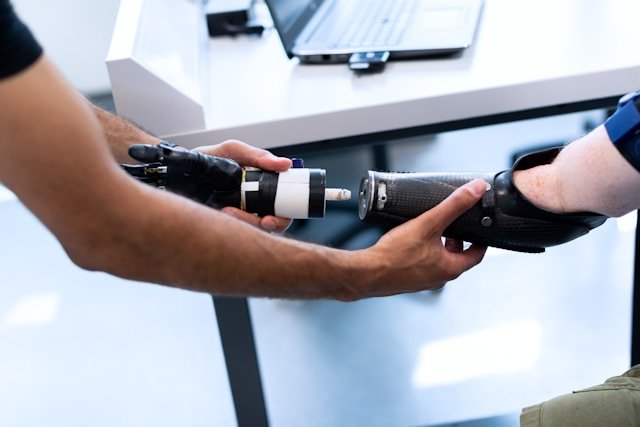
Not only does the study challenge a long-held belief about brain plasticity, but it also suggests that restoring movement or sensation to a paralyzed limb or a prosthetic controlled by brain-computer interface—the kind of work spearheaded by researchers at Pitt Rehab Neural Engineering Labs—is possible in the long-term.
The University of Pittsburgh is one of a number of institutions that is researching whether movement and sensation can be restored to paralyzed limbs or whether amputated limbs might be replaced by artificial, robotic limbs controlled by a brain interface. The newly reported study suggests that because the brain maps are preserved, it should, in theory, be possible to restore movement to a paralyzed limb or for the brain to control a prosthetic. “For brain–computer interfaces, our findings demonstrate a highly detailed and stable representation of the amputated limb for long-term applications,” the investigators concluded.
Baker further stated, “If the brain rewired itself after amputation, these technologies would fail. If the area that had been responsible for controlling your hand was now responsible for your face, these implants just wouldn’t work. Our findings provide a real opportunity to develop these technologies now.”
Schone added: “Now that we’ve shown these maps are stable, brain-computer interface technologies can operate under the assumption that the body map remains consistent over time. This allows us to move into the next frontier: accessing finer details of the hand map—like distinguishing the tip of the finger from the base—and restoring the rich, qualitative aspects of sensation, such as texture, shape, and temperature. This study is a powerful reminder that even after limb loss, the brain holds onto the body, waiting for us to reconnect.”
The post Brain’s Body Map Persists Even After Limb Amputation appeared first on GEN – Genetic Engineering and Biotechnology News.