
Introduction
Macrobrachium rosenbergii nodavirus (MrNV) is the causative agent of white tail disease (WTD) in giant freshwater prawn Macrobrachium rosenbergii, which leads to high post-larval mortality in aquaculture1,2,3,4,5. MrNV is a member of the Nodaviridae family, which broadly infects hosts ranging from fish to aquatic insects1,2,3,4,5. Furthermore, it is a non-enveloped ssRNA + virus with a T = 3 icosahedral capsid comprising 180 identical capsid protein subunits with a 26–27 nm particle size. Each capsid protein subunit contains three major domains: the protruding C-terminal P-domain, the shell-forming S domain, and the internal N-terminus RNA-binding domain. This RNA-binding domain, enriched in positively charged residues, plays a crucial role in capsid self-assembly by binding nucleic acids2,6,7. On the other hand, the P-domain at C-terminus plays a critical role in viral assembly and host binding8,9,10,11,12,13. Removing the last 27 amino acids of the P-domain markedly reduced host cell uptake of MrNV-VLPs, a process highly dependent on caveolin-mediated endocytosis14,15. Moreover, the P-domain of the capsid protein subunit has been structurally characterized as comprising a nine-stranded antiparallel β-barrel which interacts with a β-barrel of the P-domain, an adjacent subunit to form the two-fold, blade-like spike on the virus particle surface. ach β-barrel pairing produces loop regions (residues 268–275, 296–303, 322–326, and 350–355), which form the four outermost ‘pillars’ of the P-domain16,17. The C-terminal-end 27 amino acids constitute the 3rd pillar of the domain, in which its truncation did not affect the structural integrity of the entire domain, but did affect the domains’ ability to interact with the host cell surface.
Several potential binding targets for MrNV on the host cell surface have been proposed, in which the disruption of the interactions between the virus and the purported targets has resulted in a significant reduction in host cell infection. Macrobrachium rosenbergii transglutaminase (MrTG), synthesized by hemocytes, is believed to translocate to the cell surface membrane during the host innate immune response and, through its binding to cell surface integrins, mediates the viral binding and internalization process18. Our previous study also demonstrated the crucial role of surface N/O-linked fucosylated LacdiNAcs (LDNFs) on susceptible host tissues as MrNV binding molecules to initiate infection. Furthermore, the use of the broad fucose-binding Aleuria aurantia lectin (AAL), along with specific chemical and enzymatic removals of N-glycans, resulted in very significant reductions in MrNV virus-like particles (VLPs) to susceptible gill tissues, leading to the present study’s aim to disrupt the viral-host glycan fucosylated glycan interactions to reduce MrNV infection19.To achieve this goal, we exploited the relatively straightforward genetic modification and production of the self-assembling recombinant MrNV capsid proteins as icosahedral VLPs, which can be utilized as targeting nanocontainers for plasmid DNA and dsRNA delivery into insect and shrimp cells20,21. To produce MrNV-VLPs that can specifically bind specifically to fucosylated N-glycans found in susceptible cell lines such as Sf9 insect cells and host tissues, the P-domains of the capsid protein were genetically truncated to determine a stable form of P-domain-less VLPs (V250-MrNV-VLPs) that could be further modified to present a larger peptide stably. Accordingly, the V250-MrNV-VLPs’ capsid gene was further modified to include the gene of the carbohydrate-recognition domain (CRD) of the lectin CLEC17A (Prolectin), resulting in the complete replacement of the original MrNV-VLPs with the CRD sequence of a similar size.
The aim of adding the CLEC17A CRD was to generate VLPs (CLEC17A/CRD-MrNV-VLPs) with lectin-like properties to bind LDNF glycans on Sf9 cells and WTD-associated tissues in prawn. CLEC17A is a prolectin that is expressed mainly in dividing B cells found in the germinal centers of secondary lymphoid organs in humans. CLEC17A can serve as a cell adhesion molecule for fucosylated epithelial cancer cells and other fucosylated glycans of the blood group antigens, such as H1/2 and Lewis antigens21. The prolectin consists of three main regions: the carbohydrate recognition domain (CRD), the transmembrane domain (TM), and the intra-cytoplasmic domain22.
Through the application of the CLEC17A’s CRD as a complete replacement of the MrNV-VLPs’ P-domains, we aimed to create particles that mimic the prolectin’s high affinity for fucosylated glycans while exploiting the VLPs’ ability to present protruding ligands multivalently. This approach should increase the affinity of the CRD’s towards host surface fucosylated glycans while mitigating the issue of low yields in the purification of recombinant lectins or lectin conjugation through the more cost-effective expression of VLPs23,24. In this study, CLEC17A/CRD-MrNV-VLPs are stable icosahedral particles with intact CRD protrusions. These chimeric VLPs exhibit fucosylated glycan-binding activity, reduce MrNV-VLP and live virus binding to Sf9 cells, and highlight the flexibility of MrNV-VLPs as scaffolds for larger peptide domains. Given our previous studies showing that MrNV-VLPs could be used as nanocarriers of bioactive molecules21,25, these findings demonstrate the potential of MrNV-VLPs to be modified to present larger bioactive peptides such as CRDs as protruding domains, offering enhanced flexibility for the presentation of multimeric and multivalent bioactive ligands, thereby improving their potency and therapeutic applications.
Materials and methods
Design and structural prediction of chimeric VLPs
The sequence of the MrNV capsid protein (GenBank: EU150129) was used as the template to design chimeric MrNV-VLPs with replaced P-domains. Two variants were created: (1) V250-MrNV-VLPs, generated by deleting residues 251–374 of the P-domain, and (2) CLEC17A/CRD-MrNV-VLPs, created by inserting the CLEC17A CRD (residues 257–395) with a GSGGSG linker. The amino acid sequences of both the V250-MrNV-VLPs and the CLEC17A were entered into the PHYRE2 server for complex protein 3D structure prediction26. The predicted protein models of the capsid protein subunits were fitted and aligned into an icosahedral capsid structure of MrNV-VLPs using UCSF ChimeraX software27.
Preparation of recombinant MrNV-VLPs with P-domain modifications
V250-MrNV-VLPs with truncated P-domains as MrNV-VLPs variants were generated by truncating the nucleotides encoding residues E251-N371 in the MrNV capsid protein gene. Primers were designed to amplify the desired regions as described previously28. The altered MrNV capsid protein gene PCR products were then ligated into the pGEM-T Easy vector (Promega, Madison, WI), and the DNA sequence was verified by sequencing (Macrogen, Seoul, Korea). The ligated genes were digested and inserted into the pET17b expression vector (Novagen, Darmstadt, Germany) with a hexa-histidine tag sequence for downstream purifications by affinity chromatography. For CLEC17A/CRD-MrNV VLPs, a synthetic gene encoding the CRD domain was inserted in the same vector.
Expression and purification of chimeric MrNV capsid protein in E. coli BL21 (DE3)
Recombinant pET17b positive clones were transformed into E. coli BL21 (DE3) and cultured in LB broth containing 50 µg/ml carbenicillin at 37 °C. Protein expression was induced with 1 mM IPTG and incubated at 16 °C for 24 h. Cells were harvested and lysed, and the proteins purified using a bead agarose (Ni-NTA) chromatographic column (Qiagen, Germany). The eluates were further desalted with a Sephadex G25 desalting column (GE Healthcare, Piscataway, NJ). The protein expression and purification were analyzed with SDS-PAGE and immunoblotting. The proper assembly of VLPs was confirmed by negative staining and TEM analysis (Tecnai 20 FEI microscope operated at 200 kV).
Binding efficiency of CLEC17A/CRD-MrNV-VLPs to immobilized fucosylated glycoconjugates
The binding of CLEC17A/CRD-MrNV-VLPs to fucosylated antigens, including fucose and various Lewis antigens (tri-Lewis, Lewis Y (Ley), sialyl-Lewis A (sia-Lea), and sialyl-Lewis X (Sia-Lex)), was evaluated using ELISA. Plates were coated with sugar molecules and incubated with VLPs. Binding was detected using an anti-hexahistidine antibody (1:1000), followed by their corresponding secondary antibodies (1:2000) conjugated with HRP, and protein-antibody complexes were visualized with a SureBlue TMB substrate (KPL, Gaithersburg, MA). 1 N HCl solution was used to stop the reaction, and the intensity of the developed enzymatic products was detected with a spectrophotometer at 450 nm using an ELISA plate reader.
Preparation of MrNV inoculum
MrNV was isolated from infected post-larvae of Macrobrachium rosenbergii. Infected tissues were homogenized in phosphate-buffered saline (PBS) and centrifuged to remove debris as described previously29. The supernatant was filtered through 0.22 μm filters and used as inoculum in subsequent experiments.
Sf-9 cell culture and MrNV infection
Sf9 cells were cultured in serum-free medium (SF-900 III SFM) containing 1x antibiotic-antimycotic (Gibco, Grand Ireland, NY) in 25-ml flasks at 27 °C. After reaching > 90% confluence, the cells were dislodged and sub-cultured on 6-well plates at a concentration of 2 × 10⁶ cells/well and were further cultured for another day. The cells were divided into three groups: (1) control, (2) live MrNV inoculum-infected cells, and (3) CLEC17A/CRD-MrNV-VLP pre-incubated cells. Cells were incubated with MrNV for 1 h at 4 °C, and then excess binding was removed, followed by 72 h of incubation at 27 °C. Cells were then harvested for RNA extraction and immunofluorescence staining.
Inhibition assay by ELISA using MrNV-VLPs
Maxisorp plates (Nunc, Roskilde, Denmark) with 96-wells were coated overnight with Sf9 cell lysates protein at 4 °C. The wells were washed extensively with PBS before incubating with 20 µg/ml of CLEC17A/CRD-MrNV-VLPs for 1 h. The plates were again washed extensively with PBS and then incubated with wild-type MrNV-VLPs. Binding was detected using ELISA as described earlier, but anti-MrNV antibody (1:1000) was used as the primary antibody.
Indirect immunofluorescence and confocal laser microscopy
Sf-9 cells were fixed with 4% paraformaldehyde (30 min, RT) and washed with PBS. Free aldehydes were quenched with 30 mM glycine in PBS. Cells were exposed to 1:500 mouse anti-MrNV antibody in a blocking solution (0.5% BSA in PBS-Tween-20). This monoclonal antibody (a kind gift from Prof. Paisan Sithigorngul, Srinakarinwirot University, Thailand) has shown high specificity towards the MrNV capsid protein28,30. After that, cells were further incubated with 1:1000 goat anti-mouse Alexa 594 (Invitrogen) and counterstained with DAPI. Images were captured using a confocal microscope (Olympus FV1000).
Semi-quantitative reverse transcriptase-PCR (RT-PCR)
Total RNA was extracted from Sf9 cells and subjected to RT-PCR using a Superscript III One-Step RT-PCR kit (Invitrogen, Eugene, CA) with MrNV-specific primers. Three separate total RNA extractions underwent PCR amplification runs comprising 40 cycles: preheated at 95 °C for 5 min followed by cycles of 95 °C for 60 s, 60 °C for 30 s, and 72 °C for 60 s. At least three separate PCR products were visualized via agarose gel electrophoresis, and RNA expression levels were quantified using densitometric analysis in Image J31. Actin served as internal control.
Detection and quantification of MrNV infection
To confirm infection, MrNV in Sf9 was analyzed using a two-step nested RT-PCR with two sets of specific primers (forward: 5′ GATACAGATCCACTAGATGACC 3′; reverse: 5′ GACGATAGCTCTGATAATCC 3′) and (forward: 5′ GGCAGGCTACGTCACAAGT 3′; reverse: 5′ GCATGGAAAATCCACAGACC 3′), yielding two DNA fragments of 681 bp and 250 bp, respectively, in accordance with the previously established protocol32,33. The initial stage PCR products were subsequently utilized as templates for qRT-PCR to measure the level of MrNV infection and viral load in each group. The relative expressions of the MrNV genes were quantified using the 2 − ΔΔCt technique in comparison to the internal control, β-actin. The copy number of MrNV was quantified using a standard curve derived from serially diluted pGEM-T easy vectors harboring the capsid protein genes of MrNV, in accordance with the established technique previously described in Jariyapong et al.34. The data were evaluated utilizing an unpaired t test with a two-tailed distribution, and the findings were presented as mean ± standard deviation (SD) of three replicates.
Results
Structural modeling confirms feasibility of P-domain replacement of the MrNV capsid protein subunit
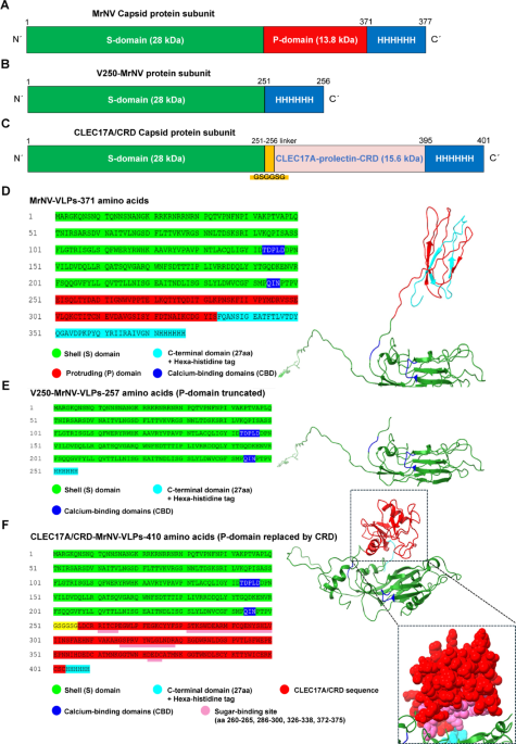
The protein layout of the MrNV, V250-MrNV and CLEC17A/CRD-MrNV capsid protein subunits are shown in Fig. 1A–C. The wild-type MrNV capsid protein subunit gene consists of a 28 kDa S-domain sequence, followed by a 13.8 kDa P-domain sequence and a hexa-histidine tag, resulting in ~ 42 kDa protein. The V250-MrNV capsid protein gene removes the sequence encoding the P-domain entirely, resulting in a 28 kDa protein. The CLEC17A/CRD-MrNV gene consists of an S-domain of 28 kDa followed by a sequence encoding the flexible linker GSGGSG subsequently followed by a 15.6 kDa CLEC17A CRD sequence. As a result, the CLEC17A CRD sequence replaces the original P-domain sequence entirely, ending with a hexa-histidine tag. This resulted in the CLEC17A/CRD-MrNV capsid protein gene predicted to encode a ~ 45 kDa protein.


Design and prediction of CLEC17A/CRD to replace the protrusion (P) domains of the capsid protein subunit gene of Macrobrachim rosenbergii nodavirus (MrNV-VLPs). The general capsid protein subunit layouts are shown: A wild-type MrNV-VLPs, B MrNV-VLPs without P-domains or V250-MrNV-VLPs, C MrNV-VLPs with their P-domains replaced by the CLEC17A CRD called CLEC17A/CRD MrNV-VLPs. D–F Shows all three capsid proteins’ amino acid sequences and PHYRE2 complex structure predictions. The inset of the last panel shows the protruding CRD domains of CLEC17A/CRD MrNV-VLPs with the possible carbohydrate-binding site colored in pink as predicted by molecular docking. The predicted icosahedral structures of all three capsids show apparent differences in the protrusions and surface topographies (G–I).
Homology modeling showed that residues 251–371 of the MrNV P-domain were successfully replaced with CLEC17A CRD residues 246–378. The resulting subunits differed structurally from wild-type MrNV and V250-MrNV-VLPs, producing new protrusions consistent with CRD domains (Fig. 1F) when compared to the original MrNV capsid (Fig. 1D) and V250-MrNV-VLPs (Fig. 1E). After structure alignment of each CLEC17A/CRD capsid protein subunit to the subunits of MrNV-VLPs (Fig. 1G) constituting the VLP structure, it was observed that the structure of the CLEC17A/CRD in the capsid’s outer leaflet with minimal structural clashing (Fig. 1I). The red ribbon in the figure highlights the structural changes in the protruding regions compared to the wild-type MrNV-VLPs (Fig. 1G), with the CRD protrusions predicted to be less prominent and less blade-like. The modification is visible in the 5-fold symmetry views, with the protruding CLEC17A/CRD present in all 180 subunits of CLEC17A/CRD-MrNV-VLPs (Fig. 1I). Nevertheless, both CLEC17A/CRD-MrNV and MrNV-VLPs were predicted to present visible yet distinct protrusions/spikes as compared to the completely smooth and P-domain-less V250-MrNV-VLPs (Fig. 1H).
The truncation of the region encoding the P-domain of MrNV capsid gene resulted in the generation of smaller icosahedral particles lacking distinct surface protrusions (V250-MrNV-VLPs)
Truncating the P-domain from the MrNV capsid gene resulted in the expression of smaller capsid proteins and in the production of smaller icosahedral-like particles with a smoother surface topography, as shown in the TEM micrographs (Fig. 2D, second panel). This observation corresponded well with the predicted icosahedral structure in Fig. 1H of the V250-MrNV-VLPs. The diameters of V250-MrNV-VLPs ranged from 20 to 22 nm, which was significantly smaller than the wild-type MrNV-VLPs (26–28 nm). Truncating the capsid gene anywhere into the second calcium-binding domain (D244-N246) resulted in truncated capsid protein expression but neither particle formation nor subunit polymerization were observed (results not shown). Despite the size reduction, the V250-MrNV-VLPs retained their structural integrity with significant topological changes from wild-type MrNV-VLPs characterized by their lacking observable protrusions. This indicated that the truncation of the P-domains in their entirety from the MrNV capsid protein still allowed the assembly and formation of icosahedral particle formation; hence, providing a platform for further modifications, as shown in the present study with the inclusion of the CRD sequence of the CLEC17A prolectin.

Expression, purification, and assembly into stable CLEC17A/CRD-MrNV VLPs. A SDS-PAGE analysis of the purity of capsid protein expression visualized by Coomassie Blue R250 staining of wild-type MrNV-VLPs, no protrusion domain (V250-MrNV-VLPs) and CLEC17A/CRD-MrNV-VLPs at the sizes ~ 42 kDa, 28 kDa and 45 kDa, respectively. The first panel of B shows the Western blot using anti-hexahistidine antibodies which reveals the purity of purification and the accuracy of the molecular weights of the various capsids. The second Western blot panel C shows the use of anti-CLEC17A/Prolectin CRD antibodies, showing the correspondence to the predicted molecular weight of the CLEC17A/CRD capsid protein of 45 kDa as compared to the 18 kDa weight of monomeric CLEC17A (Prolectin) CRDs and that of the avidin-multimerized “poly” form. D Shows the transmission electron micrographs of VLPs demonstrated by negative TEM staining. The icosahedral structures of wild-type MrNV-VLPs, V250-MrNV-VLPs, and CLEC17A/CRD-MrNV-VLPs are shown at a magnification of 100 kX. The average size of the MrNV-VLPs particles formed was approximately 26–28 nm, V250-MrNV-VLPs was approximately 20–22 nm, and CLEC17A/CRD-MrNV-VLPs was 30–32 nm. Bars = 50 nm.
CLEC17A/CRD-MrNV capsid proteins were successfully expressed and self-assembled into icosahedral VLPs (CLEC17A/CRD-MrNV-VLPs)
We previously demonstrated that MrNV-VLPs can present small peptides by replacing or extending the terminal 27 amino acids of the P-domain. Therefore, to further investigate the versatility of the MrNV-VLPs, we explored the addition of a much larger ligand in the form of the fucosylated glycan-binding carbohydrate recognition domain (CRD) from CLEC17A (Prolectin). The CRD of CLEC17A has a molecular weight (MW) of approximately 15.6 kDa, with the capsid protein subunits of CLEC17A/CRD-MrNV-VLPs having a predicted MW of approximately 45 kDa, which was slightly higher than the 42.2 kDa of wild-type MrNV-VLPs. SDS-PAGE and Western blot analysis confirmed the expression of CLEC17A/CRD-MrNV-VLPs with an MW of approximately 45 kDa (Fig. 2A, B), consistent with the predicted protein layout shown in Fig. 1C. Western blot with anti-hexahistidine (Fig. 2B) and CLEC17A CRD-specific antibodies (anti-Prolectin) further verified the presence of the CLEC17A/CRD in the particles (Fig. 2C). These results confirmed that the CLEC17A/CRD-MrNV-VLPs were successfully expressed and contained the intact CRD region of CLEC17A as a complete replacement of the original P-domains of the MrNV capsid protein subunit. Transmission electron microscopy (TEM) revealed that expressed CLEC17A/CRD-MrNV capsid protein subunits could assemble into stable icosahedral particles. Furthermore, TEM observations revealed CLEC17A/CRD-MrNV-VLPs to be icosahedral particles with a diameter of about 30–32 nm, which was slightly larger than that of the wild-type MrNV-VLPs at 26–28 nm. These observations confirmed the successful formation of stable icosahedral particles with the protruding CRD regions fully intact on CLEC17A/CRD-MrNV-VLPs (Fig. 2D panel 3).
Binding efficiency of CLEC17A/CRD-MrNV-VLPs to fucosylated antigens
ELISA experiments revealed that CLEC17A/CRD-MrNV-VLPs at a concentration of 5 µg/ml showed significant binding to immobilized fucosylated glycans, including Lewis Y and sialylated Lewis antigens (Fig. 3). The binding efficiency of CLEC17A/CRD-MrNV-VLPs was significantly higher compared to V250-MrNV-VLPs and wild-type MrNV-VLPs, confirming the ability of the CRD-modified VLPs to target fucosylated antigens with higher affinity. This finding demonstrated that CLEC17A/CRD-MrNV-VLPs possess enhanced specificity and binding potential towards both simple and complex fucosylated glycans, making them promising candidates for targeting fucosylated-glycan-expressing cells such as the MrNV-susceptible Sf9 cell line.

Binding efficiency CLEC17A/CRD-MrNV-VLPs on immobilized glycans by ELISA. CLEC17A/CRD MrNV-VLPs and V250- MrNV-VLPs were tested for the level of binding to different immobilized glycans (α-Fucose, LeY, Lea, Sia-LeY as fucose glycans and Tn as non-fucose glycan negative control, and coating buffer as buffer control). The levels of goat-anti mouse and anti-hexahistidine antibody detected indicated the amount of binding capacity and were further measured by a microplate reader at 452 nm absorbance. The data were expressed as the mean number ± SD. Statistical significance: ****p < 0.0001.
CLEC17A/CRD-MrNV-VLPs significantly reduced MrNV binding levels to Sf9 cell lysates
Figure 4 shows the significant reduction of MrNV-VLP binding to Sf9 cell lysate-coated wells at a concentration of 5 µg/ml binding by an equivalent concentration of CLEC17A/CRD-MrNV-VLPs. When lysate-coated wells were pre-incubated with 5 µg/ml CLEC17A/CRD-MrNV-VLPs, the binding of MrNV-VLPs was reduced by around 82.91%. This result suggested that CLEC17A/CRD-MrNV-VLPs are high potential compet with MrNV and/or MrNV-VLPs for binding sites in the protein lysates of MrNV-susceptible Sf9 cells, leading to our subsequent investigation of the chimeric VLPs in reducing MrNV interactions with live Sf9 cells.

Effect of CLEC17A/CRD-MrNV-VLPs on the level of binding of wild-type MrNV-VLPs to Sf9 cell lysates by ELISA. Sf9 cell lysates were coated on the plate and incubated with or without CLEC17A/CRD-MrNV-VLPs before the wells were incubated with wild-type MrNV-VLPs. The data were expressed as the mean ± SD. Statistical significance: ***p < 0.00001.
CLEC17A/CRD-MrNV-VLPs significantly reduced the level of MrNV infection and copy number in Sf9 cells
Confocal microscopy and PCR (Fig. 5) demonstrated that Sf9 cells pre-incubated with CLEC17A/CRD-MrNV-VLPs at a concentration of 5 µg/ml significantly reduced MrNV infection by purified MrNV inoculum. As shown in Fig. 5A, bottom row of panels, the red signals signifying the presence of MrNV binding and infection were significantly lowered by the pre-incubation with CLEC17A/CRD-MrNV-VLPs than in Sf9 cells incubated solely with MrNV inoculum (Fig. 5A, middle panels). Furthermore, Sf9 cells pre-treated with CLEC17A/CRD-MrNV-VLPs followed by MrNV inoculum showed a decrease of about 48.65% in MrNV RNA levels represented by the reduction in the percent intensity of the bands (Fig. 5B, left panel, agarose electrophoresis, 3rd, 6th and 9th lanes as triplicates) as opposed to cells infected by MrNV inoculum alone (Fig. 5B, left panel, agarose electrophoresis, 4th, 7th and 10th lanes). The level of MrNV replication in the cell lines were further detected by specific qRT-PCR primers (Fig. 5C), further supporting these chimeric VLPs’ protective effect against viral infection. Cells pre-treated with CLEC17A/CRD-MrNV-VLPs showed a ~ 1.73-fold reduction (~ − 71.59%) in the relative expression of MrNV RNA; representing a reduction from ~ 10^4.63 to ~ 10^2.45, corresponding to ~ 99.3% fewer viral copies.” These results suggested that pre-incubation with CLEC17A/CRD-MrNV-VLPs significantly reduced MrNV infection in Sf9 cells and could be effective as potential viral blockers of MrNV infection in susceptible cells.

Effect of CLEC17A/CRD-MrNV-VLPs on the level of MrNV infection in Sf9 cells by confocal microscopy and qRT-PCR. A Sf9 cells were cultured and incubated with CLEC17A/CRD-MRNV-VLPs before being infected with MrNV live virus for 3 days and then detected with anti-MrNV antibody. B, C Cells were collected for RNA extraction and RNA levels were detected by RT-PCR and qRT-PCR using MrNV primers; β-actin was used as the control. Scale bar = 20 μm. The data were expressed as the mean ± SD. Statistical significance: **p < 0.001, ***p < 0.0001.
Conclusion
The development of CLEC17A/CRD-MrNV-VLPs represented a promising biotechnological tool for the effective reduction of MrNV infection, with potential applications in aquaculture and virology. Their targeted disruption of viral attachment suggested future avenues for developing similar nanoparticle systems for therapeutic interventions against other fucosyl-glycosylated-binding aquatic viruses, as proposed in Fig. 6.

Schematic illustrations of the strategies to reduce MrNV infection by CLEC17A/CRD-MrNV-VLPs. A CLEC17A/CRD interaction with fucosyl-glycosylated bind and enter Sf9 cells/ susceptible host cells/tissues. B CLEC17A/CRD-MrNV-VLPs competitively bind to fucosyl-glycosylated/ on host cells to potentially decrease MrNV virus infection.
Discussion
We successfully designed chimeric MrNV VLPs in which the P-domains were replaced with the CRD of human prolectin CLEC17A. Our results confirmed that the CLEC17A/CRD-MrNV-VLPs were stable, retained their icosahedral structure, and exhibited enhanced specificity toward fucosylated glycans expressed by Sf9 cells, significantly reducing both MrNV-VLPs binding and live MrNV infection. This outcome supports and elaborates on our earlier findings, where MrNV-VLPs demonstrated their potential as robust nanocontainers and vehicles for therapeutic biomolecules such as plasmid DNA and dsRNA into insect and shrimp cells21,25. These earlier studies highlighted the structural integrity and versatility of MrNV-VLPs, establishing them as promising vectors for therapeutic interventions in aquaculture22.
Furthermore, the truncation of the P-domains from the MrNV capsid gene resulting in icosahedral yet smaller particles (V250-MrNV-VLPs) presented us with the intriguing possibility of adding larger bioactive peptides that target important virus-interacting molecules such as surface glycans. Incorporating the CRD derived from CLEC17A into MrNV-VLPs represents a strategy to enhance glycan-binding capability. While the focus here is on nodavirus interactions, the same approach may be adaptable to other systems that exploit glycan-mediated entry. In relevance to the present study, CLEC17A, or Prolectin, is a C-type lectin initially identified as a fucose-binding molecule associated with carcinoma cell adhesion through recognition of glycan epitopes, such as Lewis antigens and blood group antigens22. Incorporating the CLEC17A CRD into MrNV-VLPs strategically provided them with glycan-binding capability. Indeed, previous reports emphasize that the multivalent presentation of CRDs significantly amplifies lectin specificity and functional activity compared to monovalent interactions, making them highly effective in competitive receptor inhibition strategies7,23. Accordingly, the CLEC17A/CRD-MrNV-VLPs were demonstrated in this study to be able to bind and target the fucosylated LDNFs which we recently reported to be crucial molecules for the binding of the virus to gill tissues in prawn which are an important entry and disease associated tissue in WTD19.
In addition, in this study we showed that the chimeric capsid subunits were able to undergo self-assembly after the substitution of the original P-domains with CLEC17A-CRDs. Such structural maintenance upon replacement with larger protein domains has previously been challenging due to potential steric hindrance or protein misfolding issues7,23. Our findings suggest that the S-domain of MrNV-VLPs can accommodate significant modifications, such as CRD insertion, without compromising assembly. However, broader testing with other domains will be needed to confirm this flexibility. Additionally, previous studies have demonstrated varying degrees of success when viral protrusion domains have been genetically replaced by large foreign epitopes, ligands, or entire protein domains. For example, the HBV core antigen has been engineered to display epitopes and immunomodulatory proteins35,36, while norovirus VLPs have been modified with receptor-binding domains to generate vaccine candidates37,38. Cowpea Mosaic Virus VLPs have also demonstrated that insertion of large peptides requires careful optimization to maintain stability39. These examples highlight both the potential and the challenges of using viral scaffolds for large-domain displays. These findings suggest the potential exploitation of the flexibility and robustness of MrNV-VLP scaffolds for further biomedical and therapeutic applications.
Although Sf9 cells provide a useful model as shown in our previous study14, they may not fully recapitulate the glycosylation patterns of M. rosenbergii tissues19. Confirming efficacy in shrimp models or primary cells will therefore be essential to evaluate practical applicability. Viral entry mechanisms differ substantially between viruses, particularly with respect to the glycan structures they exploit for attachment. MrNV likely interacts with fucosylated glycans (LDNF) as shown by Somrit et al., 2020, whereas other viruses such as influenza and coronaviruses typically bind sialic acid residues on host glycoproteins40. Therefore, future work should incorporate validation in shrimp models or primary tissue cultures to better assess the efficacy and applicability of these engineered VLPs in natural aquaculture settings. The immunogenicity of CLEC17A/CRD-MrNV-VLPs in shrimp remains unknown and should be addressed in future studies. Similarly, glycomic profiling will be valuable for identifying the precise epitopes targeted by these particles. These findings should pinpoint the precise epitopes involved, thus refining the specificity and potential therapeutic utility of these engineered particles. Additionally, we continue to explore alternative expression systems, such as yeast or plant-based platforms, which further facilitate scalable and economically feasible production, enhancing the overall applicability of these nanoparticles.
Nevertheless, our inhibition assays showed that pre-incubation of Sf9 cells with CLEC17A/CRD-MrNV-VLPs significantly reduced the binding and subsequent infection of MrNV. These results provide proof-of-concept that CLEC17A/CRD-MrNV-VLPs can act as decoy particles, competing for host glycan receptors and reducing infection. Their applicability to other viral systems remains to be established. This observation supports our initial aim that CLEC17A/CRD-MrNV-VLPs can act as decoy or competitive binding particles, effectively competing for viral receptor sites on the host cells, thus impeding the virus from establishing an infection. Confocal microscopy, RT-PCR and qRT-PCR further confirmed a marked reduction in MrNV RNA in Sf9-cells treated with CLEC17A/CRD-MrNV-VLPs, demonstrating their significant protective effect. These findings have potential applications in the design of MrNV nanoparticle-based therapies. Given their ability to be genetically modified while maintaining structural stability, these chimeric VLPs may offer a versatile platform for developing targeted antiviral strategies in aquaculture, pending further in vivo validation.
Data availability
The datasets used and/or analyzed during the current study are available from the corresponding authors on reasonable request.
References
-
Bonami, J. R. et al. White tail disease of the giant freshwater prawn, Macrobrachium rosenbergii: separation of the associated virions and characterization of MrNV as a new type of nodavirus. J. Fish. Dis. 28 (1), 23–31 (2005).
-
Odegard, A., Banerjee, M. & Johnson, J. E. Flock house virus: a model system for understanding non-enveloped virus entry and membrane penetration. Curr. Top. Microbiol. Immunol. 343, 1–22 (2010).
-
Barke, D. E. et al. First report of piscine nodavirus infecting wild winter flounder pleuronectes Americanus in passamaquoddy Bay, new Brunswick, Canada. Dis. Aquat. Org. 49 (2), 99–105 (2002).
-
Castri, J. et al. Sea Bream Sparus aurata, an asymptomatic contagious fish host for nodavirus. Dis. Aquat. Org. 47 (1), 33–38 (2001).
-
Delsert, C., Morin, N. & Comps, M. Fish nodavirus lytic cycle and semipermissive expression in mammalian and fish cell cultures. J. Virol. 71 (7), 5673–5677 (1997).
-
Dasgupta, R., Selling, B. & Rueckert, R. Flock house virus: a simple model for studying persistent infection in cultured drosophila cells. Arch. Virol. Suppl. 9, 121–132 (1994).
-
Maharaj, P. D. et al. Nanoparticle encapsidation of flock house virus by auto assembly of tobacco mosaic virus coat protein. Int. J. Mol. Sci. 15 (10), 18540–18556 (2014).
-
Owens, L. et al. Macrobrachium rosenbergii nodavirus disease (white tail disease) in Australia. Dis. Aquat. Org. 85 (3), 175–180 (2009).
-
Qian, D. et al. Extra small virus-like particles (XSV) and nodavirus associated with whitish muscle disease in the giant freshwater prawn, Macrobrachium rosenbergii. J. Fish. Dis. 26 (9), 521–527 (2003).
-
Sahul Hameed, A. S. et al. Experimental transmission and tissue tropism of Macrobrachium rosenbergii nodavirus (MrNV) and its associated extra small virus (XSV). Dis. Aquat. Org. 62 (3), 191–196 (2004).
-
Sahul Hameed, A. S. & Bonami, J. R. White tail disease of freshwater Prawn, Macrobrachium rosenbergii. Indian J. Virol. 23 (2), 134–140 (2012).
-
Yoganandhan, K. et al. White tail disease of the giant freshwater Prawn Macrobrachium rosenbergii in Thailand. Dis. Aquat. Org. 69 (2–3), 255–258 (2006).
-
Zhang, Q. et al. A new nodavirus is associated with covert mortality disease of shrimp. J. Gen. Virol. 95 (Pt 12), 2700–2709 (2014).
-
Somrit, M. et al. The key molecular events during Macrobrachium rosenbergii nodavirus (MrNV) infection and replication in Sf9 insect cells. Virus Res. 223, 1–9 (2016).
-
Somrit, M. et al. C-terminal domain on the outer surface of the Macrobrachium rosenbergii nodavirus capsid is required for Sf9 cell binding and internalization. Virus Res. 227, 41–48 (2017).
-
Chen, K. F. et al. The Macrobrachium rosenbergii nodavirus: a detailed review of structure, infectivity, host immunity, diagnosis and prevention. Rev. Aquacult. 13, 2117–2141 (2021).
-
Chong, L. C. et al. Expression, purification and characterization of the dimeric protruding domain of Macrobrachium rosenbergii nodavirus capsid protein expressed in Escherichia coli. PLoS One 14, 1–12 (2019).
-
Sirikharin, R. et al. Cell surface transglutaminase required for nodavirus entry into freshwater Prawn hemocytes. Fish. Shellfish Immunol. 89, 108–116 (2019).
-
Somrit, M. et al. Macrobrachium rosenbergii nodavirus virus-like particles attach to fucosylated glycans in the gills of the giant freshwater Prawn. Cell. Microbiol. 22 (12), e13258 (2020).
-
Goh, Z. H. et al. Virus-like particles of Macrobrachium rosenbergii nodavirus produced in bacteria. J. Virol. Methods. 175 (1), 74–79 (2011).
-
Jariyapong, P. et al. Encapsulation and delivery of plasmid DNA by virus-like nanoparticles engineered from Macrobrachium rosenbergii nodavirus. Virus Res. 179, 140–146 (2014).
-
Breiman, A. et al. Carcinoma-associated fucosylated antigens are markers of the epithelial state and can contribute to cell adhesion through CLEC17A (Prolectin). Oncotarget 7 (12), 14064–14082 (2016).
-
Tobola, F. & Wiltschi, B. One, two, many: strategies to alter the number of carbohydrate binding sites of lectins. Biotechnol. Adv. 60, 108020 (2022).
-
Rekha Mol, K. R. A. A. M. H. Use of lectin-functionalized and lectin-targeted nanoparticles for multiple therapeutic applications. In Applications of Multifunctional Nanomaterials Micro and Nano Technologies (eds Thomas, S. et al.) 543–566 (Elsevier, 2023).
-
Grataitong, K. et al. Chimeric virus-like particles (VLPs) designed from shrimp nodavirus (MrNV) capsid protein specifically target EGFR-positive human colorectal cancer cells. Sci. Rep. 11 (1), 16579 (2021).
-
Kelley, L. A. et al. The Phyre2 web portal for protein modeling, prediction and analysis. Nat. Protoc. 10 (6), 845–858 (2015).
-
Pettersen, E. F. et al. UCSF chimerax: structure visualization for researchers, educators, and developers. Protein Sci. 30 (1), 70–82 (2021).
-
Wangman, P. et al. Production of monoclonal antibodies specific to Macrobrachium rosenbergii nodavirus using Recombinant capsid protein. Dis. Aquat. Org. 98 (2), 121–131 (2012).
-
Ravi, M. et al. Clearance of Macrobrachium rosenbergii nodavirus (MrNV) and extra small virus (XSV) and immunological changes in experimentally injected Macrobrachium rosenbergii. Fish. Shellfish Immunol. 28 (3), 428–433 (2010).
-
Longyant, S. et al. Monoclonal antibodies against extra small virus show that it co-localizes with Macrobrachium rosenbergii nodavirus. Dis. Aquat. Org. 99 (3), 197–205 (2012).
-
Schneider, C. A., Rasband, W. S. & Eliceiri, K. W. NIH image to imageJ: 25 years of image analysis. Nat. Methods. 9 (7), 671–675 (2012).
-
Yoganandhan, K., Sri Widada, J., Bonami, J. R. & Sahul Hameed, A. Simultaneous detection of Macrobrachium rosenbergii nodavirus and extra small virus by a single tube, one-step multiplex RT‐PCR assay. J. Fish Dis. 28 (2), 65–69 (2005).
-
Thongsum, O. et al. Submersion treatment of chimeric MrN-VLPs encapsulating therapeutic double‐stranded RNA effectively rescues prawn viral infection. J. Fish Dis. e70009. https://doi.org/10.1111/jfd.70009 (2025).
-
Jariyapong, P. et al. Hematopoietic tissue of Macrobrachium rosenbergii plays dual roles as a source of hemocyte hematopoiesis and as a defensive mechanism against Macrobrachium rosenbergii nodavirus infection. Fish Shellfish Immunol. 86, 756–763 (2019a).
-
Paul Pumpens, E. G. Hepatitis B core particles as a universal display model: a structure-function basis for development. FEBS Lett. 442 (1), 1–6 (1999).
-
O’Rourke, J. P., Peabody, D. S. & Chackerian, B. Affinity selection of epitope-based vaccines using a bacteriophage virus-like particle platform. Curr. Opin. Virol. 11, 76–82 (2015).
-
Mohsen, M. O., Augusto, G. & Bachmann, M. F. The 3Ds in virus-like particle based-vaccines: design, delivery and dynamics. Immunol. Rev. 296 (1), 155–168 (2020).
-
Lu, Y., Welsh, J. P. & Swartz, J. R. Production and stabilization of the trimeric influenza hemagglutinin stem domain for potentially broadly protective influenza vaccines. Proc. Natl. Acad. Sci. USA 111 (1), 125–130 (2014).
-
Wang, C., Beiss, V. & Steinmetz, N. F. Cowpea mosaic virus nanoparticles and empty virus-Like particles show distinct but overlapping immunostimulatory properties. J. Virol., 93(21). (2019).
-
Boonkua, S. et al. Replacing protruding domains of MrNV virus-like particles with Sialic acid binding domains enhances binding to SARS-CoV-2 susceptible cells and reduces pseudovirus infection. Sci. Rep. 15 (1), 25200 (2025).
Acknowledgements
This work was supported by Office of the Permanent Secretary, Ministry of Higher Education, Science, Research and Innovation (OPS MHESI), Thailand Science Research and Innovation (TSRI) (contract no. RGNS 64-163 and RGNS 64-213) awarded to MS and AW.
Ethics declarations
Competing interests
The authors declare no competing interests.
Additional information
Publisher’s note
Springer Nature remains neutral with regard to jurisdictional claims in published maps and institutional affiliations.
Supplementary Information
Rights and permissions
Open Access This article is licensed under a Creative Commons Attribution-NonCommercial-NoDerivatives 4.0 International License, which permits any non-commercial use, sharing, distribution and reproduction in any medium or format, as long as you give appropriate credit to the original author(s) and the source, provide a link to the Creative Commons licence, and indicate if you modified the licensed material. You do not have permission under this licence to share adapted material derived from this article or parts of it. The images or other third party material in this article are included in the article’s Creative Commons licence, unless indicated otherwise in a credit line to the material. If material is not included in the article’s Creative Commons licence and your intended use is not permitted by statutory regulation or exceeds the permitted use, you will need to obtain permission directly from the copyright holder. To view a copy of this licence, visit http://creativecommons.org/licenses/by-nc-nd/4.0/.
About this article

Cite this article
Chantunmapitak, R., Boonkua, S., Thongsum, O. et al. Chimeric virus-like particles carrying the CLEC17A carbohydrate-recognition domain significantly reduce Macrobrachium rosenbergii nodavirus infection in Sf9 cells. Sci Rep 15, 43311 (2025). https://doi.org/10.1038/s41598-025-27357-3
-
Received:
-
Accepted:
-
Published:
-
Version of record:
-
DOI: https://doi.org/10.1038/s41598-025-27357-3