Introduction
Lemon (C. limon L. Osbeck), belonging to the Rutaceae family, encompasses numerous varieties. Among these, the Lisbon and Eureka varieties are predominantly cultivated in southern Iran, particularly in Jahrom1. Historically, lemons have been used in traditional medicine for treating various conditions, including tumors. Fresh lemon fruit extract with garlic (Allium sativum L.) is recommended in Iranian traditional medicine for treating different tumors2,3. A large part of the biologically valuable compounds of this plant are found in its wastes, i.e., the peel. Lemon peel makes up about 13% of the whole fruit and 65% of its waste4,5. However, many valuable compounds, including pectin and flavonoids, are in the peel5,6. Citrus lemon peel is a valuable source of anticancer compounds, thereby underscoring the necessity for further investigation into its anticancer potential7. Therefore, it holds promise as a candidate for the treatment of various cancers, including breast cancer.
Breast cancer is one of the most common malignant diseases as well as the second cause of cancer death in women8. According to the American Cancer Society, in 2022, the incidence of invasive and non-invasive women breast cancer in this country reached 287,850 and 51,400 cases, respectively9. Several treatment options for breast cancer include surgery, chemotherapy, or hormone therapy. Despite different treatment options, recurrence and metastasis are still the main obstacles associated with high breast cancer mortality10. Besides, the high dose and long-term use of chemical drugs may cause severe damage to organs damage to normal cells and One of the major obstacles to chemotherapy is the resistance of cancer cells to drugs11. Therefore, there is a need for new therapeutic methods for this type of cancer. Targeted therapy is a form of cancer treatment that employs compounds to specifically target and inhibit the activity of genes and proteins contributing to cancer cell survival and proliferation. So far, many studies have been conducted on the genes involved in the invasion or apoptosis inhibition in breast cancer. One of the critical signaling pathways in the cancer cell, which is the target of many anti-cancer drugs, is the MAPK pathway.
The JNK2 gene belongs to the MAP kinase family, crucial for proliferation, apoptosis, and migration, and acts as an oncogene in several cancers12. SNAIL is involved in epithelial-mesenchymal transition (EMT), survival, and cancer cell migration, with antiapoptotic effects and upregulation in several cancers13. VIMENTIN is a marker for aggressive breast cancer, especially hormone-independent types, with higher levels correlating with poor prognosis14. Additionally, FAS is a tumor necrosis factor receptor family member that initiates extrinsic apoptosis when bound by Fas ligand. Studies indicate that suppression of FAS expression leads to breast tumor resistance to apoptosis15,16. Consequently, given the various causes of breast cancer and the continuous discovery of new factors, the use of modern technologies is essential to identify and treat this disease.
Today, new technologies are utilized to reduce side effects and increase the efficiency of chemotherapy agents. Nano-scale carriers, including niosomes, can cross biological barriers, protect the drug, and release its optimal amount17,18. The non-ionic nature of niosomes leads to lower toxicity and slower release, which augments the effect of the encapsulated drug. Niosomes can cross biological barriers, protect drugs, and release optimal amounts, leading to lower toxicity and improved drug efficacy. Encapsulating plant compounds in niosomes increases their resistance and stability against degradation, enhancing the bioavailability of phytochemicals19,20.
Previous studies showed that flavonoid derivatives and citrus juice inhibited the proliferation in MCF-7/6 and MDA-MB‐435 breast cancer cells as well as in rat models21,22. In another study, these flavonoids prevented the proliferation of human breast cancer cell lines (MDA-MB-231 and MCF-7) and human colon cancer cells (HT-29) in some concentrations23. The whole citrus peel extract is believed to have higher antioxidant and anti-cancer activity than isolated compounds24,25,26,27. Some studies show that the key components of Citrus limon are effectively retained in nanoparticles. This encapsulation ensures that their pharmaceutical efficacy is maintained upon entering the human body. Nanoparticles from lemon juice can inhibit the growth of various cancers by modulating related gene expression28,29. The combination therapy of chemotherapy agents with herbal compounds represents an intriguing approach for researchers aiming to develop effective and targeted treatments for cancer30,31. Thus, we focused on creating a niosomal formulation of methanolic and ethanolic extracts from citrus peels and examining their effects on breast cancer cells. Then, a comparative analysis of the anti-cancer properties of these extracts against the established chemotherapy drug, doxorubicin, was conducted, alongside an exploration of the potential synergistic effects when combined with doxorubicin. This study also assessed the expression of JNK2, SNAIL, VIMENTIN and FAS genes to identify the genes and pathways affected by lemon extract.
Results
Radical scavenging activity of the extract
Free radical scavenging activity was done from 3 to 400 µg/mL of ME relative to the standard index. The results show that the extract was the closest to the standard index regarding free radical scavenging at a concentration of 200–400 µg/mL. Also, the antioxidant activity of the extract increased in a dose-dependent manner. IC50 in this graph was calculated using the slope of the line and with the help of Prism 8 software. IC50 for ascorbic acid as positive control and standard indicator and methanol extract was 22.58 ± 2.78 µg/mL and 65.4 ± 3.61 µg/mL, respectively. In other words, the ME showed a lower antioxidant activity than ascorbic acid. Statistical analysis results in Prism 8 software for IC50 showed a significant difference between the antioxidant properties of methanolic extract and vitamin C (P < 0.01). The results of the measuring pesticide residues showed that among 78 pesticides, only 0.086 mg/kg of propiconazole was detected in the sample below the limit.
Characterization of synthesized and loaded niosomes
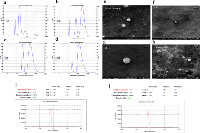
DLS analysis on blank niosome (NIO), NIO/EE, NIO/ME, and NIO/DOXO is shown in Fig. 1a-d. The mean particle size (nm), the Polydispersity Index (PDI), zeta potential (mV), and EY% is shown in Table 1. All physicochemical data are presented as mean ± standard deviation (SD) from at least three independent measurements. Furthermore, Fig. 1e-h shows the SEM image for the noisome formulation of extracts and DOXO with spherical morphology without condensation or aggregation and also shows Zeta potential diagram of blank and loaded niosomes, respectively (Fig. 1i & j). The size of niosomes remained unchanged after being stored at 4 °C for several days, as determined by the DLS test.

The physicochemical properties of synthesized niosomes. Upper left: (a): The size of blank niosomes, (b): niosome containing ethanolic extract, (c): niosome containing methanolic extract, (d): niosome containing doxorubicin measured by the DLS method. Upper Right: SEM images of blank Niosomes (e), niosome containing ethanolic extract (f), niosome containing methanolic extract (g), and niosome containing doxorubicin (h) magnification 20.000×. Below: The zeta potential of blank (i) and loaded niosomes (j) was obtained at -19.7 and − 14.7 mV, respectively.
Investigation of cell toxicity using MTT assay
Initially, the toxic effect of blank niosome, EE, ME and DOXO (without niosome formulation) was investigated in 24, 48 and 72 h on MCF-7 and MDA-MB-231 cell lines by MTT assay. The aim was to determine the appropriate concentration of niosome, extracts and drug for loading in niosome as well as the adequate treatment time. The results show that the treatment with crude extracts and the drug had significant lethal effects on both cells following 48 h. The average ratio of viable cells treated with 2µM niosomes for the two cell lines was 96.58% viable/total number of cells. Higher concentrations of niosome (≥ 2 µM) led to increasing cytotoxicity rates (Fig. 2a). With increasing concentrations of EE and ME, a decrease in viability rate and an increase in mortality were observed (Fig. 2b, c). Treatment with a range of DOXO as positive control showed a lethal effect of the drug on cells in ≥ 0.5 µM (Fig. 2d). Also, the results show that 200 µg/mL of the extracts had the same cytotoxicity as DOXO. After 48 h of treatment with EE, ME and DOXO, the death rate of MCF-7 cells was 20%, 4% and 65%, respectively. For MDA-MB-231 cells, the death rate was 4%, 4% and 45%. The IC50 values for EE and ME were 167 and 177.77 µg/mL for MCF-7 and 95 and 175 µg/mL for MDA-MB-231, respectively. According to the MTT assay results on two cell lines, 2 µM niosome was chosen for formulation in the subsequent experiments Also, 200 µg/mL of extracts and 0.5 µM of DOXO were used for niosome loading (Fig. 2a-d).

MCF-7 and MDA-MB-231 cell viability assessment using the MTT assay after treatment with blank niosome (2, 5, 0.5, and 10 μM), ethanolic and methanolic extracts (25, 50, 100, 200, and 400 μg/mL), and doxorubicin (0.5, 1, 2, 4 and 5 μM) over 24, 48, and 72 h. Data indicates that treatment with crude extracts and the drug had significant cytotoxic effects on all cells following 48 h. The error bars in the graph represent the mean ± standard deviation (SD) of three replicate experiments. The symbol * represents a significance level of P<0.05, ** represents P<0.01, *** represents P<0.001, and **** represents P<0.0001.
Analysis of synergistic effects
The synergistic effects of the drug and two extracts compared to the individual treatment with each of them were investigated.
The findings show that after 24 h of combined treatment with doxorubicin and the extracts, the decrease in cell viability was either not statistically significant or minimal compared to the 48-hour. In contrast, after 48 h, the combined treatment exhibited significantly greater cytotoxic effects than each agent alone (Fig. 3).
The data showed that following a 24-hour treatment with EE and ME alone on the MCF-7 cells exhibited an average viability rate of (85.82, 101.88) percent, while the MDA-MB-231 cells showed a rate of (89.35, 90.32) percent. The average viability of cells in the drug-only treated group for MCF-7 was (110.14%) and MDA-MB-231 (66.26%). However, when considering the combined impact of EE and ME with DOXO at different concentrations, the overall average percentage decreased to (84.94, 91.90) for MCF-7, (63.10, 68.24) and MDA-MB-231, respectively, The viability of MCF-7 and MDA-MB-231 cells after 48-hour treatment with EE and ME was found to be (88.13, 79.92%) and (94.49,112.21%) respectively. However, the viability of these cells decreased significantly to 27.93% and 52.13%, respectively, after treatment with DOXO. Interestingly, the combined treatment of EE and ME with DOXO resulted in a further decrease in viability to 31.39, 30.68% and 23.25, 21.91%, for MCF-7 and MDA-MB-231 cells respectively (Fig. 3).

Synergistic effects of individual and combinations of ethanolic (EE) and methanolic (ME) extracts with doxorubicin on the viability of MCF-7 and MDA-MB-231 following 48 h. The data were displayed as the mean ± SD in three replicates. (**** P < 0.0001, *** P < 0.001).
Evaluation of apoptotic activity
To explore whether NIO/EE, NIO/ME, and NIO/DOXO lead to apoptotic/necrotic death in cells, the two cell lines were treated at a final concentration of 2 µM niosome formulation for 48 h, and apoptotic/necrotic cell death was assessed by Annexin V and PI staining. As shown in Fig. 4, the apoptotic activity of NIO/EE, NIO/ME, and NIO/DOXO in two cell lines was significantly increased (P < 0.0001) compared to the control group. The percentage of early apoptosis in MCF-7 and MDA-MB-231 increased to 10% and 6.40%, respectively, when treated with NIO/EE. Similarly, NIO/ME and NIO/DOXO treatments resulted in early apoptosis percentages of 1.14%, 4.54%, and 1.51%, 3.92%, respectively, for the same cell lines. In contrast, the control group exhibited early apoptosis percentages of 0.84% and 0.54%, respectively. Additionally, the late apoptotic percentages for the two cell lines were as follows: NIO/EE (0.80% & 12.7%), NIO/ME (6.26% & 8.37%) and NIO/DOXO (9.0% & 9.36%) compared to the control group (0.34% & 0.067%, respectively) (Fig. 4). The results of comparing total apoptotic death with cytotoxicity in the MTT assay for extracts and doxorubicin without niosomal formulation in two cell lines are shown in Table 3. Statistical analysis of early and late apoptotic death is shown in Fig. 5. Visual observations indicated morphological changes towards apoptosis of treated cells compared to the control group. The control group cells were attached to the bottom of the slide with a normal spindle-like shape or polyhedral appearance. However, the treated cells tended to be round and wrinkled with a dense nucleus, which was out of the normal state. Cell debris was also much higher in the treated cells than in the control cells. Morphological changes in the group treated with doxorubicin were more noticeable than in both extracts. Also, the effects of NIO/ME were more evident than NIO/EE (Fig. 6).

Early and late apoptotic effects of ethanol and methanol niosomal extract (NIO/EE & NIO/ME), and Niosomal doxorubicin (NIO/DOXO) after 48 h of treatment on two cell lines (MDA-MB-231 and MCF-7). Early and late-stage apoptotic cells are shown in the graph’s upper left (Q1) and right (Q2) quadrants. The apoptotic activity of all three formulations demonstrated a significant increase in both cell lines compared to the control group. The statistical analysis of the data is shown in Fig. 5.

A statistical evaluation of early and late apoptosis after 48 h of exposure to NIO/EE, NIO/ME, and NIO/DOXO formulations was performed on two cell lines, compared to an untreated control. (a) In MCF-7 cells, NIO/EE significantly increased early apoptosis (P < 0.0001) but did not enhance late apoptosis. In contrast, NIO/ME and NIO/DOXO significantly raised late apoptosis (P < 0.0001). Early apoptosis from NIO/EE was significantly higher than the other treatments (P < 0.0001), while late apoptosis was significantly lower (P < 0.0001). (b) In MDA-MB-231 cells, niosomal formulations significantly increased both early and late apoptosis compared to the control (P < 0.0001). Additionally, early and late apoptosis levels in the NIO/EE group were significantly higher than in the NIO/ME group (P < 0.0001).

Morphology of MCF-7 and MDA-MB-231 cell lines stained with eosin-methylene blue after 48 h of treatment with niosomal ethanolic and methanolic extracts and doxorubicin (NIO/EE, NIO ME & NIO/DOXO). The cells were damaged and left the normal state, and many cell lesions were observed in them compared to the control. Red arrows indicate apoptotic cells (×320 magnification).
Migration assay
The results of the migration assay revealed that, after 48 h for MCF-7 and MDA-MB-231 the control groups exhibited a mere 18.6 & 1.4% of the scratch surface remaining (Fig. 7). However, the NIO/EE groups showed a significant increase of 127.36 & 94.8%, the NIO/ME group exhibited an 86.15 & 91.68% increase and the NIO/DOXO group demonstrated a remarkable 113 & 112.4% increase in the scratched surface (Fig. 7). It should be noted that for both control and treatment groups with No change in value, DMEM medium supplemented with 10% v/v FBS was used. And both chose a certain percentage of FBS. This data indicates a significant migration inhibition in treatment groups compared to the control group (Fig. 7).

The wound healing inhibitory potential of niosomal formulation containing ethanolic and methanolic extracts (NIO/EE, NIO/ME) as well as NIO/DOXO was evaluated on MCF-7 and MDA-MB-231 cell lines after 48 h. The results show a significant increase in the percentage of wound area in all two cell lines compared to the control group. Error bars represent the mean ± SD of three replicate experiments. The symbol * represents a significance level of P < 0.05, ** represents P < 0.01, *** represents P < 0.001, and **** represents P < 0.0001.
Quantitative RT-PCR
In this section of the investigation, the quantification of specific genes implicated in programmed cell death or pathways associated with aggressiveness was conducted following the administration of NIO/EE and NIO/ME. The qRT-PCR results revealed significant changes in the expression levels of several genes in two breast cancer cell lines, MCF-7 and MDA-MB-231, following treatment with NIO/EE and NIO/ME. The expression of both VIMENTIN (VIM) and SNAIL, key markers associated with epithelial-to-mesenchymal transition (EMT), was significantly reduced in both cell lines treated with NIO/EE and NIO/ME. This decrease suggests a potential reduction in the cells’ ability to undergo EMT, which is often linked to increased invasiveness and metastasis in cancer cells. However, the expression of SNAIL in MCF-7 cells treated with either NIO/EE or NIO/ME did not show significant changes. This inconsistency may indicate cell line-specific responses to the treatments. The expression of FAS, a gene involved in apoptosis, was significantly increased in both cell lines following treatment. However, in MDA-MB-231 cells treated with either NIO/EE or NIO/ME, the increase in FAS expression was not able, suggesting enhanced apoptotic sensitivity in these cells. The expression of the JNK2 gene, part of the JNK signaling pathway known for its role in stress response and apoptosis, showed a significant reduction after treatment with NIO/ME in both cell lines. In MCF-7 cells treated with NIO/EE, a decrease in JNK2 expression was observed, while in MDA-MB-231 cells, the expression diminished severely to an undetectable level. This drastic reduction in JNK2 expression in MDA-MB-231 cells treated with NIO/EE may indicate a more pronounced impact on the JNK signaling pathway in this cell line (Fig. 8).

Analysis of expression levels of 4 genes by RT-qPCR in two cell lines after normalization by GAPDH. The decreased expression level of VIMENTIN, SNAIL, and JNK-2 genes (***P < 0.001, ****P < 0.0001) and increased FAS expression (****P < 0.0001) is shown in all two cell lines treated with niosomal formulation containing ethanolic and methanolic extracts (NIO/EE and NIO/ME). Data are presented as mean ± SD in three replicates.
Discussion
Lemon has long been used in Iranian traditional medicine to treat various diseases32. The results of a quantitative systematic review showed an inverse relationship between the consumption of citrus fruits and the risk of breast cancer33. A study showed that the flavonoids in the extract of lemon seeds potentially prevent breast cancer cell growth and metastasis in invitro and invivo model systems34. However, they generally show low bioavailability due to poor solubility in water35. Therefore, their solubility, membrane permeability, and access to the body cells are low. To reduce these problems and achieve higher effectiveness, we used niosome-containing extracts of lemon peel to investigate their effects on breast cancer cell lines Because niosomes have many advantages, including being more compatible with the body and having a better therapeutic effect than other oil formulations, they also have a high ability to entrap hydrophilic, and hydrophobic therapeutic agents. Tu et al. also showed that the niosomal formulation of Andrographolide improves its efficacy in bioavailability and tissue distribution and thus has significant potential in targeting liver cancer tissue36. In addition, niosomes can increase the agent’s effectiveness by protecting it37 also This study highlights the efficacy of NIO/EE and NIO/ME formulations in achieving a high encapsulation efficiency. Furthermore, the niosomal formulations loaded with extracts exhibited a pronounced enhancement in anticancer effects when compared to the extracts alone across two breast cancer cell lines. This notable improvement underscores the potential of niosomes as effective delivery systems for augmenting the therapeutic efficacy of anticancer agents. The absence of agricultural pesticides in the lemon peel can be caused by not using pesticides in gardens or the long interval between spraying and measuring the pesticide residues38. Therefore, any lethal or inhibitory property of the extracts is caused by its compounds and cannot be attributed to contaminating pesticides.
The selection of MCF-7 and MDA-MB-231 cell lines in our study is grounded in their representation of two distinct and commonly studied subtypes of breast cancer: estrogen receptor-positive (ER+) and triple-negative breast cancer (TNBC), respectively The MCF-7 cell line is PR-positive, ER-positive and HER2-negative and is used to develop chemotherapeutic drugs. It expresses estrogen receptors and thereby serves as an ideal model for studying ER + breast cancer, which constitutes approximately 70–80% of all breast cancers39. MDA-MB-231, on the other hand, is a TNBC cell line lacking estrogen, progesterone and HER2 receptors. TNBC is more aggressive and has fewer targeted therapies compared to ER + or HER2 + breast cancers, making it a critical model for investigating new treatment39,40. Many studies have utilized these cell lines to compare responses between ER + and TNBC subtypes because they represent two ends of the breast cancer spectrum41. therefore, the selection of MCF-7 and MDA-MB-231 cell lines allows for a comprehensive comparison between ER + and TNBC subtypes, facilitating the identification of subtype-specific drug effects and contributing to the broader goal of personalized medicine in breast cancer treatment. Synergistic effects analysis provided a range of concentration ratios where maximum synergy was obtained. The synergistic effects results showed a significant decrease in viability compared to untreated cells and cells treated with each extract or drug alone. However, in some tests, the synergistic reduction in viability was close to the drug’s lethality. Generally, it confirms the synergistic effect of the drug and extract. Combination therapy instead of monotherapy is one of the newest cancer therapy methods42. Enhancing the therapeutic efficacy of chemotherapy drugs is one of the advantages of plant extracts. Specifically, citrus extracts have been shown to influence cancer cell proliferation and apoptosis through various mechanisms, including the regulation of signaling pathways such as JNK2343. This highlights the potential of citrus extracts to synergize with chemotherapy by modulating key signaling pathways involved in cancer progression The synergistic effects results showed higher cytotoxicity of the compounds in the EE/ME and DOXO, an approved chemotherapy drug for preventing cancer cell proliferation.
Finally, NIO/EE, NIO/ME, and NIO/DOXO formulations were synthesized, and their biological properties were evaluated. Mahoutforoush and colleagues studied the effects of doxorubicin and docetaxel, alongside methotrexate and nanostructured lipid carriers (NLCs), on MCF-7 breast cancer cells. Their findings showed that co-delivery significantly increased cytotoxicity and reduced colony formation and cell migration. In a 2023 follow-up, they investigated methotrexate combined with pennyroyal oil using PEGylated NLCs, finding that this combination greatly enhanced efficacy in MCF-7 cells. They observed that pennyroyal-NLCs and pennyroyal-NLCs/MTX induce apoptosis and autophagy by altering the expression levels of related mRNA44,45. According to the SEM results, the formulations loaded with extracts and doxorubicin kept their spherical morphology and the shapes were almost similar to each other, so at least it shows that the loading of extracts and drugs cannot change the morphology of the formulation. In a similar study by Barani et al., loading of carum and TQ in Niosome did not change the morphology of the formulation46. According to the DLS result, the larger size of loaded niosomes than blank niosomes indicates successful lading of the extracts and DOXO in niosomes (Fig. 2). Various factors can affect the enlargement of the niosome and turn it into a complex phenomenon; which can include the types of components used in this process, including the plant extract components, the type of physical properties of the wall membrane, and the metabolic effect of together, as well as factors such as pH and temperature and the method of loading are also influential. In a similar study on niosomes loaded with lycopene, the size of niosomes was measured with and without lycopene, and the results showed that the size of niosomes increased when loaded with lycopene47. In accordance with the present results, previous studies have demonstrated that loading can change the size of the formulation, too48. Additionally, the PDI values for all formulations were below 0.35, indicating a homogeneous particle size distribution suitable for biomedical applications. These results are consistent with previous reports, where PDI values below 0.3–0.4 are considered acceptable for niosomal drug delivery systems49. One of the most critical requirements of a nanocarrier is its potential for encapsulating drugs. The high surface-to-volume ratio of nanocarriers enables them to influence the intrinsic properties and bioactivity of drugs27 An ideal nanocarrier should load more drugs.
The high EY% for the synthesized niosomes indicates the successful loading of chemicals into these formulations. These formulations exhibited satisfactory stability despite being freshly made. When niosomes were stored for three days at 4 °C, the DLS test determined no change in their size. Additionally, previous studies have demonstrated that niosomes loaded with Carum, when stored at 4 °C for six months, only experienced a minimal increase of 12 nm in size46. Cells treated with the niosome formulation of the extracts showed a higher rate of apoptotic death than the control group.
Interestingly, the formulation increased the cytotoxic effect of the extracts compared to the crude extract alone (Table 3). The enhanced cytotoxicity observed in the niosome-loaded extract suggests a greater uptake of the extract by the cells compared to the extract alone.
On the other hand, NIO/DOXO has less cytotoxic effect than DOXO. It indicates the efficient function of niosome formulation for controlled DOXO release and preventing its sudden release. Most likely, the niosomal formulation of DOXO prevents the adverse side effects of chemotherapy on the patient’s body. This finding shows the advantage of using niosome for chemotherapy drug delivery in vivo. The non-ionic nature of niosomes leads to lower toxicity and slow release. These features limit their harmful reaction with the cell, enhancing the encapsulated drug’s effect50. Because doxorubicin is a pure single molecule and the extract is a mixture of dozens of molecules, the difference in the behavior of the two after loading into the niosome is expected. Doxorubicin is easily dissolved in water; therefore, its direct release into the culture medium has severe lethal effects.
Nevertheless, the niosome delivers it to the cell gradually and controlled. On the other hand, some compounds of the extract are not easily dissolved in the culture medium and, therefore, cannot enter the cell. However, due to their physical and chemical properties, niosome transport these compounds efficiently through the cell membrane.
The flow cytometry results of our study showed that the rate of apoptotic cell death significantly increased compared to the control cells.
Apoptotic cells exhibit early staining with annexin V, while late stages of cell death or necrosis are indicated by PI staining. Our findings indicate that NIO/DOXO induces late apoptotic cell death more significantly than early apoptotic death in MCF-7 cells after 48 h, compared to the extracts. This observation implies that the effects of DOXO manifest earlier than those of the NIO/ME, as evidenced by the increased incidence of late apoptotic death at the 48-hour mark. DOXO, a chemotherapeutic agent and topoisomerase inhibitor, is known to cause DNA damage, making its earlier effect relative to the extracts unsurprising.
Conversely, the NIO/EE exhibited a delayed effect compared to both DOXO and the NIO/ME. After 48 h, it resulted in only 10% of early apoptotic death and less than 1% of late apoptotic death. The disparity in the effects of the two extracts may be attributed to their differing compositions. The solubility of ethanol and methanol is not identical, and their dissolved compounds and their properties vary significantly51.
Additionally, the impact of the extracts on MDA-MB-231 cells differed from that on MCF-7 cells. The NIO/EE caused a higher mortality rate in MDA-MB-231 cells compared to the NIO/ME. The inherent differences between the two cell lines, including gene expression, invasion potential, as well as the alterations induced by continual culture, may account for these variations52,53. The findings of the wound healing assay demonstrated that the migration of two cell lines was significantly inhibited in the treated cells compared to the control. Similar to our results, Park et al. (2016) also showed that the extract taken from peels of Korean orange could inhibit migration in breast cancer cell lines54.
qPCR was performed for four genes involved in breast cancer: FAS, VIM, SNAIL and JNK2. The FAS signaling system is involved in the extrinsic pathway of apoptosis that regulates homeostasis and removes harmful cells24 which was significantly increased in expression in this study. Therefore, our data suggest that the niosome containing lemon extracts led to the induction of the extrinsic pathway of apoptosis in breast cancer cells. Previous studies also revealed that citrus nanovesicles increase cancer cells apoptotic death55. SNAIL and VIM are among the genes that induce migration and invasion behavior in malignant cells and they are essential to breast cancer metastasis and epithelial-mesenchymal transition drivers56,57. The inherent characteristic of the formulated extracts decreased the expression of these genes responsible for metastasis. The qPCR results confirm the invasion assay findings, indicating a reduction in cell invasion. Zeng LH showed that high-dose vitamin C stops the migration and invasion of breast cancer cell lines by suppressing epithelial-mesenchymal transition58. The JNK2 gene significantly contributes to cancer development; for instance, its involvement in tumorigenesis has been observed in human glioblastoma models via the activation of Akt59, . The formulation of niosomes containing the extracts has been shown to decrease the expression levels of this gene. In a similar study, Cirmi et al. 2016 stated that tangerine in citrus flavonoids inhibited JNK in breast cancer cells60. Therefore Niosomal formulations of extracts can significantly inhibit the proliferation and metastasis of breast cancer cells by inducing the expression of genes involved in apoptosis and suppressing oncogenic genes. This therapeutic strategy enhances the anticancer effects by inducing apoptosis in cancer cells. Raimondo et al. also showed that exosomal nanoparticles of lemon juice prevent the proliferation of lung and colon cancer cells as well as leukemia by inhibiting the expression of cancer genes55.
Regarding the comparative analysis of the effects of the two extracts, it can be said that, both cell lines With the effect of methanolic extract in formulation and without it, had expected lethality but according to flow cytometry and MTT results, the EE was more lethal than the ME for two cell lines, On the other hand for MCF-7, both with and without niosomes, this lethality was more pronounced. MDA-MB-231 and MCF-7 cell lines are HER2-negative, which may mean that HER2-negative cells are more sensitive to compounds based on ethanol solvent but MDA-MB-231, as a triple-negative breast cancer cell line, exhibited more promising results against ethanol-based solvent compounds compared to MCF-7, an estrogen receptor-positive (ER+) cell line.
This suggests that MDA-MB-231 may be more suitable for investigating novel treatments. The differential response between these cell lines highlights the importance of selecting appropriate models for studying breast cancer therapies, particularly for triple-negative subtypes which are often more aggressive and resistant to conventional treatments.
Conclusion
Our results showed that lemon formulations of NIO/EE and NIO/ME significantly affect breast cancer cell viability and migration compared to extracts alone. The niosome containing these extracts could trigger apoptotic death and inhibit cell migration and invasion Regarding the comparative analysis of the effects of the two extracts, it can be said that according to flow cytometry and MTT results, the EE was more lethal than the ME for MCF-7 and MDA-MB-231 cell lines, either in the niosomal state or without it. It was even observed that the percentage of cytotoxicity was more pronounced for MCF-7 cells. The biochemical properties of extracted compounds can be different in EE and ME Thus, the differences in the response of the cells to EE or ME can be attributed to different biomolecules dissolved in two extracts or to inherent differences between the cell lines. Therefore, loading phytochemicals and even common chemotherapy drugs (such as DOXO) inside niosomes increases the efficiency of anti-cancer activities. Analyzing the phytochemical compounds found in lime peel, identifying the intracellular pathways and targets of these compounds, and in vivo studies in animal models can clarify the anti-cancer effects of this plant.
Methods
Materials
Human breast cancer cell lines (MCF7, ATCC No: HTB-22 and MDA-MB-231, ATCC No: HTB-26) were bought from the National Cell Bank of Pasteur Institute (Tehran, Iran). Dulbecco’s Modified Eagle Medium (DMEM) and Fetal bovine serum (FBS) were obtained from Thermo Fisher Scientific Co (Massachusetts, USA). (DOXO was acquired from EBEWE Pharma (Austria). MTT reagent was purchased from Melford (UK). Sorbitane monostearate (Span 60), Polyoxyethylene sorbitan monostearate (Tween 60), cholesterol, dimethyl sulfoxide (DMSO), of 2,2-diphenyl-1-picrylhydrazyl (DPPH), and Chloroform were obtained from Merck (Germany). Annexin V apoptosis detection kit FITC, eBioscience™ was purchased from Thermo Fisher Scientific. RealQ Plus 2× Master Mix Green was from Amplicon (Denmark).
Preparation of ethanolic and methanolic extract of lemon peel
Citrus limon (L.) Osbeck, specifically the Lisbon variety cultivated in the Jahrom region of Fars province, was harvested in December 2020. When obtaining the fruit sample, we relied on its unique characteristics, including its egg-like shape, thick peel and pale-yellow flesh, which a botanist also verified. The herbarium number of this variety, which has already been registered in the SPCRI database, is 3.3.5.3. Since this plant is mainly a garden species and is widely cultivated, there is no need to obtain permission from government authorities to sample its fruit1. Intact fruits free from any disease-related blemishes were thoroughly washed with water. Then, the outer layer of peel, known as epicarp (flavedo), was removed using a shredder and allowed to dry in the shade. In this step, care was taken to avoid getting from the mesocarp (albedo). A total of 50 g of dried peel was wholly pulverized and extracted using a Soxhlet extractor with a solvent comprising 20% water and 80% ethanol or methanol over 10 h. Both alcohols dissolves both polar and non-polar substances, while water aids in extracting hydrophilic compounds, making this mixture ideal for the comprehensive extraction of plant compounds such as flavonoids, antioxidants and phenolic compounds61. The complete extraction of all compounds using a Soxhlet apparatus requires a duration of 10 hours62.
The extract was concentrated using a rotary device. Then, the samples were dried in an oven at 40 °C to evaporate the remaining solvent. The extract was stored in a 15 mL tube in the refrigerator in the dark until the experiment time. A dried pulverized lemon peel sample was sent to the Marjaan Khatam Co. (Tehran, Iran) to analyze the agricultural pesticide residues.
Radical scavenging activity (RSA) measurement
The antioxidant capacity of the extracts was evaluated using the RSA assay, with ascorbic acid as the reference standard. Concentrations of 3, 6.25, 12.5, 25, 50, 100, 200, 300, and 400 mg/mL were prepared for both samples and standard, each tested in six replicates. Methanol served as a blank control, and 150 µL of DPPH reagent (2 mg in 50 mL methanol) was added to three replicates, while the other three received 150 µL of blank methanol. After a 30-minute incubation, absorbance was measured at 517 nm using a BioTek ELISA reader, and the percentage of DPPH radical inhibition was calculated using Eq. (1):
$${text{RSA }}left( % right),=,{text{1}}00 times left( {{text{A}}0, – ,{text{A1}}} right)/{text{A}}0$$
(1)
A0 represents the absorption of the negative control (all components except the extract), while A1 is the sample’s absorption. Graphs for the standard and sample were created, and their slopes were determined. The slope of the IC50 line, indicating the antioxidant amount needed to reduce DPPH concentration by 50%, was then calculated.
Synthesis and loading of niosomes
The Insight Model of nanosynthesis apparatus from Riz Samane Behbood Darman Company (Mashhad, Iran, www.nanosynthesizer.ir) was used to produce niosomes via a microfluidic approach with a micromixer and microchip. the molar ratio of span, tween, cholesterol was 0.35.0.35.0.30. Stock solutions of non-ionic surfactants and cholesterol were prepared in ethanol, with a final volume of 50 mL. The mixture was ultrasonicated for one minute and heated to 45 °C for another minute, resulting in a 4 mg/mL stock solution concentration and a 100 µg/mL final formulation. Stock concentrations were set at 20 mg/mL for cholesterol, 60 × 20 mg/mL for span, and 60 × 40 mg/mL for tween. Precise volumes were measured and combined in a beaker at 45 °C. A 5 mL syringe injected the organic phase, while a 10 mL syringe delivered the aqueous phase (deionized water at 55 °C) at a combined flow rate of 12 mL/min, with a 5:1 aqueous to organic phase ratio. The optimal temperature and flow rate were selected to improve constituent solubility for better mixing and uniform solution, producing niosome with uniform size and effective encapsulation. After formulation, the sample was dialyzed against 100 ml of water for four hours to reduce ethanol content.
Evaluation of essential formulation parameters, including the mean particle size, the polydispersity index (PDI) and morphology of the synthesized niosomes were measured by Dynamic Light Scattering (DLS) (Cilas Co., France) and light microscopy. The zeta potential of the niosomes was measured using the Zetasizer Nano ZS instrument (Malvern, UK).
Examining the morphology of the formulations using SEM
The morphology of the formulations was examined using scanning electron microscopy (SEM-Quanta 250 FEG, FEI Company, USA). Each niosomal formulation was affixed to double-sided carbon tape and dried on aluminum under a vacuum. The niosome samples were coated with a thin layer of gold at the nanometer scale and placed in an argon-inert atmosphere. Finally, images of each sample were captured at various magnifications. Rastak Company conducted the preparation and photography of the samples (Tehran, Iran).
Determination of entrapment yield (EY%)
The entrapment yield of extracts and DOXO was assessed by measuring the absorbance in a Cary 50 UV-Vis spectrophotometer (Agilent Technologies, Inc. CA, USA) across a wavelength spectrum of 200–750 nm to achieve the index peak of solutions. The EE, ME and DOXO showed an index peak at wavelengths of 208, 274 and 500 nm, respectively. Calibration curves were established for the extracts and DOXO at specific concentrations at these three wavelengths. To determine the entrapment yield of the loaded formulations, they were centrifuged (at 15,000 rpm) for 30 min at 4 °C. Subsequently, an indirect approach was used to assess the EY% by calculating the difference between the total extract/DOXO added in the formulation and the residual amount in the supernatant. The supernatant solution was analyzed at the mentioned wavelengths to calculate the EY% of extracts and DOXO based on Eq. (2):
$${text{EY}}% ~=~frac{{({text{Total amount of extracts}}/{text{DOXO}} – {text{extracts}}/{text{DOXO in the supernatant}})}}{{{text{Total amount of extracts}}/{text{DOXO}}}} times 100$$
(2)
The remaining red pellet is solved in 5 mL PBS and passed through a 0.45-µm membrane filter to obtain niosomes with a better and more uniform size distribution.
Cell culture and toxicity assay
Two breast cancer cell lines were cultured in DMEM medium supplemented with 10% v/v FBS and antibiotics (65 µg/mL penicillin and 100 µg/mL streptomycin) and incubated in a humid incubator (Binder Co., Germany) with 5% CO2 at 37 °C. The culture medium was renewed every two days.
To assess the cytotoxicity of lemon peel extract, MDA-MB-231 and MCF-7 cells (15 × 103 cells/well) were cultured in a 96-well plate. The following day, the cells were exposed to varying concentrations of lemon peel extracts (25, 50, 100, 200, and 400 µg/mL). To determine the optimal dosage of niosomes with minimal cytotoxicity, 0.5, 2, 5, and 10 µM of niosomes were selected for initial screening. Concurrently, 0.5, 1, 2, 4, and 5 µM of DOXO were added as the positive control in a separate column of wells. A blank column with no cells or culture medium was allocated, and an untreated control group received only cells and culture medium. Each experimental group had three replicates for different concentrations, with treatments administered over 24, 48, and 72 h. Following the incubation period, 5 µL of sterile MTT solution (0.5 mg/mL in PBS) was added to the wells and incubated for 4 h at a 37 °C incubator. Then, 100 µL of DMSO was added, and the plate’s absorption was measured using an ELISA reader (BioTek Co., USA) at 570 nm. A concentration of the extracts that stops the growth of 50% of the cells after 48 h was considered the IC50 index, calculated using GraphPad Prism 8 software (GraphPad Software, LLC, 2018).
Evaluation of extract-drug combination effect
Synergistic effects were analyzed to evaluate the combined cytotoxic impacts of DOXO and the extracts. The MDA-MB-231 and MCF-7 cells were cultured in 96-well plates and treated with different concentrations of the drug and the extracts, specifically [(0.5 µM/200 µg/mL), (1 µM/100 µg/mL), (2 µM/50 µg/mL) and (4 µM/25 µg/mL), respectively]. These concentrations were selected according to the preliminary MTT assay results. The synergistic effect of doxorubicin and extracts on cell viability was investigated using the MTT assay. The experimental conditions, including a control group without treatment and a blank, were identical to the MTT assay protocol.
Flow cytometry analysis
To assess the apoptotic effect of extracts, MDA-MB-231 and MCF-7 cells (17 × 105 cells/well) were treated with NIO/EE, NIO/ME and NIO/DOXO at a final concentration of 2 µM of niosome formulation for 48 h in six-well plates. A control group containing non-treated cells was simultaneously cultured. Three replicates were considered for each treatment and control group. Following the treatment, cells were detached utilizing a trypsin-EDTA solution. The resulting cell suspension was centrifuged at 200× g for 5 minutes, and the cell pellet was rinsed with PBS. Subsequently, the cells were stained with Annexin V and propidium iodide (PI) in a dark environment according to the manufacturer’s instructions and analyzed with a Cyflow space flow cytometer (Partec Co., Germany). An unstained sample of live cells was used to obtain the optimum voltage setting for the channels. One sample was exclusively stained with PI, while another was stained solely with annexin to determine the quadrant border. The treated cells were stained with both Annexin and PI. Data were analyzed using FlowJo software (FlowJo, LLC, V 10.5.3 2018).
Evaluation of morphological changes using Eosin-Methylene blue staining
The cells were seeded on sterile coverslips at the bottom of 12-well plates and incubated for 24 h. Afterward, NIO/EE, NIO/ME, and NIO/DOXO were added to each well of the treatment group and placed in the incubator for 48 h. A column was also designated as an untreated control, to which only the medium was added to the cells. Three rows of repetitions were considered for each group. The slide was then rinsed with PBS and fixed with methanol. They were then stained with 1% eosin and 1% methylene blue (30 s). Finally, the coverslips were affixed to the slide with Entellan glue and were observed and photographed using an optical microscope (Leica, IL, USA).
Wound healing assay
The cell migration was measured using wound healing assay. MDA-MB-231 and MCF-7 cells (700 × 103 cells/well) were cultivated in a 24-well plate in three repetitions until reaching 90% confluency. The next day, a scratch was made in the cell monolayer using a yellow pipette tip in the middle of the well on the assumed diameter of the circle. After the removal of suspended cells through washing, images were taken simultaneously. Subsequently, the niosome formulation of the extracts and DOXO drug at a final concentration of 2 µM was added to each well. Control samples were left untreated, and their scratched area was considered as control. Forty-eight hours post-treatment, the scratched surface was photographed again. The closure rate of the scratched surface in treated and control samples was calculated using ImageJ software version 1.53 (NIH, MD, USA). Each treatment was repeated in three wells, and the average was analyzed in GraphPad Prism (version 9, 2020).
Quantitative reverse transcription polymerase chain reaction (RT-qPCR)
MDA-MB-231 and MCF-7 cells (500 × 103 cells/well) were cultured in 6-well plates for 24 h. Each experiment was conducted with three biological replicates, and the average results were analyzed. The cells were rinsed with PBS and treated with fresh media containing NIO/EE, NIO/ME and NIO/DOXO for 48 h. Control group cells remained untreated. RNA was then extracted with a total RNA purification kit (Jena Bioscience Co., Germany) according to the manufacturer’s instructions. The quality and quantity of RNA were evaluated by running a sample on agarose gel electrophoresis and reading its absorbance at 260 and 280 nm wavelengths in a spectrophotometer. One µg of total RNA was digested by DNase I and then used as a template to synthesize cDNA by Easy cDNA Synthesis Kit (Parstous Co., Iran). Two negative controls, without cDNA template and reverse transcriptase enzyme, accompanied each reaction. The first control indicates the potential contamination of the reaction components, while the second control demonstrates the possible contamination of the RNA sample with genomic DNA. Gene expression was quantified by the primers presented in Table 2. Primers were designed using the Primer-BLAST database, analyzed in Gene Runner (version 5/6), and further modified if necessary. The primers were ordered from SinaClon Company (Tehran, Iran) and kept in a freezer at -20 °C after being dissolved in a TE (Tris/EDTA) buffer.
qPCR was performed using 1 µL of synthesized cDNA, 10 µL of RealQ Plus 2× Master Mix Green (Amplicon Co, Denmark), 200 nM of each primer, and 2 µL of double-distilled water, totaling 20 µL. The reaction was conducted on a Rotor-Gene 3000 with an initial denaturation at 95 °C for 15 min, followed by 40 cycles of 95 °C for 30 s, 57 °C for 30 s, and 72 °C for 30 s, and 80 °C for 5 s. Melting curve analysis involved increasing the temperature from 72 °C to 99 °C in 1 °C increments every five seconds. Raw data were analyzed, and gene expression was quantified using LinReg software (version 2021)63. Each experiment was repeated at least three times to obtain reproducible results. The amplified products were visualized via electrophoresis on a 1% agarose gel and photographed with G BOX HR (Syngene Co, UK) after staining using GelRed solution (Biotium Co, Belgium).
Statistical analysis
The cytotoxicity results of extracts and DOXO were calculated as a completely randomized basic design with three replications as viability rates. Student t-tests and two-way ANOVAs were used to compare three or more experimental groups. The data were presented as mean ±-SD. P < 0.05 was considered statistically significant. Half maximal inhibitory concentration (IC50) and statistical analysis were calculated using GraphPad Prism 9.0 software.
Data availability
All data generated or analyzed during this study are included in this published article.
References
-
Klimek-Szczykutowicz, M., Szopa, A. & Ekiert, H. Citrus limon (Lemon) Phenomenon-A review of the chemistry, Pharmacological properties, applications in the modern pharmaceutical, food, and cosmetics industries, and biotechnological studies. Plants (Basel). 9. https://doi.org/10.3390/plants9010119 (2020).
-
Caputo, L. et al. Chemical composition and biological activities of essential oils from peels of three Citrus species. Molecules 25, 1890 (2020).
-
Motavalizadeh Ardekani, A. et al. Medical treatment of Cancer in traditional Iranian medicine. J. Islamic Iran. Traditional Med. 3, 3–18 (2012).
-
Ellouze, I. In Mediterranean Fruits Bio-wastes: Chemistry, Functionality and Technological Applications221–260 (Springer, 2022).
-
Gómez-Mejía, E., Rosales-Conrado, N., León-González, M. E. & Madrid, Y. Citrus peels waste as a source of value-added compounds: extraction and quantification of bioactive polyphenols. Food Chem. 295, 289–299 (2019).
-
Mahato, N., Sharma, K., Sinha, M. & Cho, M. H. Citrus waste derived nutra-/pharmaceuticals for health benefits: current trends and future perspectives. J. Funct. Foods. 40, 307–316. https://doi.org/10.1016/j.jff.2017.11.015 (2018).
-
Koolaji, N. et al. Citrus Peel flavonoids as potential cancer prevention agents. Curr. Developments Nutr. 4, nzaa025 (2020).
-
Tang, L. et al. Inhibition of angiogenesis and invasion by DMBT is mediated by downregulation of VEGF and MMP-9 through Akt pathway in MDA-MB-231 breast cancer cells. Food Chem. Toxicol. 56, 204–213 (2013).
-
Giaquinto, A. N. et al. Breast cancer statistics, 2022. CA Cancer. J. Clin.72, 524–541 (2022).
-
Smith, R. A. et al. Cancer screening in the united states, 2017: a review of current American Cancer society guidelines and current issues in cancer screening. Cancer J. Clin. 67, 100–121 (2017).
-
Liang, X. J., Chen, C., Zhao, Y., & Wang, P. C. Circumventing tumor resistance to chemotherapy by nanotechnology. Methods Mol. Biol. 596, 467–488 (2010).
-
Braicu, C. et al. A comprehensive review on MAPK: A promising therapeutic target in Cancer. Cancers (Basel). 11 https://doi.org/10.3390/cancers11101618 (2019).
-
Wang, Y., Shi, J., Chai, K., Ying, X. & Zhou, B. P. The role of snail in EMT and tumorigenesis. Curr. Cancer Drug Targets. 13, 963–972. https://doi.org/10.2174/15680096113136660102 (2013).
-
Wang, X. et al. Vimentin plays an important role in the promotion of breast cancer cell migration and invasion by leucine aminopeptidase 3. Cytotechnology 72, 639–647. https://doi.org/10.1007/s10616-020-00402-x (2020).
-
Bębenek, M., Duś, D. & Koźlak, J. Prognostic value of the Fas/Fas-ligand system in breast cancer. Contemp. Oncology/Współczesna Onkologia. 17, 120–122 (2013).
-
Zeng, L. et al. Hyperglycaemia confers resistance to chemotherapy on breast cancer cells: the role of fatty acid synthase. Endocr. Relat. Cancer. 17, 539–551 (2010).
-
Mozafari, M. R. J. N. T. F. O. N. Bioactive entrapment and Targeting using Nanocarrier Technologies: an introduction. Nanocarrier Technologies. 1–16 (2006).
-
Saharkhiz, S., Zarepour, A. & Zarrabi, A. A new theranostic pH-responsive niosome formulation for doxorubicin delivery and bio-imaging against breast cancer. Int. J. Pharm. 637, 122845. https://doi.org/10.1016/j.ijpharm.2023.122845 (2023).
-
Chen, Y. & Sommer, C. The role of Mitogen-Activated protein kinase (MAPK) in morphine tolerance and dependence. Mol. Neurobiol. 40, 101–107. https://doi.org/10.1007/s12035-009-8074-z (2009).
-
Cetin, E. O. et al. Preparation of ethanol extract of Propolis loaded niosome formulation and evaluation of effects on different Cancer cell lines. Nutr. Cancer. 74, 265–277. https://doi.org/10.1080/01635581.2021.1876889 (2022).
-
Bracke, M. E. et al. The citrus methoxyflavone Tangeretin affects human cell-cell interactions. Adv. Exp. Med. Biol. 505, 135–139 (2002).
-
So, F. V., Guthrie, N., Chambers, A. F., Moussa, M. & Carroll, K. K. Inhibition of human breast cancer cell proliferation and delay of mammary tumorigenesis by flavonoids and citrus juices. Nutr. Cancer. 26, 167–181. https://doi.org/10.1080/01635589609514473 (1996).
-
Morley, K. L., Ferguson, P. J. & Koropatnick, J. J. C. l. Tangeretin and nobiletin induce G1 cell cycle arrest but not apoptosis in human breast and colon cancer cells. Cancer Lett. 251, 168–178 (2007).
-
Rashedi, I., Panigrahi, S., Ezzati, P., Ghavami, S. & Los, M. Autoimmunity and apoptosis-therapeutic implications. Curr. Med. Chem. 14, 3139–3151 (2007).
-
Azman, N. F. I. N., Azlan, A., Khoo, H. E. & Razman, M. R. Antioxidant properties of fresh and frozen peels of citrus species. Curr. Res. Nutr. Food Sci. J. 7, 331–339 (2019).
-
Zia ur, R. Citrus Peel extract – A natural source of antioxidant. Food Chem. 99, 450–454. https://doi.org/10.1016/j.foodchem.2005.07.054 (2006).
-
Mishra, B., Patel, B. B. & Tiwari, S. Colloidal nanocarriers: a review on formulation technology, types and applications toward targeted drug delivery. Nanomed. Nanotechnol. Biol. Med. 6, 9–24 (2010).
-
Baldini, N. et al. Exosome-like nanovesicles isolated from Citrus limon L. exert anti-oxidative effect. Curr. Pharm. Biotechnol. 19(11), 877–885. https://doi.org/10.2174/1389201019666181017115755 (2018).
-
Raimondo, S. et al. Citrus limon-derived nanovesicles inhibit cancer cell proliferation and suppress CML xenograft growth by inducingTRAIL-mediated cell death. Oncotarget 6(23), 19514 (2015).
-
Ghanem, A. et al. Rumex Vesicarius L. extract improves the efficacy of doxorubicin in triple-negative breast cancer through inhibiting Bcl2, mTOR, JNK1 and augmenting p21 expression. Inf. Med. Unlocked. 29, 100869. https://doi.org/10.1016/j.imu.2022.100869 (2022).
-
Zeinoddini, S., Nabiuni, M. & Jalali, H. The synergistic cytotoxic effects of doxorubicin and Viola odorata extract on human breast cancer cell line T47-D. J. Cancer Res. Ther. 15, 1073–1079. https://doi.org/10.4103/jcrt.JCRT_990_17 (2019).
-
Keihanian, F., Moohebati, M., Saeidinia, A. & Mohajeri, S. A. Iranian traditional medicinal plants for management of chronic heart failure: A review. Medicine 102, e33636 (2023).
-
Song, J. K. & Bae, J. M. J. J. o. B. C. Citrus fruit intake and breast cancer risk: a quantitative systematic review. J. Breast Cancer. 16, 72–76 (2013).
-
Kim, J., Jayaprakasha, G. K., Uckoo, R. M., Patil, B. S. & J., F. toxicology, c. Evaluation of chemopreventive and cytotoxic effect of lemon seed extracts on human breast cancer (MCF-7) cells. Food Chem. Toxicol. 50, 423–430 (2012).
-
Zhao, J., Yang, J. & Xie, Y. J. I. J. O. P. Improvement strategies for the O.al bioavailability O. P.orly water-soluble flavonoids: an O.erview. Int. J. Pharm. 570, 118642 (2019).
-
Tu, Y. et al. Preparation and characterisation of Andrographolide niosomes and its anti-hepatocellular carcinoma activity. J. Microencapsul. 31, 307–316 (2014).
-
Rajera, R., Nagpal, K., Singh, S. K. & Mishra, D. N. Niosomes: a controlled and novel drug delivery system. Biol. Pharm. Bull. 34, 945–953 (2011).
-
Fantke, P., Gillespie, B. W., Juraske, R. & Jolliet, O. Estimating Half-Lives for pesticide dissipation from plants. Environ. Sci. Technol. 48, 8588–8602. https://doi.org/10.1021/es500434p (2014).
-
Park, B. J., Whichard, Z. L. & Corey, S. J. Dasatinib synergizes with both cytotoxic and signal transduction inhibitors in heterogeneous breast cancer cell lines–lessons for design of combination targeted therapy. Cancer Lett. 320, 104–110 (2012).
-
Neve, R. M. et al. A collection of breast cancer cell lines for the study of functionally distinct cancer subtypes. Cancer Cell. 10, 515–527 (2006).
-
San-Millan, I. et al. Role of lactate in the regulation of transcriptional activity of breast cancer-related genes and epithelial-to-mesenchymal transition proteins: a compassion of MCF7 and MDA-MB-231 cancer cell lines. Preprint at bioRxiv https://doi.org/10.1101/2023.03.23.533060 (2023).
-
Mokhtari, R. B. et al. Combination therapy in combating cancer. Oncotarget 8, 38022 (2017).
-
Wulandari, F., Sari, A. A., Hanifa, M. & Hidayatullah, M. H. in BIO Web of Conferences. 01002 (EDP Sciences).
-
Mahoutforoush, A., Solouk, A., Hamishehkar, H., Nazarpak, M. H. & Abbaspour-Ravasjani, S. Novel decorated nanostructured lipid carrier for simultaneous active targeting of three anti-cancer agents. Life Sci. 279, 119576 (2021).
-
Mahoutforoush, A. et al. Targeted delivery of Pennyroyal via methotrexate functionalized pegylated nanostructured lipid carriers into breast Cancer cells; A multiple pathways apoptosis activator. Adv. Pharm. Bull. 13, 747 (2023).
-
Barani, M., Mirzaei, M., Torkzadeh-Mahani, M. & Adeli-Sardou, M. J. S. r. Evaluation of carum-loaded niosomes on breast cancer cells: physicochemical properties, in vitro cytotoxicity, flow cytometric. DNA Fragmentation Cell. Migration Assay. 9, 7139 (2019).
-
Mashal, M. et al. Retinal gene delivery enhancement by lycopene incorporation into cationic niosomes based on DOTMA and polysorbate 60. J. Control Release 254, 55–64 (2017).
-
Mashal, M. et al. Retinal gene delivery enhancement by lycopene incorporation into cationic niosomes based on DOTMA and polysorbate 60. J. Control Release. 254, 55–64 (2017).
-
De Silva, L. et al. Characterization, optimization, and in vitro evaluation of Technetium-99m-labeled niosomes. Int. J. Nanomedicine 14, 1101–1117 (2019).
-
Kazi, K. M. et al. A future of targeted drug delivery systems. J. Adv. Pharm. Tech. Res. 1, 374–380. https://doi.org/10.4103/0110-5558.76435 (2010). Niosome.
-
Srivastava, V., Negi, A. S., Kumar, J. K., Gupta, M. M. & Khanuja, S. P. Plant-based anticancer molecules: A chemical and biological profile of some important leads. Bioorg. Med. Chem. 13, 5892–5908. https://doi.org/10.1016/j.bmc.2005.05.066 (2005).
-
Dai, X., Cheng, H., Bai, Z. & Li, J. Breast Cancer cell line classification and its relevance with breast tumor subtyping. J. Cancer. 8, 3131–3141. https://doi.org/10.7150/jca.18457 (2017).
-
Jiang, G. et al. Comprehensive comparison of molecular portraits between cell lines and tumors in breast cancer. BMC Genom. 17, 525. https://doi.org/10.1186/s12864-016-2911-z (2016).
-
Park, J. et al. Polysaccharides from Korean Citrus hallabong peels inhibit angiogenesis and breast cancer cell migration. Int. J. Biol. Macromol. 85, 522–529 (2016).
-
Raimondo, S. et al. Citrus limon-derived nanovesicles inhibit cancer cell proliferation and suppress CML xenograft growth by inducing TRAIL-mediated cell death. Oncotarget 6, 19514 (2015).
-
Thompson, E. W. et al. Association of increased basement membrane invasiveness with absence of Estrogen receptor and expression of vimentin in human breast cancer cell lines. J. Cell. Physiol. 150, 534–544 (1992).
-
Barrallo-Gimeno, A. & Nieto, M. A. The snail genes as inducers of cell movement and survival: implications in development and cancer. Development 132, 3151–3161 (2005).
-
Zeng, L. H. et al. High-dose vitamin C suppresses the invasion and metastasis of breast cancer cells via inhibiting epithelial-mesenchymal transition. OncoTargets Therapy. 12, 7405 (2019).
-
Antonyak, M. A. et al. Elevated JNK activation contributes to the pathogenesis of human brain tumors. Oncogene 21, 5038–5046 (2002).
-
Cirmi, S. et al. Chemopreventive agents and inhibitors of cancer hallmarks: May citrus offer new perspectives? Nutrients 8, 698 (2016).
-
Anwar, F. & Przybylski, R. Effect of solvents extraction on total phenolics and antioxidant activity of extracts from flaxseed (Linum usitatissimum L). ACTA Scientiarum Polonorum Technologia Aliment. 11, 293–302 (2012).
-
Pawliszyn, J. Extraction techniques and applications: Biological/medical and environmental/forensics. In Comprehensive Sampling and Sample Preparation, Vol. 3. 1–76. (Elsevier, Oxford, 2012).
-
Ruijter, J. M. et al. Amplification efficiency: linking baseline and bias in the analysis of quantitative PCR data. Nucleic Acids Res. 37, e45. https://doi.org/10.1093/nar/gkp045 (2009).
Acknowledgements
This work was extracted from Toktam Deylami’s Ph.D. thesis. The authors have no conflict of interest.
Ethics declarations
Competing interests
The authors declare no competing interests.
Additional information
Publisher’s note
Springer Nature remains neutral with regard to jurisdictional claims in published maps and institutional affiliations.
Rights and permissions
Open Access This article is licensed under a Creative Commons Attribution-NonCommercial-NoDerivatives 4.0 International License, which permits any non-commercial use, sharing, distribution and reproduction in any medium or format, as long as you give appropriate credit to the original author(s) and the source, provide a link to the Creative Commons licence, and indicate if you modified the licensed material. You do not have permission under this licence to share adapted material derived from this article or parts of it. The images or other third party material in this article are included in the article’s Creative Commons licence, unless indicated otherwise in a credit line to the material. If material is not included in the article’s Creative Commons licence and your intended use is not permitted by statutory regulation or exceeds the permitted use, you will need to obtain permission directly from the copyright holder. To view a copy of this licence, visit http://creativecommons.org/licenses/by-nc-nd/4.0/.
About this article

Cite this article
deylami, T., Mehdi Yaghoobi, M. & Torkzadeh-Mahani, M. Comparison of Niosomal formulation of citrus limon peel extracts and doxorubicin effects on MCF-7 and MDA-MB-231 human breast cancer cells. Sci Rep 15, 28359 (2025). https://doi.org/10.1038/s41598-025-10498-w
-
Received:
-
Accepted:
-
Published:
-
DOI: https://doi.org/10.1038/s41598-025-10498-w