
Introduction
Nanotechnology is a revolutionary frontier in modern research, providing novel solutions for biomedicine, diagnostics, environmental monitoring, and targeted medication delivery1. Nanomaterials are at the core of this transformation, with unique physicochemical qualities such as high surface area-to-volume ratios, controllable shape, and size-dependent reactivity that set them apart from their bulk counterparts2. These distinguishing characteristics have advanced the utilization of nanostructures in medicine, biosensing, and catalysis.
Metal and metal oxide nanoparticles, particularly those made of copper, zinc, silver, and gold, have attracted considerable attention owing to their exceptional biological, electrical, and catalytic properties3. Copper nanoparticles (CuNPs) are one of the most promising materials. Compared to noble metals like silver or gold, they are more plentiful, less costly, and environmentally friendly. CuNPs have potent antimicrobial, antioxidant, and catalytic properties, as well as electrical conductivity and thermal efficiency. This makes them perfect for healthcare applications, particularly in settings with limited resources, where scalability and affordability are essential4.
Nevertheless, despite their effectiveness, traditional synthesis methods such as chemical reduction, physical vapor deposition, and microwave-assisted synthesis are frequently hampered by hazardous solvents, high-energy requirements, and environmental risks. These concerns have sparked interest worldwide in creating sustainable, affordable, biocompatible and environmentally friendly synthesis routes. A biological substitute that uses natural reducing and stabilizing agents made from plants, microbes, or animal products, the green synthesis paradigm has gained traction in this regard5,3.
Ayurveda has long recognized the therapeutic potential of cow-derived products, incorporating them into agriculture, environmental practices, and traditional medicine to promote both human and spiritual well-being. Cow urine distillate (CUD), a purified biofluid prepared by distilling fresh cow urine, has drawn interest because of its therapeutic qualities and bioactive profile. Many of the organic acids, nitrogenous compounds, vitamins (A, B, C, D, and E), urea, creatinine, and different enzymes, along with water, would be found in the distillate, while some of the heavier minerals and non-volatile compounds might remain in the residue. Strong reducing, antioxidant, antimicrobial, and immunomodulatory properties are imparted by this complex composition, which makes CUD a promising agent for biomedical applications and green synthesis6,7,8,9. Compared to raw cow urine, CUD has a mild odor, greater stability, and is better tolerated in biomedical applications. Recent studies have identified several bioreductive molecules within CUD, such as phenols, allantoin, and uric acid, that facilitate the reduction of metal salts and stabilization of nanoparticles, thereby eliminating the need for hazardous reagents10,11,12,13.
Despite these promising characteristics, to our knowledge, no systematic study has been published on the green synthesis of CuNPs using CUD in a controlled and optimized environment. Furthermore, the potential of CUD-mediated CuNPs (CUD-CuNPs) to treat various pathophysiological conditions, such as microbial infections, oxidative stress, and metabolic dysregulation, has yet to be fully explored. Existing literature is often limited to preliminary or empirical observations, with insufficient rigorous characterization and biological validation, severely limiting translational relevance and clinical potential of these green nanomaterials.
To address this knowledge gap, the present study focuses on the eco-friendly synthesis of CUD-CuNPs, and their systematic optimization through a Quality-by-Design (QbD) approach. This statistical design enables the controlled manipulation of the process parameters to produce nanoparticles with the desired characteristics. The synthesized CUD-CuNPs were thoroughly characterized and evaluated for their in-vitro biological activities, including antibacterial, antioxidant, and antidiabetic effects. These were assessed using pathogen inhibition assays, free radical scavenging methods, and enzyme inhibition studies targeting α-amylase and α-glucosidase. This comprehensive evaluation highlights the therapeutic versatility of CUD-CuNPs and supports their potential use in sustainable nanomedicine, bridging the gap between traditional knowledge and modern nanotechnology.
Materials and methods
Materials
Copper (II) sulfate pentahydrate (CuSO4·5H2O) of analytical grade was obtained from Merck, India. Double-distilled water was used as the medium for all experimental procedures.
Collection and distillation of cow urine
Following aseptic collection from an Indian cow, i.e. Bos indicus, maintained in a hygienic environment, urine was immediately transferred to sterile stainless-steel containers. A glass distillation apparatus was used for continuous distillation at 40–50 °C, yielding 3–4 L of distillate over 7 to 8 h daily for eight days. The collected CUD was then freeze-dried to obtain a powder, which was stored in glass containers for further experimentation.
Production of CUD-mediated copper nanoparticles (CUD-CuNPs)
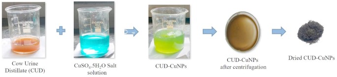
A bio-reduction method employing CUD was used to synthesize CuNPs. The process involved adding 5 mL of CUD (freeze-dried) to 50 mL of a 5 mM aqueous CuSO₄ solution, with the pH adjusted to 7.0 using 1 M NaOH (aq), resulting in a green colloidal system. After two hours of continuous stirring at 35 °C, the color change was visually tracked, and the time for this change was recorded. To confirm the formation of CUD-CuNPs, samples were periodically collected and analyzed spectrophotometrically using a UV-vis Spectrophotometer (Shimadzu, UV-2450). The nanoparticles were then isolated by centrifugation. The resulting pellet was dried at 60 °C for 12 h in an air oven, and the final brown powder was stored at room temperature for further analysis14. A detailed synthesis scheme of the CUD-CUNPs is shown in Fig. 1.

Schematic representation of CUD-CuNPs synthesis.
Statistical experimental design
To investigate the influence of various formulation factors on the size and wavelength of CUD-CuNPs particles, we employed the Box Behnken design (BBD). Twenty-seven experimental runs were generated using Design-Expert® (Version 11, Stat-Ease Inc., Minneapolis, USA). The effect of four independent variables- reductant concentration (X1), reaction pH (X2), reaction temperature (X3), and reaction time (X4), each examined at four levels on the responses of particle size (Y1), zeta potential (Y2), polydispersity index (PDI) (Y3), and surface plasmon resonance (SPR) peak wavelength (Y4) was analyzed using analysis of variance (ANOVA) based on the F Value (Table 1). The coefficient of determination (R²), was used to assess the model equation’s fit. The correlation between the fitted model and the responses was subsequently shown through contour and surface plots. These visual aids facilitated a better understanding of the relationship and interaction between the examined independent variables and the resulting dependent variables.
Using a one-factor-at-a-time (OFAT) strategy
The OFAT methodology was implemented to facilitate the determination of the most effective operational parameters for CUD-CuNPs synthesis, precisely identifying the critical values for reductant concentration, reaction pH, reaction temperature, and reaction time.
Physicochemical characterization of CuNPs
Spectrophotometric analysis, conducted using a UV–Vis spectrophotometer (Shimadzu, UV-2450) across the 200–600 nm electromagnetic spectrum, confirmed the formation of CUD-CuNPs by detecting a characteristic SPR band within the 269–275 nm range, which served as definitive spectroscopic evidence. The synthesized CuNPs were characterized using several techniques. Fourier-transform infrared spectroscopy (FTIR) was used to identify the functional groups. Particle size and zeta potential, essential for assessing nanoparticle stability, were determined using a particle size and zeta potential analyzer (NanoPlus with NanoPLuS AT). Powder X-ray diffraction (XRD) was employed to analyze the crystalline structure of the CUD-CuNPs produced within the 2θ range of 20°–80°. The crystallite size of the synthesized CUD-CuNPs was estimated using Scherrer’s equation (D = 0.9λ / β cos θ), where D is the crystallite size, λ is the X-ray wavelength (Cu Kα, 1.541862 Å), β is the full width at half maximum (FWHM), and θ is the Bragg angle. The elemental composition and distribution within the copper nanoparticles were investigated using energy-dispersive X-ray microscopy (EDAX) combined with scanning electron microscopy (SEM, Quanta FEG 250). Finally, transmission electron microscopy with selected area electron diffraction (TEM-SAED) using a Tecnai 12 G2 system provided insights into the detailed surface features of the nanoparticles.
In-vitro biological assays
Antibacterial assay
The antibacterial potential of the CUD and CUD-NPs was assessed using an agar well diffusion assay, measuring the zone of inhibition against the selected strains, Escherichia coli (MTCC 1751), Staphylococcus epidermidis (MTCC 435), Bacillus subtilis (MTCC 441), and Pseudomonas aeruginosa (MTCC 1748)15,16,17. Individual microwells received 50 µL aliquots of Mueller-Hinton (MH) broth and varying concentrations (50, 100, 200, and 400 µg/mL) of CUD-CuNPs. Standardized bacterial suspensions (10⁶ CFU/mL) of selected strains were introduced into the wells, followed by incubation at 37 °C. Ciprofloxacin (100 µg/mL) was used as the standard antibiotic against P. aeruginosa, whereas amoxicillin (100 µg/mL) served as the reference drug for E. coli, S. epidermidis, and B. subtilis. For the agar well diffusion method, microbial strains from pure cultures were subcultured on nutrient agar slants and grown at 35 °C. Once the Plate Count Agar (PCA) in Petri dishes had solidified, four 6 mm diameter wells were punched into the agar using a sterile cork borer. The spread plate method was used to inoculate the PCA plates with bacterial suspensions in normal saline. Then, a micropipette introduced 20 µL of the test drug solution at different concentrations into each well of the inoculated agar plates. A separate group of agar plates inoculated with the same four bacterial strains was the positive control and treated with standard concentrations. Following this treatment, all plates were incubated at 30 ± 2 °C for 24 h, and the diameters of the resulting growth inhibition zones were quantified.
Antidiabetic activity
The in-vitro antidiabetic potential of CUD-CuNPs was evaluated by assessing their inhibitory activity against α-amylase and α-glucosidase enzymes using established assays18,19. For the α-amylase assay, a 1% starch solution served as the substrate, whereas p-nitrophenyl-α-d-glucopyranoside (PNPG) was used for the α-glucosidase inhibition assay, with acarbose as a standard antidiabetic reference for comparison.
For the α-amylase inhibitory assay, 100 µL of CUD and CUD-CuNPs at different concentrations (10–500 µg/mL) were combined with a substrate solution of 1% starch in 20 mM phosphate buffer. The mixture was incubated at 25 °C for 10 min, followed by the addition of 100 µL of α-amylase enzyme (1 U/mL), and further incubation for 10 min at 25 °C. The reaction was terminated by adding 200 µL dinitro salicylic acid reagent, and the mixture was heated at 100 °C for 5 min. After cooling to room temperature, the absorbance of the samples was measured at 540 nm15,20.
For the α-glucosidase inhibitory assay, 100 µL of CUD and CUD-CuNPs (at concentrations ranging from 10 to 500 µg/mL) were combined with 100 mM sodium phosphate buffer and 50 µL of 5 mM p-nitrophenyl-α-d-glucopyranoside solution. The resulting mixture was incubated at 37 °C for 5 min. Subsequently, 50 µL of phosphate buffer containing α-glucosidase (1 U/mL) was added, and the mixture was incubated for 30 min. The reaction was terminated by adding 500 µL of 1 M sodium carbonate, and the absorbance was recorded at 405 nm using a spectrophotometer. Subsequently, the results were benchmarked against acarbose20,21.
Antioxidant activity
To understand the potential of CUD-CuNPs in disease treatment and management, considering the importance of antioxidants, their antioxidant properties were evaluated through different in-vitro assays: the 2,2-diphenyl-1-picrylhydrazyl (DPPH), the nitric oxide (NO), and the hydrogen peroxide (H2O2) scavenging assays20,22. These methods evaluate the ability to inhibit free radicals based on their endpoints, radical generation mechanisms, and consistency. In all assays, CUD and CUD-CuNPs were prepared at concentrations of 10, 20, 40, 60, 80, and 100 µg/mL by combining them with the appropriate reagents according to established standard procedures.
Statistical analysis
Results are presented as the mean ± standard deviation. Non-linear regression analysis determined the IC50 values for enzyme inhibitory (α-amylase and α-glucosidase) and antioxidant (DPPH, NO, and H2O2 scavenging assays) experiments. Statistical significance of the reported values was ascertained using one-way analysis of variance (ANOVA), followed by Duncan’s post-hoc test at a 5% significance level (p < 0.05). All statistical analyses were performed using STATISTICA version 12.5 software (StatSoft Inc., Hamburg, Germany)23.
Results and discussion
This study represents a significant breakthrough by effectively bridging traditional knowledge with modern nanotechnology. Using CUD for the green synthesis of CuNPs is a significant leap forward, offering a sustainable, cost-effective, and non-toxic alternative to conventional chemical methods. The research’s reliance on a systematic Box-Behnken design for process optimization is a key strength, moving the field beyond anecdotal reports and toward a data-driven, reproducible synthesis protocol. The comprehensive characterization and multifaceted biological evaluation of the CuNPs provide robust evidence of their formation and therapeutic potential, confirming their promise as antibacterial, antidiabetic, and antioxidant agents.
Proposed mechanism of CuNPs synthesis
Green synthesis of CuNPs from copper sulfate using CUD is proposed through a biological reduction process facilitated by diverse CUD biomolecules (Figure 2). CUD contains a range of natural reducing agents, including organic molecules like polyphenols and ferulic acid, and inorganic substances such as uric acid, creatinine, and various enzymes, which operate within this pathway. They achieve this by donating electrons to copper ions (Cu²⁺) from the sulfate solution, transforming them into elemental copper (Cu°). These freshly generated Cu⁰ atoms coalesced, forming stable nuclei, which subsequently expanded into nanoparticles by attracting more reduced copper or merging with other nascent particles. Simultaneously, the same biomolecules within the CUD play a dual role as effective stabilizing and capping agents. By adsorbing onto the surface of developing nanoparticles, they prevent aggregation and regulate the final dimensions and shape of the particles12,24.

Proposed mechanism for green nanoparticle synthesis of CUD-CuNPs.
Optimization of CUD-CuNPs
This study performed a BBD experiment to optimize the synthesis of CUD-CuNPs. The parameters examined as variables were reductant concentration, reaction pH, reaction temperature, and reaction duration. Green synthesis of CuNPs can occur through the bioreduction of metal ions, where metal particles interact with biomolecules present in cow urine distillate. The negatively charged regions of these biomolecules are attracted to positively charged metal ions, forming a stabilized complex via these interactions. Some constituents of CUD, such as urea and creatinine, can serve as bio-reducing, capping and/or stabilizing agents. The influence of four independent variables—reductant concentration (X1), reaction pH (X2), reaction temperature (X3), and reaction time (X4) on nanoparticle formation was systematically explored in this research using a 27-run BBD as the Design of Experiments (DOE). The objective of the experiment was to determine how these factors impacted the response variables: particle size (Y1), zeta potential (Y2), PDI (Y3), and SPR peak wavelength (Y4). The experimental matrix, which displays the tested variable levels and the corresponding measured responses, is presented in Table 2.
The particle sizes of the produced CuNPs ranged from 234.83 to 327.90 nm, with smaller sizes occurring with lower reductant concentrations and moderate pH. The zeta potential ranged from − 17.25 to -2.78 mV, demonstrating increased stability at lower pH and temperatures. PDI values (0.387–0.497) indicated considerable particle uniformity, while λmax shifts (258.18–268.97 nm) showed minimal differences in size and surface characteristics. Overall, the synthesis conditions had a substantial impact on the nanoparticle properties.
Model fitting
The quadratic model demonstrated strong adjusted R² values for all responses (Y1: particle size = 0.9853; Y2: zeta potential = 0.9965; Y3: PDI = 0.9880; Y4: λmax = 0.9531) and predicted R² values (Y1 = 0.9612; Y2 = 0.9907; Y3 = 0.9684; Y4 = 0.8852). The quadratic model is effective for analyzing and modifying nanoparticle qualities, as evidenced by strong statistical indicators, significant sequential p-values (< 0.0001), and acceptable lack-of-fit values (p > 0.05). The quadratic models performed well, with high R² values for particle size (Y1: 0.9932), zeta potential (Y2: 0.9984), PDI Y3: 0.9945), and λmax (Y4: 0.9784). In each case, the predicted R² values were in close agreement with the adjusted R² values, with differences well below the acceptable threshold of 0.2, indicating a reliable model performance. Furthermore, all models had adequate signal strength, as evidenced by high Adeq Precision values (Y1: 39.04, Y2: 81.56, Y3: 38.79, and Y4: 22.54), which exceeded the recommended minimum of 4. These statistical parameters, taken together, confirm the models’ robustness and adequacy in capturing the formulation variables’ effects and guiding further optimization. The ANOVA results (Table 3) confirmed that the quadratic models developed for all four responses- particle size, zeta potential, PDI, and SPR peak were statistically significant. The high F-values (ranging from 38.74 to 523.99) and very low p-values (p < 0.0001) suggest that the variation explained by the models is not due to random noise. The non-significant lack of fit (p > 0.05) in each case, albeit close to the level of 0.05, in a few instances (particle size: 0.0574, zeta potential: 0.0601, PDI: 0.0559), indicates that the models adequately fit the experimental data.
The analysis revealed that X₁ (Reductant concentration) significantly influenced all four responses, with strong linear effects on particle size, zeta potential, PDI, and SPR peak, along with consistent significance of its quadratic term (X₁²). This confirms its central role in nanoparticle formation and stability. X₂ (pH) significantly influenced particle size, zeta potential, and SPR peak in both linear and quadratic terms, but had no significant impact on PDI. X₃ (Temperature) had a significant linear effect on particle size and PDI, but a quadratic term was important for PDI and SPR peak, indicating moderate response sensitivity to temperature variations. X₄ (Reaction time) had a limited linear effect and was only significant for PDI. However, its quadratic form contributed to the PDI and SPR peak models, indicating its role in fine-tuning nanoparticle characteristics. The interaction term X₁X₂ (Reductant concentration × pH) was consistently significant for particle size and SPR peak, while X₁X₄ and X₂X₄ were selectively significant for PDI, indicating synergistic effects between key formulation variables on specific response outcomes.
Effect of variables on characteristics of CUD-CuNPs
The final equations in terms of coded factors provide insight into how each independent variable—X₁ (reductant concentration), X₂ (pH), X₃ (temperature), and X₄ (Reaction time)—affects the four key responses:
$$begin{gathered} ~{text{Particle}}~;{text{Size}} = {text{271}}.{text{21}} + {text{22}}.{text{49X}}_{1} + {text{24}}.{text{9}}0{text{X}}_{2} + {text{2}}.{text{88X}}_{3} + {text{1}}.{text{8}}0{text{X}}_{4} – {text{4}}.{text{24X}}_{1} {text{X}}_{2} hfill \ + {text{5}}.0{text{8X}}_{1} {text{X}}_{3} + {text{3}}.{text{3}}0{text{X}}_{1} {text{X}}_{4} – 0.{text{3575X}}_{2} {text{X}}_{3} – 0.{text{3325X}}_{2} {text{X}}_{4} – {text{1}}.{text{75X}}_{3} {text{X}}_{4} hfill \ – {text{8}}.{text{1}}0{text{X}}_{1}^{{;2}} + {text{23}}.{text{26X}}_{2}^{{;2}} – {text{3}}.00{text{X}}_{3}^{{;2}} – 0.{text{9975X}}_{4}^{{;2}} hfill \ end{gathered}$$
(1)
$$begin{aligned} Zeta~Potential & = – 10 cdot 42 + 4 cdot 49X_{1} + 2 cdot 40X_{2} – 0 cdot 0400X_{3} + 0 cdot 0117X_{4} – 0 cdot 0575X_{1} X_{2} \ & + 0 cdot 0100X_{1} X_{3} – 0 cdot 0150X_{1} X_{4} – 0 cdot 0050X_{2} X_{3} + 0 cdot 0150X_{2} X_{4} \ & – 0 cdot 0300X_{3} X_{4} – 1 cdot 90X_{1} ^{2} + 2.42X_{2} ^{2} – 0.1679X_{3} ^{2} – 0 cdot 0529X_{4} ^{2} \ end{aligned}$$
(2)
$$begin{aligned} PDI & = 0 cdot 4840 + 0 cdot 0073X_{1} + 0 cdot 0004X_{2} + 0 cdot 0025X_{3} + 0 cdot 0030X_{4} + 0 cdot 0277X_{1} X_{2} \ & + 0 cdot 0040X_{1} X_{3} + 0 cdot 0135X_{1} X_{4} – 0 cdot 0020X_{2} X_{3} – 0.0045X_{4} X_{4} – 0 cdot 0035X_{3} X_{4} \ & – 0 cdot 0614X_{1} ^{2} – 0 cdot 0014X_{2} ^{2} + 0 cdot 0052X_{3} ^{2} + 0 cdot 0037X_{2} ^{4} \ end{aligned}$$
(3)
$$begin{aligned} lambda max {text{ }} & = {text{ }}266 cdot 22{text{ }} – 3 cdot 40X_{1} + 1 cdot 28X_{2} – 0 cdot 0525X_{3} + 0 cdot 0633X_{4} + 0 cdot 9025X_{1} X_{2} \ & – 0 cdot 4925X_{1} X_{3} – 0 cdot 4225X_{1} X_{4} + 0 cdot 4900X_{2} X_{3} + 0 cdot 4925X_{2} X_{4} – 0.3050X_{{3}} X_{4} {text{ }} \ & – 2 cdot 65X_{1} ^{2} – 0.1825X_{2} ^{2} + 1.05X_{3} ^{2} + {text{ }}0 cdot 6475X_{4} ^{2} \ end{aligned}$$
(4)
The particle size increased significantly with both X₁ and X₂, as indicated by their significant positive coefficients (+ 22.49 and + 24.90). This suggests that a higher reductant concentration and pH led to the formation of larger nanoparticles. This effect could be attributed to the accelerated nucleation and subsequent agglomeration at high reductants and pH concentrations. The particle size increased moderately with the temperature and reaction time (X₃ and X₄). However, the negative quadratic term for X₁² (− 8.10) indicates that an excessive reductant concentration reduced the particle size, likely due to saturation effects or possible re-dissolution. The baseline value of the zeta potential was negative (-10.42), and became less negative as X₁ and X₂ increased. This implies that by changing the nanoparticles’ ionic environment or surface chemistry, higher reductants and pH levels may neutralize the surface charges.
The primary factors only slightly impacted PDI, a particle size distribution uniformity measure. The most significant influence came from X₁ (+ 0.0073) and its quadratic term (− 0.0614), suggesting that uniformity was enhanced by moderate reductant concentrations and adversely affected by higher concentrations. PDI was also impacted by interaction terms like X₁X₂ and X₁X₄, suggesting that pH and reaction time influenced the reductant’s effect.
X₁ (− 3.40) had the most substantial detrimental effect on SPR peak wavelength, i.e. λmax, indicating a bathochromic shift (a decrease in λmax) as reductant concentration increased. Variations in SPR behavior or particle size could explain this. On the other hand, X₂ and X₄ caused marginally higher λmax, which might be because of improved particle growth or stabilization under these circumstances. The complex and non-linear relationship between these variables and the optical properties of the synthesized nanoparticles was further highlighted by the significant quadratic terms for X₁² (− 2.65) and X₃² (+ 1.05).
The developed models were shown to be adequate based on the diagnostic plots of PDI, λmax, zeta potential, and particle size. The residuals were normally distributed, according to the normal probability plots (Figure S1). A random scatter was observed in the residuals versus run plots (Figure S2), suggesting error independence. The predicted versus actual plots (Figure S3) confirmed the model’s dependability, which showed a strong correlation between observed and predicted values.
The combined effects of the independent variables on the particle size are depicted in the interaction plots (Figure S4). Particle size increased significantly with increasing reducing agent concentration and pH (Figure S4A), with pH having a greater effect. Likewise, increasing temperature and reducing agent concentration increased particle size (Figure S4B). Longer reaction times promoted the formation of larger particles, as evidenced by the positive correlation between reducing agent concentration and reaction time (Figure S4C). The particle size significantly increased when pH and reaction time were combined (Figure S4E), especially at higher pH values.
Particle size increased due to the combined effects of temperature and pH (Figure S4D), while remaining somewhat similar at both reaction time values with increasing temperature (Figure S4F). Temperature consistently had a positive effect in all interactions. These results demonstrate that all four factors worked in concert to promote particle growth, with pH and reducing agent concentration having the most significant effects.
The zeta potential values shifted toward a lower magnitude, indicating reduced stability, as the pH increased and the concentration of the reducing agent increased, according to the interaction plots (Figure S5A). Higher temperatures and levels of reducing agents (Figure S5B), the reductant concentration, and the reaction time (Figure S5C) exhibited a similar trend, with the zeta potential becoming less negative.
In Figures S5D and S5E, Zeta potential magnitudes decreased with increasing pH, temperature and reaction times, indicating reduced electrostatic stability. A moderate destabilizing effect was observed in both figures. Notably, Figure S5F showed very little interaction between temperature and reaction time, as evidenced by the slight change in the zeta potential with these changes.
With increasing levels of reductant concentration across the pH range (Figure S6A), temperature (Figure S6B), and reaction time (Figure S6C), the PDI first increased and then decreased, as shown in the interaction plots in Figure S6, indicating a peak in the particle size variability at moderate concentrations. Similar trends were observed when the pH was increased over a range of temperatures (Figure S6D) and reaction times (Figure S6E), with higher PDI values corresponding to intermediate pH levels. On the other hand, PDI barely changed throughout the temperature–time interaction in Figure S6F, suggesting a reasonably stable size distribution within this range. These patterns imply that extreme process parameter values prefer narrower size distributions, while broader distributions were produced under moderate conditions.
The interaction plots in Figure S7 show the effect of the formulation parameters on the maximum absorbance wavelength, i.e. SPR peak (λmax). As shown in Figure S7A–C, an increase in reductant concentration at pH, temperature, and reaction time led to a consistent decrease in λmax values, suggesting a potential shift in particle size or morphology associated with nanoparticle formation. Conversely, Figure S7D–F demonstrated a slight increase or near plateau in λmax with increasing pH, temperature, and reaction time across their respective interacting variables. The same interactions were further confirmed through 3D response surface plots (Figure 3 and Figure S8-S10), thus validating the trends observed in the interaction plots.

3D response surface plots showing the effect of variables on response particle size.
Model validation
The ideal parameters for synthesising CUD-CuNPs were determined by numerical optimization using the desirability approach to be 1% reductant concentration, pH 5.23, temperature 44.71 °C, and a reaction time of 4 h. The software predicted the following values for the key responses under these optimized conditions: zeta potential of -16.72 mV, absorbance wavelength (λmax) of 269.37 nm, PDI of 0.405, and particle size of 234.83 nm. A particle size of 232.76 nm, PDI of 0.421, zeta potential of -16.45 mV, and λmax of 270.01 nm were obtained from experimental validation of the optimized formulation. With slight bias, the observed experimental values closely matched the predicted responses (-0.88% for particle size, 3.95% for PDI, -1.61% for zeta potential, and 0.24% for λmax), demonstrating the accuracy and robustness of the model for predicting and optimizing the synthesis parameters.
Optical analysis using UV-Vis absorbance spectroscopy
UV-Vis spectroscopy is a valuable approach for evaluating the fabrication and stability of CUD-CuNPs in aqueous solutions. The UV-Vis spectrum confirms the generation of CuNPs from CUD by showing a SPR absorbance band around 269–275 nm (Figure 4)10,14.

(A) UV-visible spectra of CUD-CuNPs at reductant concentration:-1% reductant concentration, pH 5.23, temperature 44.71 °C, and a reaction time of 4 h, showing an absorption peak at 269 nm (B) UV spectral overlay of different batches of CUD-CuNPs.
A visual indication of CuNPs formation in the sample is the change to the green color of the solution. As light interacts with NPs, specific wavelengths are absorbed, decreasing the intensity of those wavelengths in the light that emerges from the sample20. The reaction kinetics in the sample were monitored using UV-Vis spectroscopy, which confirmed that the reaction reached completion within 3 min, as shown by stable spectral readings without any further substantial alterations after this time. The impact of pH 5.23 on the bioreduction of metal salts to NPs was assessed using UV-Vis Spectroscopy, which showed a distinct absorbance peak, possibly resulting from the ionization of phenolic constituents within the CUD14. The study also revealed that the UV-Vis peak value decreased as the particle size increased. These findings concluded that pH 5.23 is the most effective for converting Cu²⁺ ions into CuNPs in this experiment.
Functional group analysis by FT-IR
FT-IR analysis was conducted to identify the characteristic stretching and vibration modes of the biosynthesized CuNPs. The broadband observed at 3572.17 cm⁻¹ indicates the stretching frequency of the -OH group of adsorbed water on the CuNP’s surface. Furthermore, the presence of peaks at 609.51 cm⁻¹ and 678.94 cm⁻¹ can be attributed to Cu-O vibrations, thus confirming the formation of CuNPs5,14.
In FTIR analysis, observing the shifts in the characteristic peaks of the functional groups provides crucial insights into the involvement of CUD in the synthesis and stabilization of CUD-CuNPs. Typically, the FTIR spectrum of CUD displays prominent bands indicating the presence of various biomolecules. For instance, a broad absorption band often appears 3332.99 cm− 1, attributed to O-H stretching vibrations from water, alcohols, and phenols, alongside N-H stretching from amines or amides like urea. Strong peaks in the 1600–1650 cm− 1 range are usually assigned to C = O stretching (Amide I band) from carboxylic acids or amides and C = C stretching from aromatic compounds. The 1597.06 cm− 1 and 1550.77 cm− 1 peaks are assigned to C = N stretching (Amide I band) amides, and C = C stretching from aromatic compounds, respectively. Additionally, C-H stretching vibrations are commonly observed around 2924 cm− 1, and C-O stretching from carbohydrates or alcohols might appear between 1000 and 1200 cm− 15,14.
A comparison of the FTIR spectrum of CUD with that of CUD-CuNPs often shows noticeable changes: shifts or reduced intensities in the C = O stretching region of 1600–1650 cm− 1 indicate the coordination of carbonyl or carboxylate groups with the copper surface, acting as effective capping and stabilizing agents. Furthermore, alterations in the C-O stretching bands (1050–1250 cm⁻¹) can confirm the involvement of polyols or other carbohydrate components in forming a protective layer around the nanoparticles. These collective spectroscopic changes prove that the organic constituents within the CUD are not merely a reaction medium but also play a vital role in both the bioreduction of copper ions and the stabilization of the resulting CuNP514.
The FT-IR measurement of the CUD (Figure 5B) showed absorption bands at 3332.99, 3055.24, 2924.09, 1597.06, 1550.77, 1381.03, 1111.00, 1064.71, 1018.41, 864.11, 825.53, 777.53, 709.80, and 671.23 cm⁻¹, revealing the presence of diverse functional groups (Figure 5B). The band at 3332.99 cm⁻¹ is attributed to O-H stretching in alcohols or phenols. The band indicates aromatic C = C bending at 1597.06 cm⁻¹, and aromatic C-C stretching is evident at 1550.77 cm⁻¹. The C-N stretch of aliphatic amines is observed at 1111.00 cm⁻¹, while the band suggests aromatic C-H stretching at 864.11 cm⁻¹. Further C-N stretching of aliphatic amines is also seen at 1018.41 cm⁻¹, and the band at 671.23 cm⁻¹ suggests C-Cl stretching in alkyl halides.
The FT-IR spectra of synthesized CUD-CuNPs and overlay spectra are presented in Figure 5C and D, respectively. The NPs showed prominent peaks at 3572.17, 3325.28, 1597.06, 1550.77, 1388.75, 1319.31, 1180.44, 1103.28, 1018.41, 848.68, 678.94, 609.51, 509.21, and 462.92 cm⁻¹. The peak at 3572.17 cm⁻¹ indicates O-H stretching in alcohols or phenols, and the peak represents the aromatic C = C bending mode at 848.68 cm⁻¹. The peak at 1388.75 cm⁻¹ suggests the presence of the N-O symmetric stretch in nitro compounds, while the peak indicates the C-H wag of alkyl halides at 1319.31 cm⁻¹. The peak at 609.51 cm⁻¹ corresponds to the C-Cl stretch of alkyl halides. CUD contains biomolecules that function as reducing and capping agents, thereby promoting CuNPs formation from copper ions.

(A) FTIR spectrum of CuSO4 (B) FTIR spectrum of CUD (C) FTIR spectrum of CUD-CuNPs (D) Overlay FTIR spectrum of CUD-CuNPs, CUD and CuSO4.
Microstructural and elemental analysis using SEM-EDAX and TEM techniques
Microscopic and elemental characterizations of the synthesized CUD-CuNPs were performed (Figure 6). Their morphologies were visualized using SEM images (Figure 6A), whereas the EDAX spectra were used to determine their elemental composition (Figure 6B). The spherical morphology of the NPs was consistently observed in both TEM (Figure 6C) and SAED (Figure 6D) analyses. EDAX analysis was particularly effective in identifying the elemental constituents, confirming the presence of copper, and detecting carbon, oxygen, magnesium, silicon, sulfur, potassium, and chloride. Carbon and oxygen originate from the organic biomolecules within the CUD that act as reducing and stabilizing agents, forming a capping layer. The remaining elements, Mg, Si, S, K, and Cl, are naturally occurring minerals and electrolytes inherently present in the complex biological matrix of CUD itself, thus co-existing with the synthesized CuNPs. TEM analysis of the synthesized nanoparticles revealed a spherical morphology with an average size ranging from 20 to 40 nm.

(A) SEM, (B) EDAX, (C) TEM, (D) SAED, (E) XRD analysis of CUD-CuNPs.
XRD analysis
The XRD pattern of the final CUD-CuNPs, obtained within the 2θ range of 20⁰ to 80⁰, is illustrated in Figure 6E. Diffraction peaks were observed at 29.32°, 43.4°, 47.48°, 50.5°, and 74.2°, which are strong indicators of crystalline CuNPs. Based on these XRD peaks and a Scherrer’s equation, the crystallite size of the synthesized nanoparticles was calculated to be 29.98 nm. This size falls within the typical nanoscale range, confirming the formation of nanoparticles rather than bulk material. These peak’s sharpness and clear resolution suggest that the nanoparticles possess a well-ordered atomic structure, rather than being amorphous. While the exact crystallographic planes at (111), (200) and (220) corresponding to these 2θ values are explicitly stated, these positions are highly characteristic of metallic copper (Cu0) with face-centred cubic structure. The additional peaks would indicate the presence of copper oxides, which frequently form during green synthesis or post-synthesis exposure. These data show that the green synthesis method employing CUD effectively produced CuNPs with a well-defined crystalline structure and a relatively uniform size distribution of approximately 30 nm. Further analysis, ideally by comparing these peaks to those in the literature, would definitively confirm the specific phases (e.g., pure Cu, Cu₂O and CuO) present in the synthesized material514.
Particle size analysis
The average particle size of CUD-CuNPs was 232.76 ± 15.25 nm (Figure 7A), and the PDI was 0.421 ± 0.05. The average particle size was larger than 20–100 nm, typically observed in green-synthesized CuNPs. The larger size in the present study could suggest partial aggregation during synthesis, possibly due to the characteristic biomolecule profile within the CUD. The PDI of 0.421 signifies a moderately broad size distribution, which is common in green synthesis due to the complex nature of biological extracts12,24.

(A) Particle size (B) Zeta potential analysis of the CUD-CuNPs.
Furthermore, the zeta potential of -16.45 ± 0.23 mV (Figure 7B) offers insight into the colloidal stability of the CUD-CuNPs. The negative charge is consistent with anionic biomolecules (such as carboxylic acids, phenolics, or other negatively charged functional groups) from the CUD adsorbed onto the nanoparcticle’s surface, providing electrostatic repulsion that helps prevent immediate aggregation. This implies that although the particles possess some repulsive forces, they might still be prone to slow aggregation over time or under varying environmental conditions. In cow urine-mediated synthesis, some studies have reported moderately negative zeta potentials, consistent with the capping action of its organic components12,24.
A comparison of the XRD and particle size analysis (DLS) data highlights the difference between the fundamental particle size and its behavior in solution. Based on the Scherrer equation, the individual nanocrystalline domains are approximately 30 nm. However, DLS measurements, which capture the particle’s hydrodynamic diameter in a solvent, indicate a much larger average size of 232.76 ± 15.25 nm with a high PDI (0.421). This considerable divergence and the broad size distribution suggest that the nanoparticles have formed aggregates25. The ~ 30 nm crystallites are likely bound together in larger clusters, leading to the substantial increase in size observed by DLS. Such aggregation is a common outcome for nanoparticles and can be attributed to surface chemistry and the surrounding medium26, 27. Furthermore, in most cases, the crystallite size is smaller than the particle size due to the polycrystalline nature of the particles28.
In-vitro antibacterial activity
The antibacterial properties of copper have been well known for a long time. Soluble copper compounds are highly effective against many microorganisms, including bacteria, fungi, algae, and viruses, with relatively low toxicity to humans20. Copper kills microbes by producing reactive hydroxyl radicals that can cause irreparable damage, such as oxidation of proteins, cleavage of DNA and RNA, and damage of membranes through lipid peroxidation29. Studies have shown that metal-based nano-antimicrobials exhibit considerably higher or longer-lasting biological activity than the established bioactivity of the corresponding bulk metal18. The agar well diffusion method was employed to assess the antibacterial efficacy of the synthesized CUD-CuNPs20. The bacterial strains utilized in this assay were procured from the National Collection of Industrial Microorganisms (NCIM), Pune, India and comprised the Gram-positive: S. epidermis and B. subtilis, and the Gram-negative: E. coli and P. aeruginosa.

Antibacterial activity of CUD (A to D) and CUD-CuNPs (E to H) against the selected pathogenic bacteria. (A & E) – B. subtilis; (B & F) – E. coli; (C & G) – P. aeruginosa; (D & H) – S. epidermidis. Std – Standard drug; Numbers denote concentrations used (1,2,3,4 specifies 50, 100, 200, 400 µg/mL, respectively).
The antibacterial efficacies of CUD and CUD-CuNPs against Gram-positive and Gram-negative bacteria are shown in Fig. 8, clearly illustrated by the zone of inhibition (in mm). CUD-CuNPs showed remarkable inhibitory zones against S. epidermidis and B. subtilis (25.66 ± 0.57 mm for both), with subsequent activity against P. aeruginosa (24.00 ± 1.00 mm) and E. coli (22.66 ± 0.57 mm) (Fig. 8). Consequently, the synthesised CUD-CuNPs appear to be a promising antibacterial agent with a broad spectrum of activity, performing on par with standard antibiotics (Fig. 9).
The mechanism by which CuNPs combat microbes involves the breakdown of bacterial cell walls and membranes, generation of damaging reactive oxygen species, and interference with essential proteins and DNA18,14. Scientific evidence indicates that increasing the concentration of these NPs results in size reduction to the nanoscale, thereby improving their interaction with bacterial cells30,19. This suggests that the produced CUD-CuNPs have potential for future development as antimicrobial agents.

Comparison of the antibacterial activity of CUD and CUD-CuNPs performed against selected Gram-negative and Gram-positive bacterial strains. All values are expressed as Mean ± S.D., n = 3.
In-vitro antidiabetic activity
A key strategy for the effective treatment of type 2 diabetes is to block the function of pancreatic α-amylase and intestinal α-glucosidase enzymes. Impeding the function of α-amylase and α-glucosidase results in a slower processing of carbohydrates and glucose entry into the bloodstream, consequently reducing the rise in blood sugar after meals22,20. In this research, we evaluated the ability of both cow urine and CUD-CuNPs to inhibit the actions of α-amylase and α-glucosidase enzymes.
In contrast to the CUD, which had IC₅₀ values of 33.77 ± 0.00 and 27.43 ± 0.03 µg/mL, respectively, the study’s findings showed that CUD-CuNPs had stronger inhibitory activity against α-amylase and α-glucosidase, with IC₅₀ values of 30.88 ± 1.00 and 24.94 ± 1.02 µg/mL, respectively (Fig. 10). The standard acarbose demonstrated IC50 of 41.05 ± 0.98 µg/mL and 37.39 ± 0.96 µg/mL for α-amylase and α-glucosidase, respectively.

Comparative in-vitro antioxidant (DPPH, nitric oxide and superoxide radical scavenging assay) and antidiabetic activities (α-amylase and α-glucosidase enzyme inhibitory assays) of CUD and CUD-CuNPs. All values are expressed as Mean ± S.D., n = 3.
In-vitro antioxidant activity
When oxidative stress occurs, cells experience an imbalance due to producing reactive oxygen, nitrogen, and sulfur species. Antioxidants can prevent the damage caused by reactive substances and free radicals18. NPs derived from natural sources are increasingly recognized for their potential antioxidant properties31. Consequently, researchers have been actively investigating various metal and metal oxide NPs to document and harness their notable ability to combat free radicals.
The present study examined the antioxidant potency of CUD and CUD-CuNPs using DPPH, nitric oxide, and superoxide radical scavenging assays. Promising DPPH, nitric oxide and superoxide free radical scavenging activities were detected in CUD-CuNPs and CUD compared to the standard (Fig. 10). The CUD-CuNPs showed the highest free radical scavenging activities in DPPH, nitric oxide, and superoxide radical scavenging assays, with IC50 values of 28.27 ± 1.02, 19.21 ± 1.31 and 34.04 ± 1.04 µg/mL, respectively compared to CUD (Fig. 10). Ascorbic acid achieved an IC50 of 13.12 ± 0.85 µg/mL for DPPH radical scavenging, 18.75 ± 1.25 µg/mL for nitric oxide radical scavenging, and 20.41 ± 1.05 µg/mL for hydrogen peroxide radical scavenging.
Strengths, limitations, and future perspectives
While the study is pioneering, its primary challenge lies in its exclusive focus on in-vitro findings. The absence of in-vivo data and specific cytotoxicity studies is a significant barrier to clinical translation. This study provides a foundational framework, serving as a critical first step that validates the core concept and justifies further, more complex investigations into in-vivo efficacy, toxicology, and pharmacokinetics/pharmacodynamics. Acknowledging these constraints, the present study sets the stage for a new wave of research that can propel these green-synthesised nanomaterials toward real-world applications. This will pave the way for broader applications, including potential use as an anticancer agent, antidiabetic agent, for wound healing, or in combination therapies with existing drugs.
This research directly contributes to several UN Sustainable Development Goals (SDGs) by providing a sustainable and affordable method for developing therapeutic agents. The study supports SDG 3 (Good Health and Well-being) by demonstrating that its eco-friendly CuNPs have significant antimicrobial, antidiabetic, and antioxidant properties, crucial for combating infectious and chronic diseases. The methodology aligns with SDG 12 (Responsible Consumption and Production) by using a “green synthesis” approach with CUD, reducing reliance on hazardous chemicals and high-energy processes. This makes the production process more cost-effective and scalable, especially for resource-limited areas. Finally, the use of a natural, animal-derived product to create “sustainable, affordable, biocompatible and environmentally friendly synthesis routes” supports SDG 15 (Life on Land) by minimizing environmental risks associated with conventional chemical production, as it uses a natural, animal-derived product, which contributes to a more sustainable and less environmentally harmful production process.
Conclusion
Utilizing biomolecules from natural sources in nanoparticle synthesis is advantageous for reducing and stabilizing noble metal ions. This enables the formation of nanoparticles within a stable colloidal system, achieved through an easy, affordable, and non-toxic green chemistry method applicable across various fields. This study employed an eco-friendly, easily implementable, and economical method for synthesizing CuNPs. Characterization analyses offered an in-depth understanding of the structure and crystallinity of the resulting nanoparticles, emphasizing the substantial influence of biomolecular stabilization during their formation. In addition, the synthesized nanoparticles demonstrated notably higher antidiabetic, antimicrobial, and antioxidant activities when compared to both CUD and established standard compounds. The application of CUD for the sustainable production of nanomaterials is a relatively unexplored field. This investigation marks a novel contribution by creating a new method for CU-NPs synthesis based on biomolecules derived from the CUD. However, the efficacy of CUD-CuNPs must be rigorously evaluated in experimental models.
Data availability
No datasets were generated or analysed during the current study.
References
-
Ghosh, N. et al. ‘A comparative study of CuO nanoparticle and CuO/PVA-PVP nanocomposite based on dye removal performance and antibacterial activity in wastewater treatment’, Int. J. Environ. Chem. (2022). Available at: https://doi.org/10.1080/03067319.2022.2060088
-
Nadaf, S. J., Killedar, S. G., Kumbar, V. M., Bhagwat, D. A. & Gurav, S. S. Pazopanib-laden lipid based nanovesicular delivery with augmented oral bioavailability and therapeutic efficacy against non-small cell lung cancer. Int. J. Pharm. 628, 122287 (2022).
-
Yassin, A. Y., Abdelghany, A. M., Salama, R. S. & Tarabiah, A. E. Structural, optical and antibacterial activity studies on CMC/PVA blend filled with three different types of green synthesized ZnO nanoparticles. J. Inorg. Organomet. Polym. Mater. 33 (7), 1855–1867 (2023).
-
Abdelsattar, A. S. et al. The promising antibacterial and anticancer activity of green synthesized zinc nanoparticles in combination with silver and gold nanoparticles. J. Inorg. Organomet. Polym. Mater. 33 (7), 1868–1881 (2023).
-
Arumugam, D. G., Sivaji, S., Dhandapani, K. V., Nookala, S. & Ranganathan, B. Panchagavya mediated copper nanoparticles synthesis, characterization and evaluating cytotoxicity in Brine shrimp. Biocatal. Agric. Biotechnol. 19, 101132 (2019).
-
Bhavyasree, P. G. & Xavier, T. S. Green synthesised copper and copper oxide based nanomaterials using plant extracts and their application in antimicrobial activity: review. Curr. Res. Green. Sustainable Chem. 5, 100249 (2022).
-
Dhama, S., Mahto, S., Pathak, R. N. & Singh, R. Cow urine: a traditional Indian medicine. J. Pharm. Bioallied Sci. 4 (4), 367–369 (2012).
-
Gulhane, H., Nakanekar, A., Mahakal, N., Bhople, S. & Salunke, A. Gomutra (Cow urine): a multidimensional drug review Article. Int. J. Res. Ayurveda Pharm. 8 (5), 1–6 (2017).
-
Honary, S., Barabadi, H., Fathabad, E. G. & Naghibi, F. Green synthesis of copper oxide nanoparticles using penicillium Aurantiogriseum, penicillium citrinum and penicillium waksmanii. Dig. J. Nanomater Bios. 7, 999–1005 (2012).
-
Dabhane, H. et al. Cow urine mediated green synthesis of nanomaterial and their applications: a state-of-the-art review. J. Water Environ. Nanotechnol. 6, 81–91 (2021).
-
Kaushik, R., Jain, J. & Rai, P. Therapeutic potentials of cow derived products. Int. J. Pharm. Sci. Res. 7 (4), 1383–1390 (2016).
-
Kour, H., Kumar, K., Gupta, D. & Singh, R. ‘Cow urine mediated green synthesis of nanomaterial and their applications: a state-of-the-art review’. Journal Water Environ. Nanotechnology (2024).
-
Reddy, G. H. R., Shinde, S. P., Kadam, S. B., Yadav, P. R. & Ghuge, R. B. Cow urine distillate as a bioenhancer for anti-cancer drugs. J. Ethnopharmacol. 126 (2), 369–371 (2009).
-
Pelesinuo, K. B. et al. ‘Synthesis and characterization of Mithun (Bos frontalis) urine-based antibacterial copper oxide nanoparticles’, Biomedicines, 11 1690. (2023).
-
Gawas, G. et al. ‘Process optimization for the bioinspired synthesis of gold nanoparticles using Cordyceps Militaris, its characterization, and assessment of enhanced therapeutic efficacy’, Pharmaceuticals, 16 1311. (2023).
-
Kalaskar, M. et al. Chemical composition, antioxidant, antimicrobial, and wound healing effects of trachyspermum Roxburghianum (DC.) H. Wolff essential oil: an in vivo and in Silico approach. J. Ethnopharmacol. 327, 118055 (2024).
-
Mariyammal, V. et al. Chemical profiling of aristolochia Tagala Cham. Leaf extracts by GC-MS analysis and evaluation of its antibacterial activity. J. Indian Chem. Soc. 100 (1), 100807 (2023).
-
Andrade, F. et al. Endophytic fungi-assisted biomass synthesis of ecofriendly formulated silver nanoparticles for enhanced antibacterial, antioxidant, and antidiabetic activities. J. Drug Deliv Sci. Technol. 97, 105749 (2024).
-
Husain, S. et al. Antibacterial efficacy of facile cyanobacterial silver nanoparticles inferred by antioxidant mechanism. Mater. Sci. Eng. C. 122, 1–10 (2021).
-
Mali, S. C., Dhaka, A., Githala, C. K. & Trivedi, R. Green synthesis of copper nanoparticles using Celastrus paniculatus Willd. Leaf extract and their photocatalytic and antifungal properties. Biotechnol. Rep. 27, e00518 (2020).
-
Gracias, S. et al. Fabrication of Chitosan nanocomposites loaded with biosynthetic metallic nanoparticles and their therapeutic investigation. Environ. Res. 234, 116609 (2023).
-
Halarnekar, D. et al. Eco synthesized chitosan/zinc oxide nanocomposites as the next generation of nano-delivery for antibacterial, antioxidant, antidiabetic potential, and chronic wound repair. Int. J. Biol. Macromol. 242, 124764 (2023).
-
Raj, M. S. A. et al. A comparative analysis of leaf essential oil profile, in vitro biological properties and in Silico studies of four Indian guava (Psidium Guajava L.) cultivars, a promising source of functional food. South. Afr. J. Bot. 153, 357–369 (2023).
-
Singh, L. D. et al. Synthesis and characterization of Mithun (Bos frontalis) urine-based antibacterial copper oxide nanoparticles. J. Cluster Sci. 33 (5), 2097–2107 (2022).
-
Rodriguez Loya, J., Lerma, M., Torresdey, G. & J.L Dynamic Light Scattering and its Application To Control Nanoparticle Aggregation in Colloidal Systems: A Review 1515 . 24 (Micromachines, 2024).
-
Vollath, D. Agglomeration and aggregation of nanoparticles. Nanoarchitectonics 4 (2), 45 (2023).
-
Ashraf, M. A., Peng, W., Zare, Y. & Rhee, K. Y. Effects of size and Aggregation/Agglomeration of nanoparticles on the Interfacial/Interphase properties and tensile strength of polymer nanocomposites. Nanoscale Res. Lett. 13, 214 (2018).
-
Hassanzadeh-Tabrizi, S. A. Precise calculation of crystallite size of nanomaterials: A review. J. Alloys Compd. 968, 171914. https://doi.org/10.1016/j.jallcom.2023.171914 (2023).
-
Teschke, R. & Eickhoff, A. Wilson disease: copper mediated Cuproptosis, iron related Ferroptosis, and clinical Highlights, with comprehensive and critical analysis update. Int. J. Mol. Sci. 25 (9), 4753 (2024).
-
Dias, C. et al. Biogenic synthesis of zinc oxide nanoparticles using mushroom fungus cordyceps militaris: characterization and mechanistic insights of therapeutic investigation. J. Drug Deliv Sci. Technol. 73, 103444 (2022).
-
Amalraj, S. et al. Evaluation of phytochemicals, enzyme inhibitory, antibacterial and antioxidant effects of PsydraxdicoccosGaertn. Nat. Prod. Res. 36, 5772–5777 (2022).
Ethics declarations
Competing interests
The authors declare no competing interests.
Ethics approval
The authors declare that the manuscript has not been published previously.
Additional information
Publisher’s note
Springer Nature remains neutral with regard to jurisdictional claims in published maps and institutional affiliations.
Supplementary Information
Rights and permissions
Open Access This article is licensed under a Creative Commons Attribution-NonCommercial-NoDerivatives 4.0 International License, which permits any non-commercial use, sharing, distribution and reproduction in any medium or format, as long as you give appropriate credit to the original author(s) and the source, provide a link to the Creative Commons licence, and indicate if you modified the licensed material. You do not have permission under this licence to share adapted material derived from this article or parts of it. The images or other third party material in this article are included in the article’s Creative Commons licence, unless indicated otherwise in a credit line to the material. If material is not included in the article’s Creative Commons licence and your intended use is not permitted by statutory regulation or exceeds the permitted use, you will need to obtain permission directly from the copyright holder. To view a copy of this licence, visit http://creativecommons.org/licenses/by-nc-nd/4.0/.
About this article

Cite this article
Angle, G.P., Nadaf, S., Prabhu, S. et al. Eco-synthesis, process optimisation, and therapeutic assessment of cow urine distillate derived copper nanoparticles. Sci Rep 15, 42977 (2025). https://doi.org/10.1038/s41598-025-27117-3
-
Received:
-
Accepted:
-
Published:
-
Version of record:
-
DOI: https://doi.org/10.1038/s41598-025-27117-3