
Introduction
Breast cancer remains one of the most prevalent malignancies among women worldwide, posing a significant challenge to global healthcare systems. According to the World Health Organization (WHO), approximately 2.3 million women were diagnosed with breast cancer in 2022, making it the most commonly diagnosed cancer globally1. Among the various subtypes, MCF-7 breast cancer cells, which are estrogen receptor-positive (ER+), have been extensively studied due to their clinical relevance and responsiveness to targeted therapies2. This cell line is crucial for studying breast cancer biology and testing new drugs, as ER + tumors make up about 70% of all cases3. Despite significant advancements in treatment strategies, including surgery, chemotherapy, radiotherapy, and hormone therapy, the shortcomings of these methods make the treatment of advanced and metastatic breast cancer still challenging. Challenges such as drug resistance and tumor heterogeneity limit the effectiveness of traditional therapies4,5. These limitations highlight the urgent need for innovative therapeutic approaches to enhance drug efficacy, reduce side effects, and overcome resistance mechanisms. Ongoing research and clinical trials are essential for developing new treatment strategies to enhance patients’ quality of life with this devastating disease.
Nanotechnology has emerged as a powerful tool in cancer therapy, offering promising solutions to overcome the limitations of conventional treatments. One of the key advancements in this field is the development of nanocarriers, which can significantly enhance the solubility, stability, and targeted delivery of anti-cancer drugs. This, in turn, improves their therapeutic index while minimizing adverse effects4,6. In breast cancer treatment, nanotechnology-based drug delivery systems have demonstrated improved cellular uptake, controlled drug release, and bypassing multidrug resistance mechanisms, critical for effective therapy. Various nanomaterials, including liposomes, polymeric nanoparticles, and inorganic nanocarriers, have been explored to deliver chemotherapeutic agents and natural compounds with enhanced efficacy.
Among these, mesoporous silica nanoparticles (MSNs) have garnered considerable attention as versatile nanocarriers due to their high surface area, tunable pore size, biocompatibility, and ability to encapsulate a wide range of therapeutic agents7,8. Modifying MSNs with functional nanomaterials, such as polymers and targeting ligands, boosts their therapeutic potential by improving cellular uptake and specificity9. Graphene quantum dots (GQDs), a novel class of carbon-based nanomaterials, possess excellent physicochemical properties, including high photostability, biocompatibility, and fluorescence, which can be exploited to enhance drug delivery systems10. Their zero-dimensional structure and high surface-to-volume ratio enhance interaction with biological systems, making them suitable for biomedical applications11. Incorporation of GQDs into MSNs not only facilitates targeted delivery but also contributes to synergistic anti-cancer effects through reactive oxygen species (ROS) generation, promoting apoptosis in cancer cells12,13. This multifunctionality makes GQD-modified MSNs a promising platform for breast cancer therapy.
Urolithin B (Uro-B), a metabolite derived from ellagitannins found in pomegranates and certain nuts, has attracted significant interest due to its potent anti-cancer properties14. Uro-B induces apoptosis, inhibits proliferation, and modulates critical signaling pathways such as PI3K/Akt and MAPK, which regulate cell survival and growth15. It can alter the tumor microenvironment, influencing inflammatory cytokines and promoting autophagy, which may help prevent cancer progression16,17. These attributes position Uro-B as a promising therapeutic candidate for breast cancer. However, its poor water solubility and low bioavailability limit its clinical application18. To address these challenges, nanocarrier-based delivery systems are being developed to enhance Uro-B’s solubility, stability, and targeted delivery. In this study, we developed a nanosystem comprising Uro-B-loaded MSNs modified with GQDs (GQD@MSN-Uro-B) to improve the therapeutic efficacy against MCF-7 breast cancer cells. We evaluated its cytotoxicity, cellular uptake, and underlying anti-cancer mechanisms, aiming to demonstrate the potential of this nanoplatform as an effective and selective treatment strategy for breast cancer.
Materials and methods
Chemicals
Graphite powder (≥ 99.9%), potassium nitrate (KNO₃, ≥ 99%), sulfuric acid (H₂SO₄, 98%), potassium permanganate (KMnO₄, ≥ 99%), hydrogen peroxide (H₂O₂, 30%), and dimethylformamide (DMF, ≥ 99.8%) were purchased from Sigma-Aldrich (St. Louis, MO, USA). Cetyltrimethylammonium bromide (CTAB, ≥ 99%), tetraethyl orthosilicate (TEOS, 98%), and ammonium hydroxide (NH₄OH, 28–30%) were obtained from Merck (Darmstadt, Germany). Golexir Pars Company, School of Pharmacy, Ferdowsi Campus, Mashhad, Iran, supplied Urolithin B (Uro-B, purity ≥ 98%). 1-Ethyl-3-(3-dimethylaminopropyl) carbodiimide (EDC, ≥ 98%) and N-hydroxysuccinimide (NHS, ≥ 98%) were purchased from Thermo Fisher Scientific (Waltham, MA, USA). Ethanol (analytical grade), phosphate-buffered saline (PBS), and methanol (analytical grade) were sourced from Scharlau (Barcelona, Spain).
Preparation of GQDs
Graphene oxide (GO) was synthesized from 2 g of graphite powder and 1 g of KNO3 mixed with 100 mL of 98% H2SO4 at 0 °C. KMnO4 (15 g) was gradually added, keeping the temperature below 20 °C, followed by heating at 40 °C for 24 h. After adding 50 mL of deionized water, the temperature was raised to 98 °C. Upon cooling, the mixture was diluted with 250 mL of deionized water, and 10 mL of H2O2 was added until bubbling ceased. The solution was centrifuged, washed until neutral, and dialyzed. The resulting GO was ultrasonicated using a Branson 450 Sonifier probe sonicator at an amplitude of 30% for 24 h, with temperature monitored to ensure it remained below 40 °C by pausing intermittently for cooling. After ultrasonication, the GO was centrifuged at 3000 rpm and freeze-dried for 2 days. To synthesize GQDs, 150 mg of GO was dissolved in 17 mL of dimethylformamide (DMF) and sonicated for 30 min. The mixture was heated in a Teflon-lined autoclave at 200 °C for 5 h, centrifuged, and the solvent evaporated. The solid was dialyzed in deionized water to obtain a GQD suspension19.
Synthesis of MSN
MSNs were synthesized using a sol-gel process that involves the silica precursor tetraethyl orthosilicate (TEOS) and the surfactant cetyltrimethylammonium bromide (CTAB). The procedure began by dissolving 75 mg of CTAB in 30 mL of deionized water at a temperature of 45 °C. Next, 125 µL of TEOS was added under basic conditions, which were created by adding 275 µL of ammonium hydroxide (NH4OH) to catalyze the polymerization of silica. The mixture was stirred for 2 h at a speed of 300 rpm to allow the formation of silica structures around the surfactant micelles. The particles were collected by centrifugation once the desired morphology and pore size were achieved. They were then washed with 1% acidic methanol at 60 °C for 4 h to remove the CTAB and create mesopores. After washing, the particles were dried, and finally, calcination was performed at high temperatures. This synthesis process yields highly uniform, porous silica nanoparticles suitable for various applications, including drug delivery, catalysis, and sensing.
Synthesis of GQD-NHS-EDC-MSN
To activate carboxyl groups on GQDs using EDC (1-ethyl-3-(3-dimethylaminopropyl) carbodiimide)/NHS (N-hydroxysuccinimide), a solution of GQDs was first prepared in phosphate-buffered saline (PBS) at a concentration of 2 mg/mL. EDC and NHS were dissolved in the same buffer in a separate vial to achieve final concentrations of 0.1 M and 0.05 M, respectively. The EDC/NHS solution was then added to the GQD suspension, and the mixture was gently mixed to ensure uniform distribution. The mixture was incubated at room temperature for 60 min to allow activation of the carboxyl groups. After activation, MSNs were added to the GQD solution, and the mixture was stirred for 2 h to facilitate covalent bonding between the activated GQDs and MSNs. Finally, the resulting conjugates were purified by centrifugation or dialysis to remove unreacted reagents and obtain stable GQD-MSN conjugates20.
Loading of Uro-B onto GQDs-MSN
Two methods were employed for loading Uro-B onto the nanomaterials. In Method 1, a solution of Uro-B was prepared in ethanol and mixed with GQDs-MSN to create a uniform suspension. The mixture was ultrasonicated for 30 min and stirred at 200 rpm for 24 h at room temperature to facilitate Uro-B loading onto the GQD-MSNs. After this, the mixture was centrifuged to separate unbound Uro-B, and the pellet was washed with ethanol to remove any excess drug. In Method 2, MSNs were first loaded with Uro-B, allowing the drug to fill the pores and adhere to the surface. After achieving the desired loading, the drug-loaded MSNs were coated with GQDs, ensuring that Uro-B was effectively encapsulated within the pores and on the surface of the MSNs. For clarity, the formulations were designated as GQD@MSN-Uro-B for Method 1 and MSN-Uro-B@GQD for Method 2. Additionally, the encapsulation efficiency of GQDs-MSN was calculated using the formula: (weight of Uro-B/ weight of GQDs-MSN) × 100%. Finally, the loaded GQDs-MSN were resuspended in an appropriate buffer for further characterization.
Drug release assay
The drug release profile of Resveratrol from MWCNT-Chi-FA-Resveratrol NPs was evaluated by immersing a specific amount of the formulation in 10 ml of PBS (pH 7.4) at 37 °C. Samples were taken at predetermined intervals (10, 20, 30, 40, 50, 60, and 70 h) and centrifuged at 10,000 rpm for 10 min to separate the released Resveratrol from the MWCNT-Chi-FA-Resveratrol NPs. The supernatant was then analyzed using a UV spectrophotometer at 321 nm to quantify the amount of Resveratrol released. The cumulative release percentage was calculated based on the initial drug content, allowing for assessing the release kinetics over time.
NP characterization
Given the importance of particle size in drug delivery systems (DDSs), dynamic light scattering (DLS) was employed to analyze particles’ hydrodynamic size and distribution in an aqueous solution. This technique facilitates particle size distribution and stability assessment while in suspension. Additionally, field emission scanning electron microscopy (FESEM) was used to evaluate the morphology of the particles, and Fourier-transform infrared (FT-IR)spectroscopy was performed to investigate the functional groups present21.
Cytotoxicity assay
The MTT assay was conducted to evaluate the cytotoxic effects of drug-loaded NPs on MCF7 cancer cell lines and human dermal fibroblast (HDF) cells. Initially, both cell types were seeded at a density of 1 × 10⁴ cells in 96-well plates and allowed to adhere for 24 h. The cells were treated with various concentrations of NPs (7.8, 15.6, 31.2, 62.5, 125, 250, and 500 µg/mL) for 48 h. After the treatment, 20 µL of MTT solution (5 mg/mL) was added to each well and incubated for 4 hours at 37 °C. Following incubation, the culture medium was removed, and the formazan crystals were dissolved in 200 µL of dimethyl sulfoxide (DMSO). The absorbance was measured at 570 nm using a microplate reader, and cell viability was calculated as a percentage of the control group, allowing for the assessment of the cytotoxic effects of the NPs on both cell lines. Cell viability was calculated as a percentage of the control, and IC50 values, the concentration of drug required to inhibit cell viability by 50%, were determined for each cell line.
Annexin V-FITC/PI assay
To evaluate the apoptotic effects of GQD@MSN-Uro-B NPs on MCF7 cells, the Annexin V-FITC/propidium iodide (PI) dual staining assay was performed. MCF7 cells were cultured at a density of 1 × 10⁴ cells per well for 24 h and then treated with GQD@MSN-Uro-B NPs at concentrations of 8, 10, and 12 µg/mL for 24 h. The 10 µg/mL concentration was determined as the IC50, while the 8 and 12 µg/mL concentrations were chosen to induce approximately 25% and 75% cell death, respectively. After treatment, the cells were harvested, washed with PBS, and resuspended in a binding buffer at a concentration of 1 × 10⁶ cells/mL. Subsequently, Annexin V-FITC (5 µL) and PI (5 µL) were added, and the cells were incubated for 15 min in the dark. The stained cells were analyzed using flow cytometry to quantify the percentage of early apoptotic (Annexin V+, PI-), late apoptotic (Annexin V+, PI+), and necrotic (Annexin V-, PI+) cells.
Acridine orange/propidium iodide (AO/PI) staining
To evaluate apoptosis in MCF7 cells treated with GQD@MSN-Uro-B NPs, AO/PI staining was employed. MCF7 cells were cultured at a density of 1 × 10⁴ cells per well in 6-well plates and exposed to the 8, 10, and 12 µg/mL concentrations of GQD@MSN-Uro-B NPs for 48 h. After treatment, the cells were trypsinized, washed with PBS, and resuspended in PBS with acridine orange (1 µg/mL) and propidium iodide (5 µg/mL) for 10 min at room temperature. They were then rewashed and examined under a fluorescence microscope (Carl Zeiss, Jena, Germany).
Real-time polymerase chain reaction (real-time PCR).
Real-time PCR was performed to analyze caspase 9, TNF, and P21 mRNA expression levels in MCF7 cells. MCF7 cells were cultured and treated with GQD@MSN-Uro-B NPs at 8, 10, and 12 µg/mL for 48 h. Following treatment, total RNA was extracted using an RNA extraction kit according to the manufacturer’s instructions (QIAGEN). The quality and quantity of the extracted RNA were assessed using a ThermoFisher Nanodrop. Subsequently, complementary DNA (cDNA) was synthesized from 1 µg of RNA using a reverse transcription kit (Promega). Real-time PCR was conducted using specific primers (Table 1) for caspase 9, TNF, and P21, along with a housekeeping gene (GAPDH) as a control. The PCR reactions were carried out in triplicate, and the relative expression levels of the target genes were quantified using the ΔΔCT method. The results provided insights into the regulatory roles of these genes in the apoptotic process induced by GQD@MSN-Uro-B NPs in MCF7 cells.
Antioxidant activity assay
The ABTS and DPPH assays were conducted to evaluate the antioxidant capacity of GQD@MSN-Uro-B NPs. For the ABTS assay, a stock solution of ABTS was prepared by mixing 7 mM ABTS with 2.45 mM potassium persulfate and allowing it to stand in the dark for 12 h. The resulting ABTS radical cation was diluted with PBS to an absorbance of 0.70 at 734 nm. Various concentrations of GQD@MSN-Uro-B NPs were mixed with the diluted ABTS solution and incubated for 6 min before measuring the absorbance at 734 nm. For the DPPH assay, a 0.1 mM DPPH solution was prepared in methanol. Various concentrations of the NPs were added to the DPPH solution and incubated in the dark for 30 min. The absorbance was measured at 517 nm, and the percentage of DPPH radical scavenging was calculated. The radical scavenging activity was calculated using the formula: % inhibition = (control absorbance – sample absorbance) / control absorbance × 100.
Statistical analysis
Statistical analyses were performed using SPSS software (version 22). The normality of the data was assessed with the Shapiro-Wilk test. A one-way analysis of variance (ANOVA) was utilized to compare differences among multiple groups, and Tukey’s post hoc test was applied for detailed pairwise comparisons to identify specific group differences. A significance level of p < 0.05 was set. Results are reported as means ± standard deviations (SD).
Results
This study aimed to evaluate the anti-cancer effects of GQD@MSN-Uro-B nanoparticles on the MCF7 cancer cell line while assessing their antioxidant properties.
Encapsulation efficacy and drug release assay
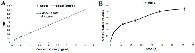
The spectrophotometric analysis assessed the encapsulation efficiency at a wavelength of 224 nm, yielding an encapsulation efficiency of 86.01% (Fig. 1A). Figure 1B illustrates the drug release profile of Uro-B from GQD@MSN NPs, showing a drug loading capacity of 24%. Also, it presents a calibration curve for Uro-B (y = 0.3797x + 0.0807, R² = 0.9994), confirming the analytical method’s reliability. Figure 1B shows Uro-B’s in vitro cumulative release over 72 h, displaying a biphasic pattern: an initial burst of about 20% in the first 10 h, followed by a sustained release, achieving 49.5% cumulative release at 48 h and leveling off at about 54.3% after 72 h.

Encapsulation Efficacy and Drug Release Assays. (A) Standard calibration curve of Urolithin B (Uro-B) with R² = 0.9994, Uro-B’s encapsulation efficiency in GQD@MSN NPs was 86.01% (spectrophotometric analysis at 224 nm). (B) In vitro cumulative release of Uro-B from GQD@MSN NPs (24% drug loading) showed an initial burst followed by sustained release over 72 h.
NP characterizations
DLS assay
DLS analysis revealed distinct characteristics for the two NP formulations. For GQD@MSN-Uro-B (Fig. 2A), the Z-average particle size was measured at 223.75 nm, with a polydispersity index (PDI) of 0.3820, indicating moderate uniformity. The mean intensity diameter was 289.52 nm, while the mean volume diameter was 334.91 nm, suggesting a range of particle sizes. The mean number diameter was recorded at 61.05 nm, and the zeta potential was found to be -22.59 ± 3.29 mV, reflecting good stability in suspension. The mean electrophoretic mobility was − 1.73 ± 0.25 mV.
In contrast, MSN-Uro-B @GQD (Fig. 2B) exhibited a larger Z-average particle size of 241.02 nm and a similar PDI of 0.3860. The mean intensity diameter was 312.90 nm, and the mean volume diameter was 367.67 nm, indicating a broader size distribution. The mean number diameter was slightly higher at 68.75 nm, with a zeta potential of -14.99 ± 5.31 mV, suggesting reduced stability compared to GQD@MSN-Uro-B. The mean electrophoretic mobility for this formulation was − 1.16 ± 0.41 mV. Both formulations possess suitable characteristics for potential biomedical applications; however, GQD@MSN-Uro-B shows stability and particle size advantages, which could improve its effectiveness in therapeutic settings.

Characterization of GQD@MSN-Uro-B and MSN-Uro-B@GQD NPs reveals distinct properties. (A) GQD@MSN-Uro-B has a Z-average particle size of 223.75 nm, a PDI of 0.3820, mean intensity/volume diameters of 289.52 nm/334.91 nm, and a zeta potential of -22.59 ± 3.29 mV, indicating good stability. (B) Conversely, MSN-Uro-B@GQD has a larger Z-average size of 241.02 nm, a similar PDI of 0.3860, mean diameters of 312.90 nm/367.67 nm, and a zeta potential of − 14.99 ± 5.31 mV, reflecting reduced stability. GQD@MSN-Uro-B shows better stability and smaller size, potentially enhancing therapeutic efficacy.
FESEM and TEM
Figure 3 provides a detailed morphological characterization of the synthesized nanoparticles. The FESEM image of GQD@MSN-Uro-B NPs (Fig. 3A), revealing well-dispersed, spherical particles with uniform size distribution and smooth surfaces, indicates successful synthesis and coating of MSNs with GQDs and Uro-B loading. The FESEM image of MSN-Uro-B@GQD (Fig. 3B), where the NPs appear slightly less uniform and more sparsely distributed, possibly reflecting surface modification or loading sequence differences. Figure 3C presents the TEM image of GQD@MSN-Uro-B NPs, confirming their spherical morphology and nanoscale size (well below 100 nm), as well as the presence of a darker core-shell structure that suggests successful encapsulation of GQDs and Uro-B within the MSN matrix.

(A) FESEM image of GQD@MSN-Uro-B NPs showing uniform, spherical morphology; (B) FESEM image of MSN-Uro-B@GQD NPs with a less uniform distribution; (C) TEM image of GQD@MSN-Uro-B NPs confirming nanoscale size and core-shell structure.
FTIR spectroscopy
As illustrated in Fig. 4, FTIR spectroscopy was employed to analyze the functional groups in various NP formulations. The spectra for GQD@MSN-Uro-B exhibited characteristic peaks at 3403.7 cm⁻¹ (O–H stretching), 2958.00 cm⁻¹ (C–H stretching), and 1641.82 cm⁻¹ (C=O stretching), among others, indicating the presence of hydroxyl, aliphatic, and carbonyl groups. The peaks at 1512.15 cm⁻¹ and 1388.88 cm⁻¹ suggest the presence of aromatic compounds and C–H bending vibrations, respectively (Fig. 2A). For MSN-Uro-B @GQD, the FTIR spectrum displayed a broader range of peaks, including significant absorptions at 3387.06 cm⁻¹ and 3301.61 cm⁻¹ (O-H stretching) and 2925.31 cm⁻¹ (C–H stretching). Notably, the peak at 1703.37 cm⁻¹ corresponds to C=O stretching. In comparison, additional peaks at 1625.84 cm⁻¹ and 1609.53 cm⁻¹ indicate the presence of aromatic structures and amide functionalities (Fig. 2B). This formulation showed a more complex spectrum, suggesting the successful incorporation of Uro-B into the MSN matrix. The MSN Blank sample revealed peaks at 3436.93 cm⁻¹ (O–H stretching) and 2941.66 cm⁻¹ (C–H stretching), with other notable peaks at 1635.66 cm⁻¹ and 1510.68 cm⁻¹, confirming the presence of surface silanol groups and residual organic moieties (Fig. 2C). The GQD spectrum demonstrated peaks at 3421.77 cm⁻¹ (O–H stretching), 2918.39 cm⁻¹ (C–H stretching), and 1693.22 cm⁻¹ (C=O stretching), indicative of the chemical structure of GQD with functional groups that may facilitate interactions with other components (Fig. 2D). In the GQD-NHS-EDC-MSN formulation, peaks at 3443.45 cm⁻¹ (O–H stretching) and 2949.83 cm⁻¹ (C–H stretching) were observed, along with a peak at 1632.65 cm⁻¹, suggesting successful conjugation of NHS and EDC groups to the MSN surface (Fig. 2E). The Uro-B spectrum displayed significant peaks at 3292.90 cm⁻¹ (O–H stretching), 3076.5 cm⁻¹ (aromatic C–H stretching), and 1701.04 cm⁻¹ (C=O stretching), confirming the presence of the drug’s characteristic functional groups (Fig. 2F). Finally, the MSN-Urolitin B formulation showed peaks at 3387.06 cm⁻¹ (O–H stretching), 3298.42 cm⁻¹ (N–H stretching), and 2929.40 cm⁻¹ (C–H stretching), indicating successful encapsulation of Urolitin B within the MSN structure (Fig. 2G). Overall, the FTIR analysis confirms the successful synthesis and functionalization of the NP formulations, highlighting the presence of key functional groups essential for their potential biomedical applications.

FTIR spectroscopy analysis of NPs reveals key functional groups in various formulations. (A) GQD@MSN-Uro-B shows peaks at 3403.7 cm⁻¹ (O–H), 2958.00 cm⁻¹ (C–H), and 1641.82 cm⁻¹ (C=O). (B) MSN-Uro-B@GQD has peaks at 3387.06 cm⁻¹ (O–H) and 1703.37 cm⁻¹ (C=O), indicating Uro-B incorporation. (C) GQD displays peaks at 3421.77 cm⁻¹ (O–H) and 1693.22 cm⁻¹ (C=O). (D) Uro-B exhibits peaks at 3292.90 cm⁻¹ (O–H) and 1701.04 cm⁻¹ (C=O). (E) MSN-Urolitin B shows peaks at 3387.06 cm⁻¹ (O–H), 3298.42 cm⁻¹ (N–H), and 2929.40 cm⁻¹ (C–H), confirming successful encapsulation. The FTIR analysis confirms NP formulations’ successful synthesis and functionalization for biomedical applications.
Cytotoxicity assay
The MTT assay results reveal significant differences in cytotoxic effects between the tested formulations on MCF7 breast cancer cells and normal HDF cells. GQD@MSN-Uro-B exhibited the most potent cytotoxicity against MCF7 cells (Fig. 5A), with an IC50 of 10 µg/mL, indicating high potency, while showing much lower toxicity toward HDF cells (IC50 = 231.2 µg/mL) (Fig. 5F), which demonstrates its selectivity for cancer cells. MSN-Uro-B@GQD also reduced MCF7 viability but with a higher IC50 of 83.19 µg/mL (Fig. 5B), indicating less potency than GQD@MSN-Uro-B. Urolithin B alone had an IC50 of 23.98 µg/mL for MCF7 cells (Fig. 5C) and over 500 µg/mL for HDF cells (Fig. 5G), suggesting that free Uro-B is less toxic to normal cells. The blank GQD@MSN formulation showed much less cytotoxicity toward both cell types, with an IC50 of 211.93 µg/mL for MCF7 cells (Fig. 5D) and greater than 500 µg/mL for HDF cells (Fig. 5H), confirming the safety of the carrier system. As a positive control, Tamoxifen demonstrated an IC50 of 15 µg/mL for MCF7 cells (Fig. 5E), which was less cytotoxic than GQD@MSN-Uro-B.

MTT assay results demonstrate various formulations’ cytotoxic effects on MCF7 breast cancer cells (A–E) and normal HDF cells (F–H). GQD@MSN-Uro-B (A) exhibited the highest cytotoxicity and selectivity toward MCF7 cells (IC50 = 10 µg/mL) with significantly reduced toxicity to HDF cells (IC50 = 231.2 µg/mL) (F). MSN-Uro-B@GQD (B) and Urolithin B alone (C) showed moderate cytotoxicity against MCF7 cells, while the blank carrier (D, H) displayed minimal toxicity to both cell types. Tamoxifen (E) served as a positive control. These results highlight the enhanced anti-cancer efficacy of the GQD@MSN-Uro-B NPs. The data is presented as mean ± standard deviation (SD), and statistical significance is denoted as * P < 0.01, ** P < 0.001, and *** P < 0.001.
Annexin V-FITC/PI assay
The results illustrated in Fig. 6 reveal an apparent dose-dependent increase in apoptotic activity in MCF7 cancer cells treated with GQD@MSN-Uro-B NPs. Untreated control cells showed the lowest apoptosis rates, with 8.33% in early apoptosis and 3.78% in late apoptosis (Fig. 6A). Treatment with 8 µg/mL of the nanoparticles resulted in a 25.5% increase in early apoptosis and a 6.21% increase in late apoptosis (Fig. 6B). At a 10 µg/mL concentration, early apoptosis rose to 33.0%. In comparison, late apoptosis increased to 20.6% (Fig. 6C). Finally, at the highest concentration tested, 12 µg/mL, early apoptosis reached 37.4%. Late apoptosis surged to 39% (Fig. 6D). These findings suggest that GQD@MSN-Uro-B NPs are highly effective at inducing apoptosis in cancer cells concentration-dependent, indicating their potential as a targeted therapeutic for cancer treatment.

Annexin V-FITC/PI Dual Staining Assay of Apoptosis in MCF7 Cells shows a dose-dependent increase in apoptosis with GQD@MSN-Uro-B NPs. (A) Untreated control cells had minimal apoptosis (8.33% early, 3.78% late). (B) At 8 µg/mL, early apoptosis increased to 25.5% and late to 6.21%. (C) At 10 µg/mL, early apoptosis was 33.0%, and late was 20.6%. (D) At 12 µg/mL, early apoptosis reached 37.4%, and late apoptosis surged to 39%. GQD@MSN-Uro-B NPs effectively induce concentration-dependent apoptosis in MCF7 cancer cells.
AO/PI staining
AO/PI staining of MCF7 cells treated with GQD@MSN- Uro-B NPs shows distinct fluorescence patterns based on dosage. The untreated control cells display uniform green fluorescence, indicating healthy cells (Fig. 7A). At 8 µg/mL, some cells reveal both green and orange/red fluorescence, indicating viable and early apoptotic cells (Fig. 7B). The 10 µg/mL concentration shows increased orange/red fluorescence, suggesting more late-apoptotic or necrotic cells (Fig. 7C). At 12 µg/mL, many cells exhibit orange/red fluorescence, indicating high levels of apoptosis and cell death (Fig. 7D).

AO/PI Staining of MCF7 Cells Treated with GQD@MSN-Uro-B NPs shows that the fluorescence patterns vary with NP dosage. (A) Untreated control cells show uniform green fluorescence, indicating health. (B) At 8 µg/mL, some cells display green and orange/red fluorescence, indicating viable and early apoptotic cells. (C) The 10 µg/mL concentration shows increased orange/red fluorescence, signifying more late-apoptotic or necrotic cells. (D) At 12 µg/mL, many cells exhibit orange/red fluorescence, indicating high levels of apoptosis and cell death.
Gene expression assay
Treatment of MCF7 cancer cells with GQD@MSN-Uro-B NPs resulted in a significant increase in the TNF biomarker, with expression rising from 0.92 ± 0.008 at 8 µg/mL to 1.13 ± 0.01 and 1.14 ± 0.012 at 10 and 12 µg/mL, respectively (Fig. 8A; P < 0.05). Additionally, the apoptosis-related biomarker caspase-9 (CAS-9) showed a dose-dependent increase, rising from 0.85 ± 0.008 at 8 µg/mL to 2.56 ± 0.01 and 3.3 ± 0.012 at 10 and 12 µg/mL (Fig. 8B; P < 0.001). Similarly, the cell cycle regulator p21 increased from 0.87 ± 0.008 at 8 µg/mL to 1.32 ± 0.012 at 12 µg/mL (Fig. 8C). These findings suggest that GQD@MSN-Uro-B NPs effectively modulate key biomarkers related to apoptosis and cell cycle regulation in MCF7 cells, indicating potential as an anti-cancer therapeutic.

Real-time PCR analysis in MCF7 cells treated with GQD@MSN-Uro-B NPs revealed significant increases in key biomarkers. (A) TNF expression rose from 0.92 ± 0.008 at 8 µg/mL to 1.14 ± 0.012 at 12 µg/mL. (B)The apoptosis marker caspase-9 increased from 0.85 ± 0.008 at 8 µg/mL to 3.3 ± 0.012 at 12 µg/mL, indicating a dose-dependent pattern. (C) p21, a cell cycle regulator, also rose from 0.87 ± 0.008 to 1.32 ± 0.012 at the same concentrations (P < 0.001). GQD@MSN-Uro-B NPs are promising anti-cancer therapies that modulate apoptosis and cell cycle biomarkers. The data is presented as mean ± standard deviation (SD), and statistical significance is denoted as * P < 0.05 and *** P < 0.001.
Antioxidant capacity assay
The antioxidant capacity of GQD@MSN-Uro-B NPs was evaluated based on their ability to scavenge ABTS and DPPH free radicals (Fig. 9). The results indicated a significant concentration-dependent increase in scavenging activity for both assays (P < 0.001). Specifically, the ABTS assay demonstrated a 76% inhibition at a concentration of 2000 µg/mL, while the DPPH assay showed a 33.93% inhibition at the same concentration. These findings confirm that GQD@MSN-Uro-B nanoparticles exhibit notable antioxidant properties, particularly against ABTS radicals.

The antioxidant capacity of GQD@MSN-Uro-B NPs was evaluated using ABTS and DPPH assays. Results showed a concentration-dependent increase in scavenging activity, with a 76% inhibition in the ABTS assay and a 33.93% inhibition in the DPPH assay at 2000 µg/mL. This indicates that GQD@MSN-Uro-B NPs have significant antioxidant properties, especially against ABTS radicals. The data is presented as mean ± standard deviation (SD), and statistical significance is denoted as * P < 0.01 and *** P < 0.001.
Discussion
Targeted cancer therapies have significantly advanced over conventional treatments by concentrating on specific molecular targets that promote tumor growth22. Targeted therapies enhance treatment efficacy while reducing side effects by utilizing drug delivery systems (DDSs). Technologies such as nanoparticles, liposomes, and conjugated antibodies enable direct delivery to cancer cells, improving drug bioavailability and controlled release. This approach addresses drug resistance by engaging altered pathways in cancer cells and supports personalized medicine, allowing for tailored treatments and potential combinations for better outcomes. As research progresses, the integration of targeted therapies into clinical practice holds great promise for improving patient prognosis and quality of life23.
The study presents compelling evidence regarding the anti-cancer potential of GQD@MSN- Uro-B nanoparticles, showcasing their effectiveness against the MCF7 cancer cell line through various assays. The unique combination of these two components in GQD@MSN-Uro-B NPs leverages their strengths while addressing limitations such as poor solubility and stability of free urolithins. DLS analysis indicates that GQD@MSN-Uro-B has superior stability and smaller particle size than MSN-Uro-B @GQD, which may enhance its therapeutic efficacy. FTIR spectroscopy confirmed the successful functionalization of the NPs. The cytotoxicity assays reveal a significant concentration-dependent effect, with GQD@MSN-Uro-B exhibiting a lower IC50 (10 µg/mL) value against MCF7 cells while being less harmful to normal HDF cells, suggesting its selective targeting of cancer cells while sparing normal cells. It is necessary to explain that in the GQD@MSN-Uro-B formulation (Method 1), Uro-B is loaded onto pre-formed GQD-modified MSNs, allowing for better interaction with GQDs and the MSN matrix. This results in a more homogeneous distribution of Uro-B, facilitating efficient and sustained drug release. The unique properties of GQDs may enhance cellular uptake and increase cancer cell cytotoxicity through improved membrane interaction and increased ROS generation. In contrast, the MSN-Uro-B@GQD formulation (Method 2) loads Uro-B into MSNs before coating with GQDs, which can hinder immediate drug release and reduce direct interaction with cancer cells. The GQD coating may also slow drug release and limit ROS generation, decreasing Uro-B bioavailability at the target site. Consequently, GQD@MSN-Uro-B shows improved drug release, cellular uptake, and greater cytotoxicity against MCF-7 breast cancer cells compared to MSN-Uro-B@GQD, emphasizing the significance of nanoparticle design and drug loading sequence in optimizing therapeutic efficacy. In addition, the selectivity of GQD-modified MSNs for MCF-7 breast cancer cells, as opposed to non-cancerous cells, stems from several factors. GQDs’ small, lamellar structure allows them to enter cancer cell nuclei, disrupting DNA activity and inducing apoptosis selectively in cancer cells. Functionalizing GQDs with targeting ligands enhances their uptake by cancer cells that overexpress specific receptors. Additionally, cancer cells show higher metabolic activity and enhanced endocytosis, facilitating the internalization of GQD-modified MSNs. The mesoporous silica framework acts as a biocompatible carrier, promoting drug loading and controlled release, while GQDs generate ROS and target the nucleus. These combined features enable GQD@MSN nanocomposites to effectively kill MCF-7 cancer cells while minimizing toxicity to normal cells. Apoptosis assays further support these findings, demonstrating a marked increase in early and late apoptosis in treated cells. Additionally, gene expression analysis shows upregulation of critical biomarkers associated with apoptosis and cell cycle regulation, reinforcing the NPs’ role in inducing cancer cell death. We selected Caspase 9, p21, and TNF for gene expression analysis due to their roles as key markers in apoptosis and cell cycle regulation, which are essential for the anti-cancer effects of our nanoplatform. Caspase 9 is crucial in the intrinsic apoptotic pathway, indicating apoptosis in response to stress or chemotherapy. p21, a cyclin-dependent kinase inhibitor, is vital for G1/S checkpoint arrest, inhibiting cancer cell proliferation following DNA damage. TNF is a pro-inflammatory cytokine that triggers extrinsic apoptosis and influences the tumor microenvironment. Our findings are consistent with prior studies that have shown the ability of urolithins to modulate apoptosis-related genes, including BAX and BCL-2, thereby tipping the balance toward cell death in cancerous tissues24. The antioxidant capacity of GQD@MSN-Uro-B is also noteworthy, as it effectively scavenges free radicals, adding another layer to its therapeutic profile. Oxidative stress is a well-established contributor to cancer progression, and the ability of these nanoparticles to scavenge free radicals adds a complementary mechanism to their anti-cancer activity. The results suggest that GQD@MSN-Uro-B NPs may create an antioxidant protection for healthy tissue, warranting further exploration in clinical settings. Overall, the use of GQDs to modify mesoporous silica nanoparticles is supported by their unique physicochemical properties, which greatly improve the delivery of Uro-B. GQDs demonstrate remarkable fluorescence, allowing for real-time tracking in biological systems, and possess high stability, ensuring prolonged circulation in the bloodstream. Additionally, their significant drug-loading capacity enables efficient encapsulation of Uro-B, thereby facilitating targeted therapy. Furthermore, the sequence in which GQDs are attached to MSNs is crucial in determining the surface chemistry and particle aggregation. For example, adding GQDs to MSNs may optimize surface interactions, enhance drug loading efficiency, and reduce aggregation. Conversely, attaching MSNs to GQDs could change the surface properties and potentially impact the overall effectiveness of the drug delivery system. This highlights the importance of GQDs in improving the therapeutic potential of Uro-B-loaded nanoparticles.
Previous studies have explored urolithins’ role in cancer therapy, particularly Uro-B25. Urolithins are secondary metabolites formed from ellagic acid and ellagitannin, primarily found in foods such as pomegranates, berries, and nuts, through the action of gut microbiota26. These compounds are categorized into three metabotypes based on the specific urolithins produced: metabotype A, which produces Uro-A; metabotype B, which produces Uro-A, IsoUro-A, and Uro-B; and metabotype 0, which makes none27. Urolithins are noted for their enhanced bioavailability compared to ellagic acid and ellagitannin, achieving micromolar concentrations in plasma and various tissues, including the breast, colon, and prostate. Their anti-cancer effects are significant, as they regulate the expression of oncogenes, cell cycle mediators, tumor suppressors, and growth factor receptors, thereby contributing to their potential in cancer prevention and therapy16,28,29. Additionally, urolithins exhibit anti-obesity, antimicrobial, and anti-inflammatory properties, highlighting their therapeutic relevance.
Urolithins, particularly Uro-B, have demonstrated significant anti-cancer effects in various cancer types, including prostate cancer. Research indicates that Uro-B induces apoptosis and inhibits proliferation in prostate cancer cell lines such as LNCaP and DU145. This dose-dependent effect leads to cell cycle arrest at the S and G2/M phases. Additionally, Uro-B treatment reduces prostate-specific antigen (PSA) and androgen receptor expression, inhibiting their interaction and transcriptional activity, which are crucial for tumor progression30,31,32. Notably, Urolithin enhances the effects of bicalutamide in androgen-dependent cell lines, suggesting its potential to overcome resistance in castration-resistant prostate cancer (CRPC)25,33. Overall, these findings position Uro-B as a promising candidate for prostate cancer treatment, warranting further clinical investigation.
In addition to its effects on prostate cancer, Urolithins exhibit significant anti-cancer properties in breast cancer, particularly in hormone-dependent cell lines. Both Uro-B and Uro-A show potent antiproliferative effects by inhibiting the proliferation of MCF-7aro cells, which overexpress aromatase34. Their ability to inhibit aromatase positions them as potential therapeutic agents targeting estrogen synthesis, a critical factor for breast cancer growth. Furthermore, Uro-B demonstrates notable antiestrogenic effects, with lower IC50 values in binding assays to estrogen receptors (ERα and ERβ) compared to many phytoestrogens, indicating its effectiveness in blocking estrogen signaling35. In studies involving MDA-MB (estrogen-negative) and MCF-7 (estrogen-positive) cells, Uro-B exhibited decreasing antiproliferative potential, reinforcing its role in combating breast cancer growth. While phase II metabolites of Urolithins showed reduced activity, the aglycones retained significant effects, underscoring the importance of Uro-B in breast cancer prevention and treatment strategies36.
Moreover, Urolithins show promising anti-cancer effects against endometrial cancer by inhibiting cell proliferation and potentially preventing metastasis37. Uro-B effectively reduces the proliferation of endometrial cancer cells, similarly to Uro-A, through an ERα-dependent mechanism. At a concentration of 10 µM, Uro-B promotes cell cycle arrest at the G2/M phase by modulating key cell cycle regulators37. Uro-A and Uro-B also influence gene expression related to cancer progression, acting as estrogen agonists. Notably, Uro-A decreases the activity and mRNA levels of Rac1 and PAK1, which are vital for cytoskeletal dynamics and cancer cell migration, indicating a preventive role against metastasis38. These findings collectively highlight the therapeutic potential of Urolithins, particularly Uro-B, in endometrial cancer treatment.
Urolithins also exhibit significant anti-cancer effects against hepatocellular carcinoma (HCC) through various mechanisms. Uro-B effectively inhibits HCC cell proliferation, inducing cell cycle arrest at the G0/G1 phase in HepG2 cells and the S phase in Bel7402 cells39. This antiproliferative effect is linked to the induction of apoptosis, evidenced by reduced Bcl-2 expression40. In vivo studies demonstrate that Uro-B suppresses tumor growth in xenograft mouse models at 40 mg/kg. Additionally, Uro-B phosphorylates β-catenin, preventing its nuclear-to-cytoplasmic translocation and thus inactivating the Wnt/β-catenin signaling pathway, which is critical in cancer progression40. These findings underscore Uro-B’s potential as a therapeutic agent for HCC.
The anti-cancer capabilities of Uro-B extend to colorectal cancer, where it induces apoptosis and cell cycle arrest41. Specifically, Uro-B (30 µg/mL) promotes apoptosis in HT-29 colon cancer cells by activating caspase 3 and upregulating p21, leading to G2/M phase arrest42. Furthermore, Uro-B inhibits mucin-type O-glycosylation in colon cancer cells, which correlates with decreased podoplanin (PDPN) expression and reduced cell migration and invasion43. While Uro-B enhances the expression of detoxifying enzymes, it also induces CYP1B1, which may present undesirable effects in cancer therapy44. These multifaceted mechanisms highlight Uro-B’s potential as a therapeutic agent in combating colorectal cancer.
Uro-B exhibits promising anti-cancer effects against bladder cancer, particularly in UMUC3 cell lines, characterized by cell cycle checkpoint abnormalities leading to genetic instability. Uro-B, Uro-A, and Uro-C reduce cell viability, demonstrating the most significant activity by inducing G2/M phase arrest45. This arrest is attributed to Uro-A’s ability to inactivate the cyclin B1/cdc2 kinase complex, which is crucial for the G2/M transition46. Furthermore, Uro-A has been shown to decrease the phosphorylation of p-Akt and ERK 1/2, indicating its potential to inhibit the PI3K/Akt and ERK pathways, vital for cancer cell proliferation and survival47,48. These findings suggest that Uro-B could be a valuable therapeutic agent in managing bladder cancer. Furthermore, Uro-B shows promise as a potential treatment for osteosarcoma by exhibiting antimetastatic, antiproliferative, and apoptotic effects on the MG-63 OS cell line. The study revealed that Uro-B induces late apoptosis, necrosis, and G2/M cell cycle arrest while increasing the expression of the tumor suppressor gene p53 and the pro-apoptotic gene Bax. Additionally, Uro-B inhibits cell migration and metastatic behavior by downregulating MMP-2 and MMP-9 expression. These findings suggest that Uro-B may serve as a novel, nontoxic natural compound for osteosarcoma therapy, although further in vivo studies are needed24. The molecular targets of Uro-B, such as BAX/BCL-2 and cell cycle regulators, play a critical role in mediating apoptosis and cell cycle arrest in cancer cells. The enhanced absorption and delivery efficiency of GQD@MSN-Uro-B NPs significantly amplifies the bioavailability of Uro-B, allowing for more effective modulation of these targets. By facilitating targeted delivery, GQD@MSN NPs ensure that higher concentrations of Uro-B reach the intended sites of action within the cells, thereby promoting the activation of pro-apoptotic pathways and inhibiting anti-apoptotic mechanisms. This innovative formulation not only highlights the therapeutic potential of Uro-B but also underscores the importance of nanoparticle carriers in enhancing the efficacy of cancer treatments.
This study presents several limitations that warrant consideration for future research. While the anti-cancer effects and antioxidant properties of GQD@MSN-Uro-B NPs were evaluated in vitro, further investigations are needed to assess their pharmacokinetics and biodistribution in vivo. Understanding how these nanoparticles behave in a biological system will provide crucial insights into their therapeutic efficacy and safety profiles. Additionally, conducting experiments in xenograft tumor models could offer a more comprehensive understanding of the potential clinical applications of GQD@MSN-Uro-B in breast cancer treatment. These studies would help elucidate the nanoparticles’ mechanisms of action, optimize dosing regimens, and evaluate their long-term safety and effectiveness, ultimately bridging the gap between laboratory findings and clinical practice.
Conclusion
In conclusion, this study highlights the potential of GQD@MSN-Uro-B NPs as a targeted anti-cancer therapy with multifaceted mechanisms of action, including apoptosis induction and antioxidant activity. Combining the unique properties of GQDs and Uro-B, these nanoparticles demonstrate enhanced stability, selectivity, and efficacy against cancer cells while sparing normal tissues. These findings contribute to the growing body of evidence supporting the use of functionalized nanoparticles in oncology and warrant further exploration in preclinical and clinical settings to realize their full therapeutic potential.
Data availability
The datasets used and/or analysed during the current study available from the corresponding author on reasonable request.
References
-
Arnold, M. et al. Current and future burden of breast cancer: Global statistics for 2020 and 2040. Breast 66, 15–23 (2022).
-
Ozyurt, R. & Ozpolat, B. Molecular mechanisms of anti-estrogen therapy resistance and novel targeted therapies. Cancers 14 (21), 5206 (2022).
-
Comşa, Ş., Cimpean, A. M. & Raica, M. The story of MCF-7 breast cancer cell line: 40 years of experience in research. Anti-Cancer Res. 35 (6), 3147–3154 (2015).
-
Afzal, M. et al. (eds) Nanomedicine in Treatment of Breast cancer–A Challenge To Conventional Therapy. Seminars in cancer Biology (Elsevier, 2021).
-
Sledge, G. W. et al. Past, present, and future challenges in breast cancer treatment. J. Clin. Oncol. 32 (19), 1979–1986 (2014).
-
Emeihe, E. V., Nwankwo, E. I., Ajegbile, M. D., Olaboye, J. A. & Maha, C. C. Revolutionizing drug delivery systems: Nanotechnology-based approaches for targeted therapy. Int. J. Life Sci. Res. Arch. 7 (1), 040–58 (2024).
-
Rosenholm, J. M., Mamaeva, V., Sahlgren, C. & Lindén, M. Nanoparticles in targeted cancer therapy: Mesoporous silica nanoparticles entering preclinical development stage. Nanomedicine 7 (1), 111–120 (2012).
-
Gary-Bobo, M. et al. Cancer therapy improvement with mesoporous silica nanoparticles combining targeting, drug delivery and PDT. Int. J. Pharm. 423 (2), 509–515 (2012).
-
Chang, D. et al. Polydopamine-based surface modification of mesoporous silica nanoparticles as pH-sensitive drug delivery vehicles for cancer therapy. J. Colloid Interface Sci. 463, 279–287 (2016).
-
Iannazzo, D., Ziccarelli, I. & Pistone, A. Graphene quantum dots: Multifunctional nanoplatforms for anti-cancer therapy. J. Mater. Chem. B. 5 (32), 6471–6489 (2017).
-
Carbonaro, C. M. et al. Graphene Quantum Dots and Carbon Nanodots: Modeling of zero-dimensional Carbon Nanomaterials 411–482 (Elsevier, 2024).
-
Yang, D. et al. Design and investigation of core/shell gqds/hmsn nanoparticles as an enhanced drug delivery platform in triple-negative breast cancer. Bioconjug. Chem. 29 (8), 2776–2785 (2018).
-
Qin, Y. et al. Graphene quantum Dots induce apoptosis, autophagy, and inflammatory response via p38 mitogen-activated protein kinase and nuclear factor-κB mediated signaling pathways in activated THP-1 macrophages. Toxicology 327, 62–76 (2015).
-
Chen, P., Guo, Z., Chen, F., Wu, Y. & Zhou, B. Recent advances and perspectives on the health benefits of urolithin B, a bioactive natural product derived from ellagitannins. Front. Pharmacol. 13, 917266 (2022).
-
Remadevi, V. et al. Urolithin A, induces apoptosis and autophagy crosstalk in oral squamous cell carcinoma via mTOR/AKT/ERK1/2 pathway. Phytomedicine 130, 155721 (2024).
-
Rogovskii, V. S. The therapeutic potential of urolithin A for cancer treatment and prevention. Curr. Cancer Drug Targets. 22 (9), 717–724 (2022).
-
Shen, C-K. et al. Inhibitory effects of urolithins, bioactive gut metabolites from natural polyphenols, against glioblastoma progression. Nutrients 15 (23), 4854 (2023).
-
Hu, Y. et al. Liposomes encapsulation by pH driven improves the stability, bioaccessibility and bioavailability of urolithin A: A comparative study. Int. J. Biol. Macromol. 253, 127554 (2023).
-
Zhang, S., Zhang, H., Liu, Q. & Chen, S. Fe–N doped carbon nanotube/graphene composite: Facile synthesis and superior electrocatalytic activity. J. Mater. Chem. A. 1 (10), 3302–3308 (2013).
-
Zadeh, E. S. et al. Smart pH-responsive magnetic graphene quantum Dots nanocarriers for anti-cancer drug delivery of Curcumin. Mater. Chem. Phys. 297, 127336 (2023).
-
Namvar, F. et al. Green synthesis, characterization, and anti-cancer activity of hyaluronan/zinc oxide nanocomposite. OncoTargets Therapy 4549–4559. (2016).
-
Zhong, L. et al. Small molecules in targeted cancer therapy: Advances, challenges, and future perspectives. Signal. Transduct. Target. Therapy. 6 (1), 1–48 (2021).
-
Adepu, S. & Ramakrishna, S. Controlled drug delivery systems: Current status and future directions. Molecules 26 (19), 5905 (2021).
-
Tajvar Nasab, N., Jalili-Nik, M., Afshari, A. R., Rezaei Farimani, A. & Soukhtanloo, M. Urolithin B inhibits proliferation and migration and promotes apoptosis and necrosis by inducing G2/M arrest and targeting MMP‐2/‐9 expression in osteosarcoma cells. J. Biochem. Mol. Toxicol. 37 (12), e23486 (2023).
-
Al-Harbi, S. A., Abdulrahman, A. O., Zamzami, M. A. & Khan, M. I. Urolithins: the gut based polyphenol metabolites of ellagitannins in cancer prevention, a review. Front. Nutr. 8, 647582 (2021).
-
Cerdá, B., Tomás-Barberán, F. A. & Espín, J. C. Metabolism of antioxidant and chemopreventive ellagitannins from strawberries, raspberries, walnuts, and oak-aged wine in humans: Identification of biomarkers and individual variability. J. Agric. Food Chem. 53 (2), 227–235 (2005).
-
Espín, J. C. et al. Iberian pig as a model to clarify obscure points in the bioavailability and metabolism of ellagitannins in humans. J. Agric. Food Chem. 55 (25), 10476–10485 (2007).
-
El-Wetidy, M. S. et al. Urolithin A induces cell cycle arrest and apoptosis by inhibiting Bcl-2, increasing p53-p21 proteins and reactive oxygen species production in colorectal cancer cells. Cell. Stress Chaperones. 26, 473–493 (2021).
-
Totiger, T. M. et al. Urolithin A, a novel natural compound to target PI3K/AKT/mTOR pathway in pancreatic cancer. Mol. Cancer Ther. 18 (2), 301–311 (2019).
-
Sánchez-González, C., Noé, V. & Izquierdo-Pulido, M. Walnut polyphenol metabolites, urolithins A and B, inhibit the expression of the prostate-specific antigen and the androgen receptor in prostate cancer cells. Food Funct. 5 (11), 2922–2930 (2014).
-
Sánchez-González, C., Ciudad, C. J., Izquierdo-Pulido, M. & Noé, V. Urolithin A causes p21 up-regulation in prostate cancer cells. Eur. J. Nutr. 55, 1099–1112 (2016).
-
Stanisławska, I. J., Piwowarski, J. P., Granica, S. & Kiss, A. K. The effects of urolithins on the response of prostate cancer cells to non-steroidal antiandrogen bicalutamide. Phytomedicine 46, 176–183 (2018).
-
Dahiya, N. R. et al. A natural molecule, urolithin A, downregulates androgen receptor activation and suppresses growth of prostate cancer. Mol. Carcinog. 57 (10), 1332–1341 (2018).
-
Adams, L. S., Zhang, Y., Seeram, N. P., Heber, D. & Chen, S. Pomegranate ellagitannin–derived compounds exhibit antiproliferative and antiaromatase activity in breast cancer cells in vitro. Cancer Prev. Res. 3 (1), 108–113 (2010).
-
Larrosa, M., González-Sarrías, A., García-Conesa, M. T., Tomás-Barberán, F. A. & Espín, J. C. Urolithins, ellagic acid-derived metabolites produced by human colonic microflora, exhibit estrogenic and antiestrogenic activities. J. Agric. Food Chem. 54 (5), 1611–1620 (2006).
-
Ávila-Gálvez, M. Á., Espín, J. C. & González-Sarrías, A. Physiological relevance of the antiproliferative and estrogenic effects of dietary polyphenol aglycones versus their phase-II metabolites on breast cancer cells: A call of caution. J. Agric. Food Chem. 66 (32), 8547–8555 (2018).
-
Mc Cormack, B. et al. The ellagic acid metabolites urolithin A and B differentially affect growth, adhesion, motility, and invasion of endometriotic cells in vitro. Hum. Reprod. 36 (6), 1501–1519 (2021).
-
Mc Cormack, B. A. et al. Effect of urolithins A and B on ectopic endometrial growth in a murine model of endometriosis. Food Funct. 12 (20), 9894–9903 (2021).
-
My, L. et al. Urolithin B suppresses tumor growth in hepatocellular carcinoma through inducing the inactivation of Wnt/β-catenin signaling. J. Cell. Biochem. 120 (10), 17273–17282 (2019).
-
Wang, Y. et al. In vitro antiproliferative and antioxidant effects of urolithin A, the colonic metabolite of ellagic acid, on hepatocellular carcinomas HepG2 cells. Toxicol. In Vitro. 29 (5), 1107–1115 (2015).
-
Wang, L. et al. The gut microbiota metabolite urolithin B prevents colorectal carcinogenesis by remodeling microbiota and PD-L1/HLA‐B. Oxidative Med. Cell. Longev. 2023 (1), 6480848 (2023).
-
Cho, H. et al. Chemopreventive activity of ellagitannins and their derivatives from black raspberry seeds on HT-29 colon cancer cells. Food Funct. 6 (5), 1675–1683 (2015).
-
Liu, F. et al. Inhibition of polypeptide N-acetyl-α-galactosaminyltransferases is an underlying mechanism of dietary polyphenols preventing colorectal tumorigenesis. Bioorg. Med. Chem. 27 (15), 3372–3382 (2019).
-
Gonzalez-Sarrias, A. et al. Dissimilar in vitro and in vivo effects of ellagic acid and its microbiota-derived metabolites, urolithins, on the cytochrome P450 1A1. J. Agric. Food Chem. 57 (12), 5623–5632 (2009).
-
Liberal, J., Carmo, A., Gomes, C., Cruz, M. T. & Batista, M. T. Urolithins impair cell proliferation, arrest the cell cycle and induce apoptosis in UMUC3 bladder cancer cells. Investig. New Drugs. 35 (6), 671–681 (2017).
-
Vicinanza, R., Zhang, Y., Henning, S. M. & Heber, D. Pomegranate juice metabolites, ellagic acid and urolithin a, synergistically inhibit androgen-independent prostate cancer cell growth via distinct effects on cell cycle control and apoptosis. Evid. Based Complement. Altern. Med. 2013 (1), 247504 (2013).
-
Chang, F. et al. Involvement of PI3K/Akt pathway in cell cycle progression, apoptosis, and neoplastic transformation: A target for cancer chemotherapy. Leukemia 17 (3), 590–603 (2003).
-
Knowles, M. A. & Hurst, C. D. Molecular biology of bladder cancer: New insights into pathogenesis and clinical diversity. Nat. Rev. Cancer. 15 (1), 25–41 (2015).
Acknowledgements
We sincerely thank the Vice Chancellor of Research of Islamic Azad University of Neyshabur for supporting and facilitating this research.
Funding
This research received no specific grant from any funding agency in the public, commercial, or not-for-profit sectors.
Ethics declarations
Competing interests
The authors declare no competing interests.
Additional information
Publisher’s note
Springer Nature remains neutral with regard to jurisdictional claims in published maps and institutional affiliations.
Rights and permissions
Open Access This article is licensed under a Creative Commons Attribution-NonCommercial-NoDerivatives 4.0 International License, which permits any non-commercial use, sharing, distribution and reproduction in any medium or format, as long as you give appropriate credit to the original author(s) and the source, provide a link to the Creative Commons licence, and indicate if you modified the licensed material. You do not have permission under this licence to share adapted material derived from this article or parts of it. The images or other third party material in this article are included in the article’s Creative Commons licence, unless indicated otherwise in a credit line to the material. If material is not included in the article’s Creative Commons licence and your intended use is not permitted by statutory regulation or exceeds the permitted use, you will need to obtain permission directly from the copyright holder. To view a copy of this licence, visit http://creativecommons.org/licenses/by-nc-nd/4.0/.
About this article

Cite this article
Nezhadhossein, B., Afshar, A.S., Homayouni Tabrizi, M. et al. Efficacy of urolithin B-Loaded mesoporous silica nanoparticles modified with graphene quantum Dots against MCF-7 breast Cancer cells. Sci Rep 15, 25473 (2025). https://doi.org/10.1038/s41598-025-10904-3
-
Received:
-
Accepted:
-
Published:
-
DOI: https://doi.org/10.1038/s41598-025-10904-3