Introduction
Silybum marianum, generally named Milk Thistle, is a botanical species belonging to the genus Leucanthemum1. Silybum marianum, as an herb, facilitates the process of detoxification2. Silymarin represents an extract derived from the seeds of Milk Thistle, which are known to contain a suite of biologically active flavonolignan compounds. The primary isomeric forms of flavonolignan in silymarin include silybin, isosilybin, silydianin, and silychristin, with silybin being the most pharmacologically potent. Because of its structure, silybin is a tiny, highly functionalized molecule resistant to reduction. Silybin comprises five hydroxyl functional groups, which are categorized based on their chemical characteristics into phenolic, secondary, and primary alcoholic groups3. The lipid composition of Silymarin is characterized by critical phospholipids that exhibit a substantial concentration of unsaturated fatty acids, including oleic, linoleic, and palmitic acids4. The lipid component additionally encompasses α-tocopherol, which modulates signal transduction pathways, gene expression, and cellular functionality to mitigate the likelihood of oncogenesis5. Silymarin possesses considerable pharmaceutical attributes encompassing antioxidant, anti-inflammatory, anti-fibrotic, and immune system-modulating activities6. However, because of its significant phase II metabolism, limited permeability across intestinal epithelial cells, low water solubility, and quick excretion in bile and urine, it has poor oral bioavailability7,8. Therefore, it becomes necessary to introduce a robust formulation to overcome these challenges.
In the past, different nanotechnology-based approaches have been employed to enhance the bioavailability and solubility of different phytochemicals, and also offer robust formulation against environmental, chemical, and physical degradation9,10. When compared to traditional biomaterials, nanostructured biomaterials offer several benefits, including their eco-friendly and cost-effective properties, enhanced bioavailability, better cellular interactions, and tailored functionalities11. They are distinguished by their nanoscale architecture and size12,13,14. Nanocrystals possess a robust, typically crystalline drug core within the nanometer dimension spectrum, accompanied by a stabilizing outer layer. NCs are frequently synthesized in aqueous solutions or non-aqueous solvent systems that are stabilized through the use of surfactants, polymers, or a combination of both. NCs improve the solubility and bioavailability of poorly soluble drugs and have several other benefits, including sustained release action, dose reduction, and tissue targeting15. Thus, the formation of silymarin NCs could enhance silymarin’s solubility and boost its herbal properties.
Improper antibiotic consumption has resulted in the development of multidrug-resistant (MDR) bacteria. Bacterial resistance, antibiotic complications, and the lack of new antibiotic agents necessitate achieving a new, efficient, alternative, and safe antibacterial drug16. Many studies have been done to investigate silymarin’s antibacterial effects. El-Sapagh et al. conducted a comprehensive evaluation of the efficacy of various extracts derived from S.marianum seeds in combating multidrug-resistant (MDR) bacteria associated with wound infections. Their findings indicated that the ethanol extract of S. marianum seeds exhibited a significant bacteriostatic performance against both gram-positive and gram-negative bacterial strains16. According to de Oliviera et al. assessment of silymarin and silybin’s antibacterial activity, silibinin exhibited notable activity against E. coli, with an MIC of 64 µg/mL (minimum inhibitory concentration). These substances show promising performance for working in concert with antibiotics17. As a result, silymarin NCs may be a natural alternative to conventional antibiotic medications, enhancing the antibacterial properties of silymarin.
Furthermore, silybin has demonstrated significant antitumor properties, as evidenced by not only the reduction of tumor cell adhesion, migration, intracellular glutathione (GSH) concentrations, and total antioxidant capacity (T-AOC) but also by the enhancement of the apoptotic index, caspase that silymarin has a strong anticancer impact while also protecting normal cells18. So far, no reported research has been conducted to prepare aqueous soluble silymarin nanocrystals using solvent evaporation and evaluate their antibacterial and cytotoxicity effects. Therefore, this study aims to prepare silymarin NCs and characterize them through several evaluations, including XRD, FESEM/TEM, EDX, FT-IR, UV-Vis, and lastly assess their antibacterial performance and cytotoxicity effects on the MDA-MB-231 cell line.
Experimental
Material
The bacterial strains used in this research comprise Escherichia coli, classified as gram-negative, and Staphylococcus aureus, which is categorized as gram-positive, were prepared by the infectious diseases department of the hospital, whereas the MDA-MB-231 cell line was acquired from the Cell Bank located in Tehran, Iran. Silybum marianum powder was obtained from the local market.
Characterization
The crystal nature of the silymarin NCs was determined through XRD patterns (D8 ADVANCE, Germany). Also, the morphology and size of silymarin NCs were assessed through FESEM (HITACHI, Japan) and TEM (LEO 910 Model, ZEISS, Germany) images. The purity of silymarin NCs was evaluated via EDX/Mapping (JCM electron microscope operating at 10 kV). Moreover, FT-IR analysis (Shimadzu) was utilized to identify the functional groups existing in the silymarin NCs. The optical properties of silymarin NCs were evaluated using UV-Vis analysis (CE 9500, CECIL, England).
Preparation of silymarin nanocrystals
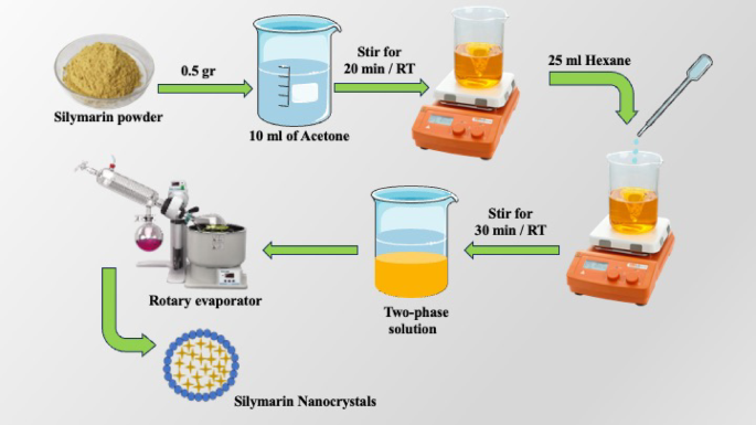
To prepare silymarin nanocrystals, first, 0.5 g of Silybum marianum (fruits) powder was dissolved in 10 mL of acetone. Then the resulting solution was stirred for 20 min at room temperature until the powder was completely dissolved in acetone. In continuation, 25 mL of hexane was added dropwise to the above solution, and the resulting mixture was stirred for 30 min at 25 °C. The resulting solution became almost two-phase (light brown phase at the bottom and colorless phase at the top). In the next step, the solvent was removed with a rotary evaporator device19, and brown aqueous soluble silymarin NCs were obtained. The synthesis schematic of silymarin NCs is shown in Fig. 1.

Synthesis of silymarin nanocrystals.
Assessment of antibacterial activity
The antibacterial properties of silymarin NCs were assessed using the diffusion agar methodology against Escherichia coli, classified as gram-negative bacteria, and Staphylococcus aureus, which is categorized as gram-positive20,21. Initially, 1 mg of silymarin NCs was agitated in 1 mL of sterile distilled water to establish a homogenous solution.
Furthermore, to assess the antibacterial attributes of silymarin NCs against Escherichia coli and Staphylococcus aureus strains, an appropriate amount of bacteria was cultivated for 24 h in a broth medium, and the resultant suspension was calibrated to a 0.5 McFarland standard22. A sterilized swab was then placed into the bacterial suspension and applied to the surface of the agar plate23. Following this, 100 µL aliquots of silymarin NCs were administered into the wells formed within the agar plates. Ultimately, the dimension of the inhibition zone was quantified after a 24-hour incubation period. The antibiotics kanamycin served as positive controls, while sterile distilled water acted as the negative control. In conclusion, the antibacterial efficacy of silymarin NCs was interpreted.
Cytotoxicity
Cell culture
In vitro, the cytotoxicity of silymarin NCs was assessed by employing the MTT test in conjunction with the MDA-MB-231 cell line. Dulbecco’s Modified Eagle Medium (DMEM), which contained 10% fetal bovine serum (FBS) and 1% streptomycin and penicillin, was employed as the culture medium for the MDA-MB-231 line24.
MTT test
The cellular density was established at 5000 cells for the cancer MDA-MB-231 line within each well of 96-well plates, which underwent incubation for 24 h under conditions of 5% CO2, and 95% humidity at 37 °C. Cell suspensions were subsequently amalgamated with varying concentrations of silymarin NCs (0–1000 µg/mL). Following this, 100 µL of the resulting sample mixture was carefully dispensed into each well of the 96-well plate, which was maintained in a 95% humidity, 5% CO2 environment and incubated at 37 °C for a total duration of 24 h.
In the subsequent step, 40 µL of MTT (aq.) was supplemented to each well, and the mixture was incubated for a further 4 h. Ultimately, 80 µL of DMSO was introduced into each well, and the absorbance was measured using a plate reader at a wavelength of 570 nm25. The viability (%) of the silymarin NCs was calculated utilizing Eq. (1). The MTT test was deployed to evaluate cell viability in each well after intervals of 24 h. Each assay was conducted in triplicate.
$$Cell~viability~left( % right)=frac{{O{D_{treatment}}}}{{O{D_{control}}}}~ times ~100$$
(1)
Statistical section
Statistical investigation of this study was completed by the application of IBMSPSS® and Prism® software of variance (ANOVA), while the p-value < 0.0001 was determined significant.
Results and discussion
UV–Vis/bandgap
The optical identification of produced silymarin NCs was performed utilizing UV–Vis spectrophotometry in the range of 200–800 nm. UV–Vis spectra of silymarin NCs demonstrated absorbance bands at two wavelengths of 230 nm and 285 nm (Fig. 2a), which are ascribed to its flavonoid components26. The linear extrapolation technique was utilized to ascertain the bandgap of silymarin NCs. This methodology entails the analysis of the correlation between (αhν)² and hν, as a derivative from Eq. (2) (Tauc equation)27, where α denotes the absorption coefficient, hν, and Eg signifies the photon and band gap energies, respectively. The band gap energy for silymarin NCs was determined to be 3.7 eV (Fig. 2b).
$${left( {alpha hnu } right)^2}~=Aleft( {hnu – ~Eg} right)$$
(2)

UV–Vis a chart and bandgap energy, b of silymarin NCs.
XRD
The structure and nature of the silymarin NCs were analyzed using the X-ray diffractometry technique. The XRD pattern underwent assessment at a 2theta range extending from 10 to 80°10. As shown in Fig. 3, the XRD pattern of the prepared silymarin NCs exhibited a singular broad peak at 2θ = 21.32°, indicating that the silymarin nanocrystals possess an amorphous phase28.

XRD pattern of silymarin NCs.
FT-IR
FT-IR analysis determined the structural attributes and functional groups of silymarin and silymarin NCs. FTIR spectra were acquired in the range of 4000–400 cm−1, and the resulting graphical representations are displayed in Fig. 4. For bioactive materials authentication, the initial step involved the analysis of the silymarin spectrum. This spectrum revealed extensive bands at 3452 cm−1 (indicative of O–H stretching), 2925 cm−1 (CH group), 1639 cm−1 (representing –C=O stretching associated with reactive flavonolignan), 1511 cm−1 (reflecting the stretching vibration of the aromatic C=C ring), 1365 cm−1 (corresponding to –C–C stretching), 1271 cm−1 (of polyols C–O stretching), 1083 cm−1 (demonstrating the stretching of the benzopyran ring), 1032 cm−1 (related to the stretching of the C–O group), and 824 cm−1 (indicative of out-of-plane –C–H bending of the alkene). The silymarin spectrum was congruent with existing literature29,30. The FTIR spectra of silymarin NCs and silymarin powder resemble each other, suggesting that silymarin was successfully incorporated into the formulation.

FT-IR spectra of Silymarin and Silymarin NCs.
Mapping/EDX/FESEM
As illustrated in Fig. 5a–c, the silymarin NCs exhibited a spherical morphology. At the same time, the elemental constituents, including carbon (C), oxygen (O), and nitrogen (N), were approved via EDX analysis (Fig. 5g). Furthermore, the use of Mapping images substantiated the elements’ uniform and homogeneous distribution in synthesized silymarin NCs (Fig. 5d–f).

FESEM (a–c), mapping (d–f), and EDX (g) of silymarin NCs.
TEM
The morphological characteristics and dimensions of silymarin NCs were assessed utilizing TEM and PSA analyses. The TEM imagery (Fig. 6a) of silymarin NCs revealed a predominantly spherical morphology31. The PSA histogram (Fig. 6b) of silymarin NCs indicated a mean size of approximately 23.14 nm.

TEM (a) and PSA of silymarin NCs (b).
Antibacterial evaluation of silymarin nanocrystals
The agar diffusion approach was utilized to assess the antibacterial potency of silymarin NCs. The study focused on Escherichia coli as gram-negative bacteria and Staphylococcus aureus as gram-positive bacteria. The formation of inhibition zones demonstrated the antibacterial efficacy of silymarin NCs in comparison with the kanamycin antibiotic. The diameters of the inhibition zones are exhibited in Table 1, while the result of the antibacterial efficacy of silymarin NCs is illustrated in Fig. 7. While there hasn’t been a definitive mechanism provided for the antibacterial properties of silymarin NCs, one of the methods employed by nanoparticles to eliminate pathogens is the generation of ROS and free radicals32.

Antibacterial efficacy of silymarin NCs.
Cytotoxicity evaluation of silymarin nanocrystals
The cytotoxicity of silymarin NCs (0–1000 µg/mL) was assessed on a cancerous MDA-MB-231 cell line for 24 h using an MTT assay (Fig. 8). The findings indicated that silymarin NCs exhibited minimal toxicity toward the MDA-MB-231 cells. Furthermore, the IC50 value of silymarin NCs was determined to be 420.3 µg/mL. The findings demonstrated a concentration-dependent inhibition on the viability of MDA-MB-231 cells. This implies that silymarin NCs possess a relative safety profile for potential biological applications.

Cytotoxicity efficacy of silymarin NCs.
Conclusion and future work
In the present investigation, our foremost aim was to realize the eco-friendly synthesis of aqueous soluble silymarin NCs. The characterization of the silymarin NCs was performed through a comprehensive suite of analyses, including XRD, FESEM/TEM, EDX, FT-IR, and UV–Vis. The XRD analysis of NCs indicated the presence of amorphous structures. The spherical shape of the silymarin NCs was substantiated by the TEM and FESEM images, while the TEM analysis of silymarin NCs exhibited a homogeneous distribution via a size of about 23.14 nm.
FTIR spectra confirmed the occurrence of distinct functional groups in both silymarin and its nanocrystals. Additionally, UV–Vis spectrophotometry of silymarin NCs revealed significant absorption bands within the wavelength range of 230 to 285 nm. Silymarin NCs exhibited significant cytotoxicity effects on the cancerous MDA-MB-231 cell line (IC50 = 420.3 µg/mL). Furthermore, it became apparent that silymarin NCs possessed noteworthy antibacterial activity. They revealed superior efficacy against harmful strains such as Staphylococcus aureus (ATCC) and Escherichia coli (ATCC) compared to clinical strains. Collectively, these findings emphasize the potential of silymarin NCs and elucidate the promising applications of these nanomaterials within biomedical sciences. In conclusion, future research could examine the potential modifications to the nanocrystal formulation that could enhance its antibacterial qualities and lessen its cytotoxicity. To completely comprehend the therapeutic potential and safety profile of these NCs in clinical applications, in vivo research will also be essential.
Data availability
The datasets used and/or analyzed during the current study are available from the corresponding author, Majid Darroudi, upon reasonable request via e-mail at darroudim@mums.ac.ir & majiddarroudi@gmail.com.
References
-
Elhassaneen, Y. A. E. A. & Mahran, M. Z. Potential protective effects of milk Thistle (Silybum marianum L.) seeds against benzo [a] Pyrene-Induced hepatic and nephritic injuries in rats: biochemical and histopathological studies. Alex. Sci. Exch. J. 45 (1), 131–152 (2024).
-
Bjørklund, G. et al. Medicinal plant-derived phytochemicals in detoxification. Curr. Pharm. Des. 30 (13), 988–1015 (2024).
-
Abenavoli, L. et al. Milk Thistle (Silybum marianum): a concise overview on its chemistry, pharmacological, and nutraceutical uses in liver diseases. Phytother. Res. 32 (11), 2202–2213 (2018).
-
Wang, X., Zhang, Z. & Wu, S. C. Health benefits of Silybum marianum: phytochemistry, pharmacology, and applications. J. Agric. Food Chem. 68 (42), 11644–11664 (2020).
-
Yassin, N. Y. et al. Silybum marianum total extract, silymarin, and Silibinin abate hepatocarcinogenesis and hepatocellular carcinoma growth via modulation of the HGF/c-Met, Wnt/β-catenin, and PI3K/Akt/mTOR signaling pathways. Biomed. Pharmacother. 145, 112409 (2022).
-
Erfanian, S. S., Ansari, H., Javanmard, S. H., Amini, Z. & Hajigholami, A. The hepatorenal protective effects of Silymarin in cancer patients receiving chemotherapy: a randomized, placebo-controlled trial. BMC Complement Med. Ther. 24 (1), 329 (2024).
-
Ahmad, U. et al. Silymarin: an insight to its formulation and analytical prospects. Acta Physiol. Plant. 37, 1–17 (2015).
-
Balasubramani, V. et al. Optimizing oxygen vacancy concentrations in ceo₂ thin films for enhanced photodetector sensitivity. Opt. Mater. 159, 116625 (2025).
-
Sibu, G. A., Gayathri, P., Akila, T., Marnadu, R. & Balasubramani, V. Manifestation on the choice of a suitable combination of MIS for proficient Schottky diodes for optoelectronic applications: a comprehensive review. Nano Energy. 125, 109534 (2024).
-
Akila, T. et al. Augmented photovoltaic performance of Cu/Ce-(Sn: Cd)/n-Si Schottky barrier diode utilizing dual-doped Ce-(Sn: Cd) thin films. Opt. Mater. 149, 115133 (2024).
-
Sibu, G. A., Balasubramani, V., Alodhayb, A. N., Muthuramamoorthy, M. & Kaliyamurthy, J. Inspection and comparative analysis of light and thermal response dynamics of Cu/V₂O₅/n-Si and Cu/La-V₂O₅/n-Si MIS diodes. J. Alloys Compd. 1010, 177168 (2025).
-
Shi, J., Votruba, A. R., Farokhzad, O. C. & Langer, R. Nanotechnology in drug delivery and tissue engineering: from discovery to applications. Nano Lett. 10 (9), 3223–3230 (2010).
-
Mussin, A. et al. PLLA nanosheets for wound healing: embedding with iron-ion-containing nanoparticles. Nanomanufacturing 3 (4), 401–415 (2023).
-
Tiwari, A. K. et al. Innovative investigation of zinc oxide nanoparticles used in dentistry. Crystals 12 (8), 1063 (2022).
-
Rabinow, B. E. Nanosuspensions in drug delivery. Nat. Rev. Drug Discov. 3 (9), 785–796 (2004).
-
El-Sapagh, S. et al. Effects of Silybum marianum L. Seed extracts on multi drug resistant (MDR) bacteria. Molecules 29 (1), 64 (2024).
-
Rakelly de Oliveira, D. et al. In vitro antimicrobial and modulatory activity of the natural products Silymarin and Silibinin. Biomed. Res. Int. 2015 (1), 292797 (2015).
-
Koltai, T. & Fliegel, L. Role of Silymarin in cancer treatment: facts, hypotheses, and questions. J. Evid. Based Integr. Med. 27, 2515690X211068826 (2022).
-
Gayathri, P. et al. Enhancing photovoltaic applications through precipitating agents in ITO/CIS/CeO2/Al heterojunction solar cell. Inorg. Chem. Commun. 168, 112866 (2024).
-
Atef, N. M., Shanab, S. M., Negm, S. I. & Abbas, Y. A. Evaluation of antimicrobial activity of some plant extracts against antibiotic-susceptible and resistant bacterial strains causing wound infection. Bull. Natl. Res. Centre. 43 (1), 144. https://doi.org/10.1186/s42269-019-0184-9 (2019).
-
Das, D. C., De, S., Bhattacharya, S. & Das, M. Antibacterial activity and phytochemical analysis of Cardanthera difformis Druce leaf extracts from West bengal, India. Int. J. Phytomed. 5 (4), 446 (2013).
-
Singh, P. et al. Synthesis of chromium oxide nanoparticles and tuning to optimize magnetic and bactericidal properties. J. Inorg. Organomet. Polym. Mater. (2025).
-
Bhardwaj, A. K. et al. Direct sunlight enabled photo-biochemical synthesis of silver nanoparticles and their bactericidal efficacy: photon energy as key for size and distribution control. J. Photochem. Photobiol. B. 188, 42–49 (2018).
-
Jabeen, N. et al. Silymarin nanocrystals-laden chondroitin sulphate-based thermoreversible hydrogels: a promising approach for bioavailability enhancement. Int. J. Biol. Macromol. 218, 456–472 (2022).
-
Shafiee, M., Sabouri, Z., Jalili, A. & Darroudi, M. Eco-friendly synthesis of pure lignin nanoparticle and silver-doped lignin nanoparticle using Fig tree bark for photocatalytic degradation of Rhodamine B dye and assessment of their anticancer and antibacterial performance. Inorg. Chem. Commun. 170, 113399 (2024).
-
Upendra, S. & Ashish, B. Simultaneous estimation of quercetin and silymarin: method development and validation. Int. J. Pharm. Biol. Arch. 4 (3), 527–531 (2013).
-
Vidhya, P. et al. Fabrication of crack-free PbS thin films by jet nebulizer spray pyrolysis technique for enhancing optoelectronic applications: an effect of Ce3 + doping concentrations. Surf. Interfaces. 42, 103292 (2023).
-
Shriram, R. G. et al. Phytosomes as a plausible nano-delivery system for enhanced oral bioavailability and improved hepatoprotective activity of Silymarin. Pharmaceuticals 15 (7), 790 (2022).
-
Dalal, P. & Rao, R. β-Cyclodextrin nanosponges for enhanced anti-melanoma potential of Silymarin with functions of anti-oxidant, anti-inflammatory, and anti-tyrosinase. Results Chem. 6, 101006 (2023).
-
Abdullah, A. S. et al. Preparation and characterization of silymarin-conjugated gold nanoparticles with enhanced anti-fibrotic therapeutic effects against hepatic fibrosis in rats: role of MicroRNAs as molecular targets. Biomedicines 9 (12), 1767 (2021).
-
Tu, L. et al. Study on the preparation of stabilizer-free silymarin nanocrystals and its oral absorption mechanisms. Int. J. Pharm. X. 8, 100292 (2024).
-
Bhardwaj, A. K. et al. Power and time-dependent microwave-assisted fabrication of silver nanoparticles decorated cotton (SNDC) fibers for bacterial decontamination. Front. Microbiol. 8, 330 (2017).
Acknowledgements
This project was financially supported by the Vice-Chancellor for Research (Grant no 4031305), Mashhad University of Medical Sciences. This study is based on the MS thesis of Mr. Iman Sirvani.
Funding
This project was financially supported by the Vice-Chancellor for Research (Grant no 4031305), Mashhad University of Medical Sciences.
Ethics declarations
Competing interests
The authors declare no competing interests.
Consent for publication
Not applicable.
Ethical approval
For this type of study, ethical approval was not required.
Consent to participate
Not applicable.
Additional information
Publisher’s note
Springer Nature remains neutral with regard to jurisdictional claims in published maps and institutional affiliations.
Rights and permissions
Open Access This article is licensed under a Creative Commons Attribution-NonCommercial-NoDerivatives 4.0 International License, which permits any non-commercial use, sharing, distribution and reproduction in any medium or format, as long as you give appropriate credit to the original author(s) and the source, provide a link to the Creative Commons licence, and indicate if you modified the licensed material. You do not have permission under this licence to share adapted material derived from this article or parts of it. The images or other third party material in this article are included in the article’s Creative Commons licence, unless indicated otherwise in a credit line to the material. If material is not included in the article’s Creative Commons licence and your intended use is not permitted by statutory regulation or exceeds the permitted use, you will need to obtain permission directly from the copyright holder. To view a copy of this licence, visit http://creativecommons.org/licenses/by-nc-nd/4.0/.
About this article

Cite this article
Sirvani, I., Sabouri, Z., Mostafapour, A. et al. Efficient synthesis of aqueous soluble Silymarin nanocrystals from Silybum marianum and assessment of their antibacterial and cytotoxicity insights. Sci Rep 15, 35529 (2025). https://doi.org/10.1038/s41598-025-19501-w
-
Received:
-
Accepted:
-
Published:
-
DOI: https://doi.org/10.1038/s41598-025-19501-w