Introduction
Supramolecular nanostructures such as micelles1,2 liposomes3,4, and polymeric nanoparticles5,6 have been extensively investigated as innovative platforms for the delivery of a wide range of active pharmaceutical ingredients (APIs). The success of these nanosystems is related to their ability to improve APIs bioavailability and the biodisponibility, by changing its pharmacokinetic and pharmacodynamic profiles, while also providing physical protection against in vivo degradation7,8. However, the inherent anionic nature of liposomes and polymeric micelles generally hinders the encapsulation of negatively charged molecules, such as nucleic acid-based biopharmaceuticals (e.g., siRNA, miRNA, and gene-silencing agents)9. As a result, these APIs are often adsorbed onto the outer surface of cationic liposomes, forming lipoplexes10, which do not shield them from enzymatic degradation in vivo. In this context, the development of alternative nanostructures capable of efficiently deliver biotechnological active principles is of particular interest11,12. Hydrogel (HG) nanoparticles, also named nanogels (NGs), have recently been proposed as potential nanosized injectable delivery systems for negative molecules13. Structurally, NGs are formed of an interior hydrogel-like network (core) stabilized by an external surfactant coating (shell). This supramolecular structuration allows preserving the inner interconnected hydrogelated network from which they derive from14,15. NGs can be prepared for mechanical submicronization and stabilization of the macroscopic HG by top-down approach14,16. Recently a small library of multicomponent peptide-based HGs, prepared by mixing in a 1/1 mol/mol ratio Fmoc-diphenylalanine (Fmoc-FF, Fmoc = fluorenylmethoxycarbonyl) gelator with cationic amphiphilic peptides (CAPs), was described17. Primary peptide sequences were designed as functional charge modifiers of anionic Fmoc-FF matrices, introducing positive charges for matrix/API electrostatic interaction. A common cationic hexapeptide (GK)3 sequence, alternating glycine (G) and lysine (K) residues, was decorated at its N-terminus with an alkyl chain, differing for their length from C8 to C18. Multiscale structural and morphological characterization of these matrices highlighted the guiding gelation rule of Fmoc-FF and revealed a partial immobilization of CAPs in gel matrix17.
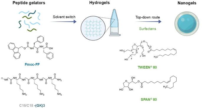
Applying a top-down strategy, Fmoc-FF/C16-(GK)3 and Fmoc-FF/C18-(GK)3 NGs externally stabilized by polysorbate 80 (TWEEN 80, Polyethylene glycol sorbitan monooleate) and sorbitan monooleate 80 (SPAN 80, Sorbitan monooleate) have been here formulated (see Fig. 1) and fully characterized. In particular, the NGs size, surface charge and shelf stability were assessed by Dynamic Light Scattering (DLS). Circular Dichroism (CD), and Fourier Transform Infrared (FT-IR) spectroscopies were carried out to evaluate similarities or differences in secondary structure of NGs and the corresponding HGs17. Moreover, NGs organization was further investigated using Small-Angle X-ray Scattering (SAXS) technique. Different formulative routes were applied to decorate Fmoc-FF/C16-(GK)3 and Fmoc-FF/C18-(GK)3 NGs with the negatively charged AlexaFluor 430 (succinimidyl ester), used as model molecule. AlexaFluor 430 was either encapsulated within the NG core or adsorbed onto surfactant shell. Comparison of the encapsulation efficiency percentage (EE%) and the encapsulation ratio percentage (ER%), obtained with the different procedures and varying the amount of AlexaFluor 430, allowed the identification of a general preparation protocol, supporting the proposed rule of NGs as potential platforms for negatively charged APIs delivery.

Chemical formulas, schematic representation of Fmoc-FF and Cn-(GK)3 peptides and multicomponent HGs and NGs. The chemical formula of TWEEN 80 and SPAN 80 surfactants are also reported.
Results and discussion
Formulation and cytotoxicity assessment of empty NGs
Fmoc-FF have gained attention due to its ability to form well-ordered nanostructures (such as micelles and HGs) through non-covalent interactions, driven by hydrogen bonding and electrostatic forces. Fmoc-FF has also been explored in combination with different molecular elements (e.g. other peptide sequences, proteins or polymers) to develop multicomponent systems18,19,20,21. It was observed that the inclusion of both hydrophilic and hydrophobic components into Fmoc-FF HG is fully compatible with the commonly used “solvent-switch” gelation procedure. This approach involves dissolving the dipeptide monomers at a high concentration (100 mg/mL) in an organic solvent (DMSO, ethanol, or 1,1,1,3,3,3- hexafluoro-isopropanol), followed by stock dilution with water. Dilution triggers gel formation resulting in a three-component system (peptide/solvent/water)22,23. Using this method, Fmoc-FF/Cn-(GK)3 1/1 mol/mol gel matrices were formulated and analyzed (n from 8 to 18 carbon atoms). The corresponding NGs were formulated according to the top-down procedure, previously optimized for preparation of pure Fmoc-FF NG14. This protocol is based on the submicronization of the corresponding macroscopic HG by homogenization and tip-sonication. The resulting nanoparticles are composed of an inner core, externally stabilized by a TWEEN 80 and SPAN 80 surfactant coating, added at 53/47 w/w (total number of mol = 3.0 ∙10− 5 mol) to have a hydrophilic-lipophilic balance (HLB) of 1014.
NGs preparations was initially evaluated by Dynamic Light Scattering (DLS) analysis at scattering angle of θ = 173°. Appling the top-down protocol, capric (C10-), lauric (C12-) and myristic C14-(GK)3 containing NGs resulted as unstable and polydisperse preparations (polydispersity index PDI > 0.350), with mean diameter (d) dimensions incompatible with potential injectability (see Figure S1 as exemplificative example). On the contrary, Fmoc-FF/C16-(GK)3 and Fmoc-FF/C18-(GK)3 shows a monomodal DLS profile (Fig. 2A), indicating a homogeneous population of aggregates (PDI = 0.202 and 0.243 for C16 and C18-(GK)3 containing systems, respectively). These results suggest that the length of the CAPs alkyl chain can affect the NG formation. Consistent with this observation, previous studies showed that macroscopical matrices of C10-, C12-, and C14- peptides led to the formation of more rigid HGs, meanwhile alkyl chain elongation causes a notable reduction in the storage modulus (G′ = 794 Pa for C16 and 73 Pa for C18). This difference may have a rule in micronization step, favoring NGs formation for palmitic (C16) and stearic (C18) mixed matrices.
Apparent translational diffusion coefficients are reported in Table 1. As well-known at infinite dilution, the mean diameter (d), may be evaluated by using the Stokes–Einstein–Sutherland (SES)24. The d of Fmoc-FF/C16-(GK)3 and Fmoc-FF/C18-(GK)3 is 139 and 102 nm, respectively. These values are smaller than the analogue one of pure Fmoc-FF (d ~ 187 nm), advising that the intercalation of the amphiphilic peptides into the Fmoc-FF causes a contraction of the nanoparticles. No significant variation in formulation size can be detected up to one month (Fig. 2B and C), thus indicating good stability features. This evidence can be correlated to the positive values found for the zeta potential (ζ) of both the NGs systems (+ 38 and + 51 mV for C16- and C18-derivatives, respectively), preventing flocculation/sedimentation for the lyophilic colloids NG.

DLS characterization. (A) Intensity profiles of C16-(GK)3 (blue line) and C18-(GK)3 NGs (green line). Correlation coefficient over time for (B) C16-(GK)3 and (C) C18-(GK)3 containing NGs.
To assess the potential capability of these NGs to be used in vivo as delivery systems, their cytotoxicity profiles were evaluated in vitro on HEK-293 cells. Cells were incubated with different concentrations of NGs for 72 h at 37 °C and cell viability evaluated by MTT assay. As clearly shown in Figure S2, no significant toxicity was observed for NGs at all the tested concentrations17.
Structural characterization of NGs by CD, FT-IR and SAXS
To thoroughly inspect the formulation features, the NGs secondary structure was evaluated using CD spectroscopy. Being optically active, peptides and their aggregates exhibit differential absorption of circularly polarized light. Consequently, CD spectra of peptides and proteins are predominantly associated with the excitation transitions of amide groups, which vary as a function of secondary structure. From the inspection of Fig. 3A and B, it can be observed that both the NG spectra are characterized by two leading signals. The first positive band, located in the 220–230 nm region (218 and 222 nm for C16-(GK)3 and 220 nm for C18-(GK)3 HG and NG, respectively), is generally attributed to β-sheet supramolecular organization of peptides25,26. The dichroic inversion of this signal, along with its increased intensity in the NG formulations compared to the HGs, may be attributed to an overall difference in the chiral environment of the NGs. However, it is worth noting that this three-dimensional environment does not alter the fundamental β-sheet organization of the NGs. The second broad peak, corresponding to the distinctive signature of the Fmoc moiety, appears slightly red-shifted in the NG formulations (269 and 268 nm for C16-(GK)3 and C18-(GK)3, respectively) compared to the corresponding HG ones (265 and 264 nm)17. The bathochromic effect may result from a different dielectric constant within the NG core, surrounded by surfactants with respect to the macroscopic HG. It is worthwhile noting that the two samples differ in their physical state, and thus, scattering phenomena could also contribute to the revealed spectral shifts27.

Secondary structure characterization. CD spectra of HGs and NGs of (A) C16-(GK)3 and (B) C18-(GK)3. (C) FT-IR deconvolution for C16-(GK)3 (blue line) and C18-(GK)3 (green line) NGs containing formulations.
The FT-IR analysis carried out on samples supported the assignment of the CD secondary structure. The FT-IR spectra of NGs were deconvoluted in the Amide I absorbance region (1700 –1600 cm− 1, Fig. 3C), which, controlled by C-O and C-N stretching vibrations, is the most sensitive spectral region for the analysis of peptides and protein’s secondary structure28. The presence of a prevalent C = O stretching band at ∼1640 cm− 1 reinforces the β-sheet organization, as do the weighted percentages of each secondary structure functions (Table S1). As expected, no significant differences can be detected between C16- and C18-(GK)3 containing formulations.
Peptide-based NGs were further characterized by SAXS measurements. These provide information on the form factor, i.e. on the shape and dimensions of the fibrils that underpin the NG structures. The SAXS data for the Fmoc-FF NG, or the mixed NGs containing alternatively C16-(GK)3 or C18-(GK)3 show similar behavior, with an intensity scaling at low wavenumber q, I ~ q− 2, which is characteristic of layered structures29. The data can be well fitted using a model for nanotape structures based on bilayers (solid lines shown in Fig. 4, fit parameters listed in SI Table S2). The bilayer electron density profile is represented by three Gaussian functions, two representing the ‘headgroups’ and the central one representing the inner hydrophobic layer30. Here, the inner part of the bilayer will comprise the Fmoc units as well as potentially the phenylalanine residues, while the ‘headgroups’ are the charged C-termini and the lysine residues in the case of the NGs containing the cationic lipopeptides. The effective half thickness of the bilayer is reduced from 77.7 Å for the Fmoc-FF NG to 42.0 Å or 31.3 Å for the mixed NGs with C16-(GK)3 or C18-(GK)3 respectively. The data suggest that the addition of lipopeptides leads to better defined and thinner bilayers, whereas Fmoc-FF nanotapes seem to comprise multilayers since the thickness from the SAXS data fitting is much larger than two molecular lengths. The data for Fmoc-FF show a less well- defined high q form factor maximum which was modelled by incorporating a contribution to the fitted SAXS data of a monomer form factor represented as a generalized Gaussian coil with radius of gyration 10 Å, this was not required to fit the data for the two NGs containing lipopeptides. The SAXS data thus indicate that all Fmoc-FF NGs contain nanotape fibrils, and that these are better defined in the NGs containing the lipopeptides, i.e. the molecules are fully aggregated, and the layer thickness corresponds to interdigitated bilayers, considering the molecular length of the lipopeptides.

SAXS data for NGs as indicated. Open symbols: measured data, solid lines: form factor fits described in the text. For ease of visualization, the data for C16-(GK)3 containing NG are shifted by division by a factor of 5 and that for C18-(GK)3 containing NG by division by a factor of 25, and for all data sets only every 5th data point is plotted.
Formulation and characterization of alexafluor 430 filled NGs
The NGs formulation were evaluated about their potential use as drug delivery platforms. AlexaFluor 430, was selected as model dye for NGs analysis, due to its water-soluble anionic nature and its spectroscopic features (λabs = 430 nm). The versatility formulative procedure, as the specific NGs structure, allows a different dye localization (Fig. 5), alternatively core-included (encapsulation route) or added within the shell via electrostatic interactions (adsorption strategy).

Schematic representation for encapsulation or adsorption strategies of AlexaFluor 430 in NGs formulations.
Increasing amounts of AlexaFluor 430 (0.05 ÷ 3.0 mmol/L concentration range) were alternatively encapsulated into the core (formulations indicated as NGA ÷ NGE) or adsorbed onto the NGs shell (formulations indicated as NGF ÷ NGK). The procedure of encapsulation was easily accomplished by preparing the corresponding AlexaFluor 430 filled HG (0.05 ÷ 3.0 mmol/L concentration range), which consequently underwent the top-down protocol. No syneresis effects were evidenced for both C16 and C18 containing dye matrices, thus suggesting an efficient localization into the fibrillary gel network for all the tested concentrations. The self-supporting properties for all the samples were verified via the inverted test tube method (data not shown). As is well known, the inclusion of additional chemical entities in HGs matrices can deeply alter the gel mechanical response as a consequence of interactions, solubility, molecular weight in relation to the supramolecular gel mesh size31. Previous rheological studies on empty 0.5 wt % HGs of Fmoc-FF/Cn-(GK)3 estimated a G’ of 794 and 73 Pa for Fmoc-FF/C16-(GK)3 and Fmoc-FF/C18-(GK)3 HGs matrices, respectively. The inclusion of the AlexaFluor 430 in the HGs causes a significant decrease in G’ value < 10 Pa for all the tested concentrations without a specific trend, as deduced by dynamic oscillation strain testes (Figure S3, Table 1). These evicedences indicate an impact on the rheological features of the matrices, leading to a viscous-like behaviour. More specifically, it seems that the inclusion of AlexaFluor 430 induces an increase in the mash size via repulsive interaction between supramolecular fibers, with a consequent decrease of G’ according to the theory of rubber elasticity32. All these matrices were efficiently transformed into the corresponding NGs preparations.
An AlexaFluor 430 absorption route was also performed, assuming a favourable dye-shell electrostatic interaction pathway. To achieve absorption (0.05 ÷ 3.00 mmol/L concentration range), preformed empty NG formulations were incubated with lyophilized dye powders overnight under stirring at room temperature. Purification of free dye was performed via SEC (size-exclusion chromatography), with consequent analytical quantification of unretained AlexaFluor 430 via UV-vis spectroscopy.
Spectroscopic features of adsorbed and loaded dye NGs were studied in comparison with free dye and nude formulation (Fig. 6, C16-containing system). As expected, AlexaFluor 430 shows a max λabs at 430 nm. However, a blue shift phenomenon is detected for both NGs preparations (λabs = 423 nm and λabs = 413 nm for adsorbed and encapsulated Alexa preparation, respectively). The hypsochromic shift can be a consequence of hydrogen bonding and solvent exposure33.

UV-vis (A) and fluorescence (B) spectra of AlexaFluor 430 and NGs formulations.
On the other hand, the blue shift in the NGs preparation can be attributed to a more hydrophilic environment for encapsulated dye in the NG core compared to the shell surfactant compartment. The same effect is verified in fluorescence spectral emission. The blue shift of the emission peak can be attributed to solvent polarity and relaxations decreasing for chromophores excited states in polar solvents. These phenomena stabilize the ground state, increasing the transition energy gap, thus causing the emission peak to shift to shorter wavelengths34.
The structural parameters of all the encapsulated/absorbed formulations, including size and ζ- potential, were assessed by DLS analyses (Table 2). In addition, intensity profiles and correlation functions of C16- and C18- containing formulations are grouped in Figure S4–S7.
All the formulations, except C16-NGK and C18-NGK (those with a higher amount of adsorbed AlexaFluor 430) were found stable and further characterized. The intensity profiles of NGs indicate the prevalence of a monodisperse population for all the experimental conditions, with a mean diameter ranging between 123 and 163 nm. A lower PDI is associated with samples prepared with the adsorption method with respect to encapsulation route ones. As observed for nude formulations, no substantial size variations were detected up to one month, thus corroborating the good stability for NG systems. This evidence can be again correlated to the positive values found for the ζ-potential (ζ), for both encapsulated and adsorbed systems, thus preventing flocculation/sedimentation for the AlexaFluor 430 containing NGs.
The amount of loaded (encapsulated/adsorbed) dye for each formulation was again analytically estimated by UV-vis spectroscopy, measuring the absorbance of free AlexaFluor 430, after its separation from NGs by SEC.
The loaded dye amount for each formulation is reported as encapsulation efficiency (EE%) and encapsulation ratio (ER%), and is collected in Table 3. From the comparison of the ER% and EE% values of both C16 and C18 NG formulations, it can be observed a dependence on the loading methodology (encapsulation versus adsorbtion). Specifically, in the set of encapsulating formulations (NGA-NGE), a linear trend of the ER% and EE% as a function of the initial AlexaFluor 430 amount was found (see Fig. 7A). In contrast, no significant variation can be detected for NGF-NGK samples.
Dye release from NGs formulations
The in vitro AlexaFluor 430 release was evaluated using a dynamic dialysis approach in water up to 120 h. Two different NGs systems (formulations C and G, Table 2) for palmitic and stearic containing components were selected, to evidence the impact of both Cn-peptide component and dye decoration strategy. Reported as cumulative percentage, release curves are shown in Fig. 7B. The dye released was evaluated via fluorescence spectroscopy, using a calibration curve in the same experimental conditions. As presumable, the dye release kinetics is primarily affected by Alexa Fluor 430 decoration approach, with final percentages of ~ 97 and ~ 26% released after 120 h for adsorbed and encapsulated formulations, respectively. An AlexaFluor 430 sustained and constant release profile was observed for the adsorbed formulation, with complete dye release by 120 h. The extremely slow-release profile for encapsulating NGs can be attributed to a delayed water exchange between internal NG core and bulk solution, thus limiting the dye diffusion. No substantial differences are detectable for C16- and C18- containing samples, reinforcing the structural homology between NGs.

(A) Encapsulation efficiency of NGs as a function of the amount of AlexaFluor 430 expressed in µg. (B) Cumulative percentage dye release for C16-(GK)3 (blue) and C18-(GK)3 containing NGs. Encapsulating and adsorbing formulations are reported as filled and empty symbols, respectively.
Materials and methods
Materials
Lyophilized Fmoc-FF powder was purchased from Bachem (Bubendorf, Switzerland). TWEEN 80, SPAN 80, n-hexane and oil mineral were purchased from Sigma-Aldrich, Fluka (Bucks, Switzerland) or LabScan (Stillorgan, Dublin, Ireland) and were used as received unless otherwise stated. AlexaFluor 430 ([9-[6-(2,5-dioxopyrrolidin-1-yl)oxy-6-oxohexyl]-8,8-dimethyl-2-oxo-4-(trifluoromethyl)pyrano[3,2-]quinolin-6-yl]methanesulfonate) as purchased from Molecular Probes (Eugene, Oregon, USA). Absorbance measurements on the AlexaFluor 430 solutions were carried out on a nanodrop 2000c spectrophotometer (Thermo Fisher Scientific Inc., Wilmington, DE, USA) equipped with a 1.0 cm quartz cuvette (Hellma) using as molar absorptivity (ε) the value of 17,740 mol− 1∙L∙cm− 1 at 430 nm. A MICCRA D-9 homogenizer (MICCRA GmbH, Germany) and a tip sonicator Branson SFX150, (Branson, Germany) were used for homogenization and tip sonication formulative steps.
Peptide synthesis and NGs formulation procedures
Peptide synthesis, purification and their chemical characterization was already reported17. C16-(GK)3/Fmoc-FF and C18-(GK)3/Fmoc-FF (1/1 mol/mol) based NGs were formulated according to the top-down procedure, using their respective HG (400 µL, 0.5 wt%) prepared via solvent switch method (DMSO/H2O) as previously described18. 1.6 mL of an aqueous, filtered solution of TWEEN 80/SPAN 80 with a HLB of 10 (53/47, w/w ratio, total content of 3.0 ∙10− 5 mol) were added to the preformed HGs. The resulting suspensions were homogenized at 25,000 min− 1 for 5 min and then tip sonicated for 5 min at 9 W.
Formulation of alexafluor 430 containing NGs
Increasing concentrations of the AlexaFluor 430 dye (0.05, 0.25, 0.50, 0.75, 1.0 and 3.0 mmol/L) were alternatively encapsulated (see procedure A) or absorbed on NGs (see procedure B), prepared according to the top-down methodology above described. The amount of the encapsulated/adsorbed dye was quantitatively determined as difference by measuring the absorbance at λ = 430 nm via UV-vis spectroscopy (ε430 = 17740 mol− 1∙L∙cm− 1). Quantification was performed after exclusion chromatography purification of formulations on a gel column (Sephadex G50 column).
Procedure A: encapsulation
360 µL of the dye water solution, at different concentrations, was added to 40 µL of peptides stock solution (100 mg/mL) in DMSO. The suspension was vortexed for a few seconds and left at room temperature until the complete gel formation. Then the HG underwent to the top-down methodology above described to obtain NGs.
Procedure B: adsorption
Lyophilized powder of the dye was dehydrated with peptide NGs at room temperature for 24 h under a gentle stirring for 24 h.
DLS measurements
The DLS measurements, in triplicate, were carried out on all the aqueous samples. Hydrodynamic radii (rH), diffusion coefficients (D) and zeta potential (ζ) of all the peptide NG formulations were measured by DLS using a Zetasizer Advance- Malver Light Scattering Technology (Malvern Panlytical, Westborough, MA, USA). Instrumental settings for the measurements were a backscatter detector at 173° in automatic modality, room temperature, and disposable sizing cuvette as cell.
Cell cultures and in vitro NGs toxicity assay
HEK-293 (human embryonic kidney) cells were cultured in were cultured in Dulbecco’s modified Eagle Medium (DMEM) (Gibco, Thermo Fisher Scientific Waltham, Massachusetts, US), supplemented with 10% Fetal Bovine Serum (FBS) (Gibco), 2 mM Glutamine, 100 U mL⁻¹ Penicillin, and 100 µg mL⁻¹ Streptomycin (Gibco) at 37 °C in a 5% CO2. Cell confluence was maintained at 60–80%, and subculturing was performed at a 1:3 ratio twice a week using 25 cm2 flasks (Nunc, Thermo Fisher Scientific, US). HEK-293 (human embryonic kidney) cells were seeded in 96-well plates at a density of 5.0 × 10⁵ cells per well35. Formulations of C16 and C18 NGs were dissolved in cultured medium. Cells were incubated in the presence of different dilutions of NGs, ranging from 1:160 to 1:320000, for 72 h.
Cell viability was measured by the 3-(4,5-Dimethylthiazol-2-yl)-2,5-diphenyltetrazolium bromide (MTT) assay according to the manufacturer’s instructions. Briefly, 20 mL of 5 mg/mL MTT solution in PBS was added to each well and then incubated for 4 h at 37 °C, 5% CO2. Cell viability dependent absorbance of the dark blue formazan crystals was measured in a spectrophotometer (λmax = 570 nm). All the analyses were performed in triplicate. Cell survival was expressed as percentage of viable cells in the presence of HG or NGs, compared to control cells grown in their absence.
CD spectroscopy
For CD spectroscopy, NGs were placed in 0.1 mm quartz cells and Far-UV CD spectra were collected through a Chirascan spectropolarimeter equipped with a thermal controller. Spectra were recorded from 290 to 190 nm. 0.5 nm step, 1.0 nm bandwidth, 1 s collection time per step were used as experimental settings for the measures. Each spectrum was obtained by averaging three scans and corrected for the blank.
FT-IR spectroscopy
A JascoFT/IR 4100 spectrometer was used for acquiring, in an attenuated total reflection (ATR) mode, spectra of all the peptide-based xerogels, using a Ge single-crystal at a resolution of 4 cm− 1. A total of 120 scans of each sample were recorded with a rate of 2 mm∙s− 1 against a KBr background. After collection in transmission mode, amide I deconvolutions (in the 1600–1700 cm− 1 region) were automatically returned as emission by Jasco SSE instrument integrated software using a method of principal component regression (PCR).
SAXS experiments
SAXS experiments were performed on beamline B2136 at Diamond (Didcot, UK). The sample solutions were loaded into the 96-well plate of an EMBL BioSAXS robot and then injected via an automated sample exchanger into a quartz capillary (1.8 mm internal diameter) in the X-ray beam. The quartz capillary was enclosed in a vacuum chamber, to avoid parasitic scattering. After the sample was injected into the capillary and reached the X-ray beam, the flow was stopped during the SAXS data acquisition. Beamline B21 operates with a fixed camera length (3.9 m) and fixed energy (12.4 keV). The images were captured using a PILATUS 2 M detector. Data processing was performed using dedicated beamline software ScÅtter.
Rheological characterization
Rheological measurements were performed at 25 °C by using a rotationally controlled stress rheometer Modular Advanced Rheometer System (Haake Mars) from ThermoScientific equipped with a 15.0 mm flat-plate geometry (P35-Ti). A freshly prepared HG sample (1.0 mL) with different concentrations of dye were used for measurements using a gap of 1.00 mm. Strain (0.1–100%) sweeps were conducted to identify the regime of linear viscoelasticity. Final analyses are reported as G′ and G″ (shear loss or viscous modulus) in Pascal [Pa].
AlexaFluor 430 release
Release of AlexaFluor 430 from four different NG formulations was studied using 1.0 mL of each purified formulation using dialysis bags (Float-A-Lyzer 3.5-5 kDa cut-off, Spectra/Por, USA) in 10 mL of pure water (under stirring at 37° C). At each selected time-point (30 min, 1, 2, 4, 8 24, 48, 72, 96 and 120 h), 2.0 mL of solution were collected and replaced with an equal amount of water. GloMax Multimode Microplate Reader (Promega Corporation, Milan, Italy) was used to quantify the released dye, using fluorescence filters (λex = 428 nm; λem = 500–550 nm) and an AlexaFluor 430 calibration curve. The amount of the released dye was reported as released percentage with respect to the total quantity encapsulated or adsorbed.
Conclusions
Peptide-based materials (including HGs and NGs) are emerging as alternative chemically different APIs delivery platforms, including small molecules and diagnostic agents. De-novo designed peptide elements can be efficiently included in formulation, modulating the cargo features and affecting its chemical responsiveness, topological properties, morphology, or electrostatic nature. Considering this prospective, multicomponent Fmoc-FF NGs were developed and studied. Fmoc-FF, including cationic amphiphilic hexapeptide sequence as functional charge modifiers, underwent a top-down strategy, producing C16- and C18-(GK)3 containing NGs. These systems were efficiently stabilized by TWEEN and SPAN surfactant shell, with no colloidal instability up to one month and an internal supramolecular β-sheet arrangement. The formation of tri-dimensional supramolecular networks via peptide self-assembling avoids the use of crosslinking agents, usually employed in polymeric nanogels formulation37. AlexaFluor 430, selected as model anionic fluorescent dye, was alternatively encapsulated in (core) or adsorbed on (shell) NGs compartments, thus evidencing the NGs versatility as nanoplatform. The EE% and ER% values were found to be comparable (~ 0.1% < EE% < ~ 1.8% and ~ 17% < ER% < ~ 89% for amount of dye ranged between 67 and 267 µg) with conventional liposomal formulations (e.g. Doxil and Miocet)38 and higher than some polymeric NGs based on 2-(diisopropylamino)ethyl methacrylate or dextran39,40.
The decoration methodology was shown to influence carrier behaviour, resulting in different release kinetics and regimes. This formulative study supports and reinforce the potential of peptide-based NG systems as tunable, modular, and biocompatible platforms for the targeted delivery of negatively charged APIs, including biotechnology-derived drugs such as siRNA, miRNA, and DNA.
Their physicochemical properties (including high biocompatibility, reproducibility, injectability, and ease of surface functionalization) are expected to enhance API stability and bioavailability, often compromised by enzymatic degradation and limited cellular uptake. Moreover, peptide NGs can be engineered to respond to biological stimuli (e.g., pH, enzymes, or redox conditions), enabling site-specific or on-demand drug release, particularly beneficial in cancer therapy and regenerative medicine. However, the current scarcity of well-characterized peptide-based NG examples in the literature41,42,43 limits comprehensive comparisons in terms of drug loading efficiency, performance, and in vitro behaviour. This highlights the need for further systematic studies to expand the understanding of peptide nanogels’ capabilities, optimize their design, and explore their clinical translation.
Data availability
The datasets generated during and/or analysed during the current study are available from the Corresponding Author on reasonable request.
References
-
Tanbour, R., Martins, A. M., Pitt, W. G. & Husseini, G. A. Drug delivery systems based on polymeric micelles and ultrasound: A review. Curr. Pharm. Des. 22, 2796–2807. https://doi.org/10.2174/1381612822666160217125215 (2016).
-
Madegard, L. et al. Bioorthogonal drug release from nanometric micelles in living cells. Chem. Eur. J. 29, 202301359. https://doi.org/10.1002/chem.202301359 (2023).
-
Eisermann, J. et al. The effect of reactive oxygen species on respiratory complex I activity in liposomes. Chem. Eur. J. 30, 202402035. https://doi.org/10.1002/chem.202402035 (2024).
-
Ringhieri, P. et al. The influence of liposomal formulation on the incorporation and retention of PNA oligomers. Colloid Surf. B. 145, 462–469. https://doi.org/10.1016/j.colsurfb.2016.05.034 (2016).
-
Beach, M. A. et al. Polymeric nanoparticles for drug delivery. Chem. Rev. 124, 5505–5616. https://doi.org/10.1021/acs.chemrev.3c00705 (2024).
-
Jin, B., Hu, L. & Li, X. Mesogenic ordering–driven self–assembly of liquid crystalline block copolymers in solution. Chem. Eur. J. 30, 202400312. https://doi.org/10.1002/chem.202400312 (2024).
-
Zhang, C., Ahmadreza, H. & Masoud, N. H. T. Nanoparticle-enhanced drug delivery systems: an up-to-date review. J. Mol. Liq. 424, 126999. https://doi.org/10.1016/j.molliq.2025.126999 (2025).
-
Eugster, R. & Luciani, P. Liposomes: bridging the gap from lab to pharmaceuticals. Curr. Op Coll. Interf Sci. 75, 101875. https://doi.org/10.1016/j.cocis.2024.101875 (2025).
-
Castillo, B. et al. A fresh look at the potential of cyclodextrins for improving the delivery of SiRNA encapsulated in liposome nanocarriers. ACS Omega. 7, 3731–3737. https://doi.org/10.1021/acsomega.1c06436 (2022).
-
Vaidya, S., Mohod, A., Eedara, A. C., Andugulapati, A. B. & Pabbaraja, S. Synthesis and characterization of a new cationic lipid: efficient SiRNA delivery and anticancer activity of survivin-siRNA lipoplexes for the treatment of lung and breast cancers. ChemMedChem 18, 202300097https://doi.org/10.1002/cmdc.202300097 (2023).
-
Gupta, A., Andresen, J. L., Manan, R. S. & Langer, R. Nucleic acid delivery for therapeutic applications. Adv. Drug Deliv Rev. 178, 113834. https://doi.org/10.1016/j.addr.2021.113834 (2021).
-
Bartkowski, M., Zhou, Y., Mustafa, M. N. A., Eustace, A. J. & Giordani, S. CARBON DOTS: bioimaging and anticancer drug delivery. Chem. Eur. J. 30, 202303982. https://doi.org/10.1002/chem.202303982 (2024).
-
Vashist, A. et al. Recent advances in nanogels for drug delivery and biomedical applications. Biomater. Sci. 12, 6006–6018. https://doi.org/10.1039/D4BM00224E (2024).
-
Rosa, E., Diaferia, C., Gallo, E., Morelli, G. & Accardo, A. Stable formulations of peptide-based nanogels. Molecules 25, 3455. https://doi.org/10.3390/molecules25153455 (2020).
-
Rosa, E. et al. Hybrid PNA-peptide hydrogels as injectable CEST-MRI agents. J. Mater. Chem. B. 12, 6371–6383. https://doi.org/10.1039/D4TB00358F (2024).
-
Ricci, M. et al. Chitosan-based nanogels containing Ln3+ chelates (Ln = Gd, Dy) as T1 and T2 MRI probes. Eur. J. Inorg. Chem. 27, 202300675. https://doi.org/10.1002/ejic.202300675 (2024).
-
Rosa, M. et al. Inclusion of cationic amphiphilic peptides in Fmoc-FF generates multicomponent functional hydrogels. ACS Appl. Bio Mater. 8, 488–502. https://doi.org/10.1021/acsabm.4c01409 (2025).
-
Vitale, M. et al. Hydroxyapatite-decorated Fmoc-hydrogel as a bone-mimicking substrate for osteoclast differentiation and culture. Acta Biomater. 138, 144–154. https://doi.org/10.1016/j.actbio.2021.11.011 (2022).
-
Hanae, A. et al. Self-assembly and hydrogel formation ability of Fmoc-dipeptides comprising α-methyl-L-phenylalanine. Polym. J. 52, 923–930. https://doi.org/10.1038/s41428-019-0301-5 (2020).
-
Raeburn, J. et al. Fmoc-diphenylalanine hydrogels: Understanding the variability in reported mechanical properties. Soft Matter. 8, 1168–1174. https://doi.org/10.1039/d2sm90141b (2012).
-
Rosa, E. et al. Multicomponent hydrogel matrices of Fmoc-FF and cationic peptides for application in tissue engineering. Macromol. Biosci. 22, 2200128. https://doi.org/10.1002/mabi.202200128 (2022).
-
Dudukovic, N. A. & Zukoski, C. F. Mechanical properties of self-assembled Fmoc-diphenylalanine molecular gels. Langmuir 30, 4493–4500. https://doi.org/10.1021/la500589f (2014).
-
Diaferia, C., Rosa, E., Morelli, G. & Accardo, A. Fmoc-diphenylalanine hydrogels: optimization of Preparation methods and structural insights. Pharmaceuticals 15, 1048. https://doi.org/10.3390/ph15091048 (2022).
-
Baer, A. et al. The Stokes–Einstein–Sutherland equation at the nanoscale revisited. Small 20, 2304670. https://doi.org/10.1002/smll.202304670 (2024).
-
Iyer, A. et al. Terminal truncated α-synuclein fibrils contain strongly twisted β-sheets. J. Am. Chem. Soc. 139, 15392–15400. https://doi.org/10.1021/jacs.7b07403 (2017).
-
Xiang, S. et al. Self-assembled, hemin-functionalized peptide nanotubes: an innovative strategy for detecting glutathione and glucose molecules with peroxidase-like activity. Nano Convergence. 10 https://doi.org/10.1186/s40580-023-00356-8 (2023).
-
Raman, C. V. The molecular scattering of light in liquids and solids. Nature 108, 402–403. https://doi.org/10.1038/108402b0 (1921).
-
Sadat, A. & Joye, I. A. Peak fitting applied to fourier transform infrared and Raman spectroscopic analysis of proteins. Appl. Sci. 10, 5918. https://doi.org/10.3390/app10175918 (2020).
-
Hamley, I. W. Small-Angle Scattering: theory, instrumentation, Data and Applications (ed John Wiley & Sons) (Wiley, 2021).
-
Pabst, G., Rappolt, M., Amenitsch, H. & Laggner, P. Structural information from multilamellar liposomes at full hydration: full q-range fitting with high quality x-ray data. Phys. Rev. E. 62, 4000–4009. https://doi.org/10.1103/physreve.62.4000 (2000).
-
Rosa, E. e al. Incorporation of PEG Diacrylates (PEGDA) generates hybrid Fmoc-FF hydrogel matrices. Gels 8, 831https://doi.org/10.3390/gels8120831 (2022).
-
Canal, T. & Peppas, N. A. Correlation between mesh size and equilibrium degree of swelling of polymeric networks. J. Biomed. Mater. Res. 23, 1183–1193. https://doi.org/10.1002/jbm.820231007 (1989).
-
Wenbo, L. et al. Effects of different solvents on ultraviolet absorption and fluorescence intensity of 2,4dichlorophenol, 2,4,6-trichlorophenol and pentachlorophenol. J. Phys.: Conf. Ser. 012103, (2021).
-
Xuan, J., Chen, L. & Tian, J. Generalized solvent effect on the fluorescence performance of spiropyran for advanced quick response code dynamic anti-counterfeiting sensing. Int. J. Mol. Sci. 26, 1531. https://doi.org/10.3390/ijms26041531 (2025).
-
Cesaro, E. et al. Exploring a peptide nucleic acid-based antisense approach for CD5 targeting in chronic lymphocytic leukemia. Plos One. 17, e0266090 (2022).
-
Cowieson, N. P. et al. Beamline B21: high-throughput small-angle X-ray scattering at diamond light source. J. Synchrotron Radiat. 27, 1438–1446. https://doi.org/10.1107/S1600577520009960 (2020).
-
Vinogradov, S. V., Kohli, E. & Zeman, A. D. Cross-linked polymeric nanogel formulations of 5’-triphosphates of nucleoside analogues: role of the cellular membrane in drug release. Mol. Pharm. 2, 449–461. https://doi.org/10.1021/mp0500364 (2005).
-
Ibrahim, M., Abuwatfa, W. H., Awad, N., Sabouni, S. R. & Husseini, G. A. Encapsulation, release, and cytotoxicity of doxorubicin loaded in liposomes, micelles, and metal-organic frameworks: a review. Pharmaceutics 14, 254;10.3390/pharmaceutics14020254 (2022).
-
Spencer, D. S. et al. Cytocompatibility, membrane disruption, and SiRNA delivery using environmentally responsive cationic nanogels. J. Contr Rel. 332, 608–619. https://doi.org/10.1016/j.jconrel.2021.03.004 (2021).
-
Naeye, B. et al. Hemocompatibility of SiRNA loaded dextran nanogels. Biomaterials 32, 9120–9127. https://doi.org/10.1016/j.biomaterials.2011.08.015 (2011).
-
Rosa, E. et al. Peptide-based hydrogels and nanogels containing Gd(III) complexes as T1 relaxation agents. Pharmaceuticals 15, 1572. https://doi.org/10.3390/ph15121572 (2022).
-
Gallo, E. et al. Peptide based hydrogels and nanogels for delivery of doxorubicin. Inter J. Nanomed. 16, 1617–1630. https://doi.org/10.2147/IJN.S296272 (2021).
-
Gallo, E. et al. Fmoc-FF hydrogels and nanogels for improved and selective delivery of dexamethasone in leukemic cells and diagnostic applications. Sci. Rep. 14, 9940. https://doi.org/10.1038/s41598-024-60145-z (2024).
Acknowledgements
C.D. M.R. and E.R. acknowledge the grant CN00000041 “National Center for Gene Therapy and Drugs based on RNA Technology” (concession number 1035 of 17 June 2022-PNRR MUR – M4C2 – Investment 1.4 Call “National Centers”,financed by EU- NextGenerationEU), project codeMUR: CN00000041−CUP UNINA: E63C22000940007. C.D.as the principal investigator acknowledges the project PRIN_2022TSLMHR titled “Biomaterials from peptide self-assembling generated by mimicking protein amyloid-like structures” – Ministero dell’Università e della Ricerca -NextGenerationEU (European Union).
Ethics declarations
Competing interests
The authors declare no competing interests.
Additional information
Publisher’s note
Springer Nature remains neutral with regard to jurisdictional claims in published maps and institutional affiliations.
Supplementary Information
Rights and permissions
Open Access This article is licensed under a Creative Commons Attribution-NonCommercial-NoDerivatives 4.0 International License, which permits any non-commercial use, sharing, distribution and reproduction in any medium or format, as long as you give appropriate credit to the original author(s) and the source, provide a link to the Creative Commons licence, and indicate if you modified the licensed material. You do not have permission under this licence to share adapted material derived from this article or parts of it. The images or other third party material in this article are included in the article’s Creative Commons licence, unless indicated otherwise in a credit line to the material. If material is not included in the article’s Creative Commons licence and your intended use is not permitted by statutory regulation or exceeds the permitted use, you will need to obtain permission directly from the copyright holder. To view a copy of this licence, visit http://creativecommons.org/licenses/by-nc-nd/4.0/.
About this article

Cite this article
Rosa, M., Rosa, E., Castelletto, V. et al. Evaluation of cationic peptide-based nanogels as delivery systems for negatively charged molecules: a formulative study. Sci Rep 15, 36875 (2025). https://doi.org/10.1038/s41598-025-20945-3
-
Received:
-
Accepted:
-
Published:
-
DOI: https://doi.org/10.1038/s41598-025-20945-3