
Introduction
Nanomaterial (NM) synthesis by various approaches has become an important area of nanotechnology due to their superior physicochemical properties, compared to their bulk counterparts1. These NMs have applications in diverse fields such as medicine, electronics, material science, catalysis, and environmental remediation2. Among numerous NMs synthesized, magnetic iron oxide nanoparticles (IONPs) have attracted researchers due to their superparamagnetic behavior, biocompatibility, and chemical stability3,4. These superparamagnetic IONPs have garnered significant attention in diverse fields such as medicine, biology, electronics, and materials science, owing to their inherent multifunctional properties5,6,7. Their small size, superparamagnetism, and low toxicity make IONPs highly useful for biomedical applications such as biomedical imaging, drug delivery, magnetic hyperthermia, and therapeutic applications.
In the realm of electronics, the memristor has emerged as a promising technology with potential applications in neuromorphic computing, memory storage, and logic circuits8,9. Memristors, or memory resistors, exhibit a unique resistance-switching behavior that enables them to mimic synaptic functions for artificial neural networks10. Incorporating IONPs into memristor devices presents exciting opportunities to enhance their performance by leveraging magnetic and electronic properties, with significant implications for neuromorphic computing and memory storage11. However, the integration of IONPs synthesized through green methods into memristor technology remains an underexplored area with immense potential.
Traditional methods for synthesizing magnetic IONPs often involve harsh chemicals and energy-intensive processes, raising concerns about environmental sustainability and human health. In response to these challenges, green synthesis has emerged as a promising alternative, leveraging biological systems and environmentally benign reagents to produce nanoparticles (NPs) with reduced ecological impact12,13,14. By utilizing plant extracts, microorganisms, or other natural resources, green synthesis offers a sustainable route to IONP synthesis with improved biocompatibility and surface functionality. Among all biological methods, green synthesis has become popular due to its availability, simplicity, rapidity, eco-friendliness, and cost-effectiveness12,15. It involves plant extracts as reducing agents to synthesize IONPs in an eco-friendly and non-toxic manner. This method leverages the natural compounds found in plants, such as terpenoids, polysaccharides, phenols, and flavonoids, to facilitate the formation of stable IONPs. This green synthesis approach produces biocompatible and multifunctional IONPs4,13,16. Several studies have reported the green synthesis of IONPs using plants14,17,18,19,20. Averrhoa carambola L., known as star fruit, is a perennial species belonging to the Oxalidaceae family. Valued for its edible fruit, the carambola holds significant commercial importance and is widely cultivated in southern China, Southeast Asia, India, and northern South America21,22,23. Several studies have demonstrated that crude extracts or bioactive compounds from A. carambola show anti-oxidant, anti-hyperglycemic, anti-obesity, anti-hyperlipidemia, anti-cancer, anti-inflammatory, cardioprotective, anti-hypertensive, and neuroprotective activity21,23,24,25. Extracts from leaves and fruit have also been employed as a reducing agent for the biogenic synthesis of silver (Ag)26, zinc (Zn)27, Ag/ZnO28, and copper oxide NPs29.
The present study reports the novel, cost-effective green synthesis of biocompatible, superparamagnetic, and multifunctional IONPs using an aqueous leaf extract of the A. carambola. Structural, magnetic, and morphological investigations of the green-synthesized IONPs were performed using several characterization techniques such as X-ray diffraction (XRD), Fourier transform infrared spectroscopy (FTIR), vibrating sample magnetometer (VSM), transmission electron microscopy (TEM), and a particle size analyzer. The green synthesized IONPs were used to investigate biocompatibility and anticancer activity by a cytotoxicity assay. In addition, the suitability of these IONPs (Fe3O4) was tested for non-volatile memory and neuromorphic computing applications. To the best of our knowledge, this is the first report on the green synthesis of biocompatible and multifunctional Fe3O4 NPs using A. carambola for their successful application in electronic devices.
Experimental section
Materials
Ferric chloride, ethanol, sodium hydroxide, etc., were obtained from Sigma Aldrich (Bangalore, India). Dulbecco’s phosphate-buffered saline (DPBS), trypsin-EDTA solution, 3-(4, 5-dimethylthiazolyl-2)−2, 5-diphenyltetrazolium bromide reagent, dimethyl sulfoxide (DMSO), and Dulbecco’s modified Eagle medium with high glucose (DMEM-HG) supplemented with 10% fetal bovine serum (FBS) were purchased from Hi-Media (Mumbai, India). Sterile double-distilled water (SDDW) was used for the preparation of all the chemicals and reagents.
Collection of plant materials and Preparation of leaf extract
The leaves of the A. carambola plant were collected from Chinchwade, Tahsil Kale (16°72’28. 0 N and 74°05’64.4E), Kolhapur district, Maharashtra, India. The leaves of A. carambola used in this study were obtained from the farm of one of the authors, where no specific permissions or licenses were required. As the plant material originated from a cultivated source and not from a wild population, voucher deposition in a public herbarium was not deemed necessary. This species is widely cultivated, taxonomically well-characterized, and easily recognizable, minimizing the likelihood of misidentification. The collected leaves were transported to the laboratory immediately after collection and used for further processing. For the preparation of the extract, leaves were properly washed with tap water to remove any adherent dirt, followed by rinsing with distilled water, and allowed to sun-dry for 72 h before being ground to prepare a fine powder. Five grams of leaf powder was added to 100 mL of double-distilled water (DDW), which was then boiled for 1 h. After cooling to room temperature, the mixture was filtered through Whatman-1 filter paper. The filtrate was collected and used as an extract for the synthesis of NPs29.
Synthesis of iron oxide nanoparticles
For the synthesis of IONPs, 20 mL of leaf extract was used as a reducing agent, and 0.1 M ferric chloride (40 mL) was added dropwise. The pH of the solution was adjusted to 9 with sodium hydroxide. The reaction was then kept at 80 °C for incubation with constant stirring at 400 rpm for 60 min. The completion of the reaction was visually observed by tracking the change in the color of the solution from initial reddish brown to black within 60 min, and by checking its attraction towards a magnet, indicating the formation of IONPs. After cooling down to room temperature, the solution was centrifuged at 8000 rpm for 10 min. The remaining residue was collected and washed three times with DDW. After washing several times, the residual solid IONPs were dried in an oven at 80 °C for 2 h. The product obtained was further calcinated at 300° C for 2 h. The green-synthesized IONPs were then characterized using various characterization techniques30.
Device fabrication
First, the fluorine-doped tin oxide (FTO) substrates were cleaned in a sonication bath for 20 min using acetone and ethanol in a 1:1 ratio. Then the substrates were cleaned using distilled water multiple times. The Doctor blade method was used for the fabrication of the device. A 0.3 g sample of IONP powder was dispersed in 10 mL DMF for 1 h using a probe sonicator. Then, 10 mL of acetonitrile was added to it and kept for 20 min in a probe sonicator. The slurry was finely ground. The slurry was deposited on the FTO substrate using the doctor blade method. The coated substrates were dried in a vacuum at 80 °C for 2 h to remove the binder. The thickness of the active switching layer (Fe3O4) was approximately 1 μm. The circular Ag top electrodes (~ 500 nm) were deposited using the thermal evaporation technique to make the final Ag/Fe3O4/FTO device.
Characterization
To study the crystalline nature of Fe3O4 NPs, XRD patterns were recorded (Bruker AXS Analytical Instruments Pvt. Ltd., Germany). Fourier transform infrared (FTIR) spectra were recorded on a Bruker Alpha (Shimadzu, Japan) spectrophotometer in the range of 500 to 4000 cm− 1. The particle size and zeta potential of IONPs were determined using a dynamic light scattering (DLS) system (Malvern Panalytical Zetasizer Ultra). The magnetic properties of the IONPs were investigated using a VSM (Lakeshore VSM 7410, Lakeshore Cryotronics, Inc.). The magnetization curve (at room temperature) and the values of the magnetic parameters, such as coercivity (Hc), saturation magnetization (Ms), and saturation remanence (Mr) were obtained from the hysteresis loop. The morphology of Fe3O4 NPs was studied using TEM (JEM-2100 200 kV JEOL Tokyo, Japan). The electrical measurements of the device were carried out using a source measurement unit (Keithley SMU 2602B) and a probe station.
Study of biocompatibility and cytotoxicity
To study the biocompatibility of IONPs towards normal cells and anticancer activity against cancer cells, HEK293 and MDA-MB-231 cell lines were used, respectively. To accomplish this, cells were cultured in T-25 flasks, trypsinized, and aspirated into a 5 mL centrifuge tube, and cell pellets were obtained by centrifugation at 300x g. The cell count was adjusted using DMEM-HG medium, such that 200 µL of suspension contained approximately 10,000 cells. In each well of a 96-well microtiter plate, 200 µL of the cell suspension was added, and the plate was incubated at 37 °C and in a 5% CO2 atmosphere for 24 h. After 24 h, the spent medium was aspirated. A stock solution of 1000 µg/mL of IONPs was prepared, from which 200 µL aliquots were added to achieve final concentrations of 31.55, 62, 125, 250, 500, and 1000 µg/mL. The plate was then incubated at 37 °C and in a 5% CO2 atmosphere for 24 h. After 24 h, the plate containing media was aspirated, and 200 µL of medium containing 10% MTT reagent was added to each well to adjust the final concentration to 0.5 mg/mL, and the plate was incubated at 37 °C, in a 5% CO2 atmosphere for 3 h. Next, the culture medium was completely removed without disturbing the crystals formed, and 100 µL of solubilization solution (DMSO) was added. The plate was gently shaken in a gyratory shaker to solubilize the formed formazan. The absorbance was measured using a microplate reader (BK-EL10A, Biobase, China) at a wavelength of 570 nm and at 630 nm. The percentage cell viability was calculated using the formula (1) after subtracting the background and the blank, and the IC50 (the concentration of the IONPs needed to inhibit cell growth by 50%) was calculated from the dose-response curve for the cell line.
$$:%:cell:viability=frac{OD:570-630:left(treatedright)}{OD:570-630:left(controlright)}times:100$$
(1)
Results and discussion
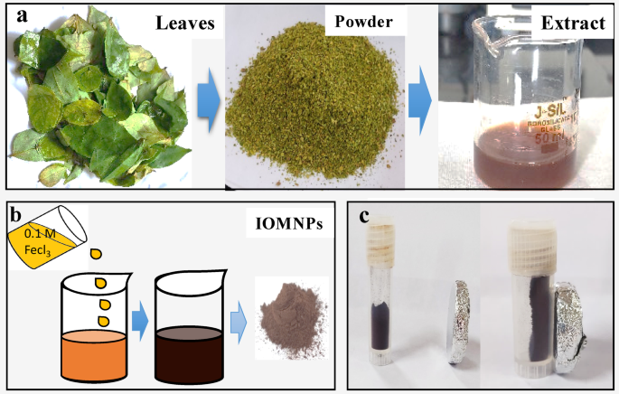
Averrhoa carambola leaf extract was used as a reducing agent for synthesizing IONPs. Figure 1a and b show a schematic representation of the synthesis of IONPs using the green synthesis method. The change in the color of the reaction after 24 h of incubation and the attraction of purified IONPs towards an externally applied magnetic field (Fig. 1c) confirmed the synthesis of IONPs. The synthesized IONPs were subsequently subjected to further characterization. The plausible mechanism involved in this green synthesis could be the involvement of phytochemicals. The Averrhoa carambola leaf extract is rich in secondary metabolites (phytochemicals). It can reduce Fe3+ mainly because it is rich in phenolic compounds (flavonoids, proanthocyanidins, gallic/phenolic acids, and tannins). The secondary metabolites, such as polyphenols/phenolic acids (gallic acid, simple phenolics) are involved in the reducing process. The phenolic OH groups contribute significantly to the reducing power by donating electrons to Fe3+ ions or forming Fe-phenolate complexes. The flavonoids and proanthocyanidins are also effective at reducing Fe3+ because the neighboring hydroxyls stabilize the resulting radical. Studies have shown epicatechin/proanthocyanidins as abundant antioxidants in star fruit. The tannins and other high-molecular polyphenols also contribute both reducing power and iron-binding (chelating) capacity31,32.

Schematic representation of Averrhoa carambola extract-mediated IOMNs (Fe3O4) synthesis process: (a) Preparation of extract from leaves, (b) Synthesis of IOMNPs, (c) Demonstration of the magnetic attraction of IOMNPs toward a magnet.
To confirm the phase, crystallinity, size, and structure of the synthesized NPs, XRD pattern analysis was carried out. Several sharp diffraction peaks were observed in the XRD spectrum, indicating the crystalline nature of the IONPs (Fig. 2a). Diffraction peaks with 2θ values of 30.16°, 35.45°, 43.25°, 57.07°, and 62.72° corresponding to the (hkl) values (200), (311), (400), (511), and (440), respectively, confirm the crystalline nature with a cubic structure of the synthesized IONPs. The XRD data matches well with the JCPDS card 00–003-0863, corresponding to Fe3O4 (Magnetite). The average crystallite size was calculated using the Debye–Scherrer’s equation (:(D:=:klambda:/beta:costheta:)), and it was found to be 15 nm.

(a) XRD pattern, (b) FTIR Spectrum, (c) VSM analysis, (d) Particle Size, (e) Zeta Potential of IONPs.
An FT-IR spectrum was recorded, suggesting the presence of functional groups on the surface of IONPs (Fig. 2b). It shows stretching vibrations at different peak locations. The peak at 3400 cm− 1 corresponds to the stretching of the -OH bond of phenolic compounds, whereas the peak at 2923 cm− 1 is due to symmetric stretching of C-H. Alkenyl C = C and aromatic C-O are related to the peaks at 1630 cm− 1 and 1266 cm− 1, respectively. The strong peaks at 633 cm− 1 and 585 cm− 1 are attributed to the Fe-O bond in Fe3O4. The FT-IR spectrum analysis and its peaks clearly showed that organic bioactive compounds and secondary metabolites were present in the extract, and they were involved as reducing agents14,18,33,34.
The VSM analysis was carried out at room temperature to check the magnetic properties of the synthesized IONPs. The saturation magnetization (Ms), remanence magnetization (Mr), and coercivity (Hc) are the magnetic parameters used to study the magnetic behaviour of the IONPs. Figure 2c shows the measured hysteresis loops of the IONPs when exposed to an external magnetic field, indicating magnetic behavior with an Ms of 0.320 emu, an Mr of 7.4 × 10− 4 emu, and an Hc of 3.68 Oe. The small Mr and Ms values and the closed hysteresis loop indicated the superparamagnetic nature of the green-synthesized IONPs. Superparamagnetic IONPs are suitable for applications in the biomedical field, such as hyperthermia and anticancer therapeutics. A similar high degree of superparamagnetic nature by green-synthesized IONPs was also previously reported in the literature35,36,37.
Size distributions were determined by DLS in an aqueous solution (Fig. 2d). The size of the IONPs was in the range of 70 nm to 200 nm, with a mean hydrodynamic diameter of 178 nm. The stability of the IONP solution was investigated based on the Zeta Potential. It measures the electrostatic potential on the surface of NPs, and it is associated with the electrophoretic mobility of the colloidal suspension. The zeta potential study revealed a negative charge of −8.1 mV, indicating the moderate stability and dispersibility of the IONPs (Fig. 2e)35.
Imaging by TEM showed IONPs with approximately spherical sizes in the range of 6–20 nm in diameter (Fig. 3a-c). Particle size analysis was done using ImageJ software on a representative set of TEM images, and a histogram was generated showing the particle size distribution (Figure S1). The selected area electron diffraction (SAED) pattern of IONPs has concentrated rings of small bright spots, indicating a nanocrystalline nature (Fig. 3d). All the rings correspond to the Fe3O4 and support the XRD analysis.

Transmission electron microscopy (TEM) images of IONPs (a) at 50 nm, (b) at 20 nm, (c) at 10 nm, and (d) SAED pattern of IONPs.
Assessment of biocompatibility and cytotoxicity was studied by using an MTT assay. It involves the conversion of MTT into purple formazan by mitochondrial enzymes of living cells. Dehydrogenase enzymes present in metabolically active cells reduce yellow tetrazolium MTT to generate reducing equivalents such as Nicotinamide adenine dinucleotide (NADH) and Nicotinamide adenine dinucleotide phosphate (NADPH). The resulting intracellular purple formazan can be solubilized and quantified by spectrophotometric means. The assay measures the cell proliferation rate and, conversely, when metabolic events lead to apoptosis or necrosis, the reduction in cell viability38,39.
The effect of the IONPs was studied on the viability of normal cell lines (HEK293) and cancerous cell lines (MDA-MB-231). In the present case, doses at concentrations of 31.25, 62.5, 125, 250, 500, and 1000 µg/mL were used to study the effect. Figure 4a shows the % cell viability of normal and cancerous cells after 24 h. When normal cells were treated with the IONPs, there was no major change in cell viability. More than 60% of cells were viable at a concentration of 500 µg/mL. A further increase in concentration to 1000 µg/mL showed 47% cell viability, indicating IONPs are biocompatible with normal cell lines. In contrast, the % cell viability of the cancer cell line was decreased by 57% at 500 µg/mL, whereas at 1000 µg/mL, ~ 70% of cells were killed, and the IC50 (concentration of the test drug needed to inhibit cell growth by 50%) for MDA-MB-231 was 979.78 µg/mL. The obtained results showed that IONPs are biocompatible up to a 0.5 mg/mL concentration. IONPs in the range of 0.5 to 1 mg/mL can be used for anticancer therapeutics formulations. Figure 4b and d show an image of the untreated normal cell line (HEK293) and a cancerous cell line (MDA-MB-23), respectively. Figure 4c and e show images of the normal cell lines (HEK293) and cancerous cell lines (MDA-MB-231) treated with extract, indicating the cytotoxic effect of IONPs on cancerous cells. In recent years, biocompatible superparamagnetic IONPs have been widely reported for several biomedical applications, including drug delivery, gene therapy, and hyperthermia40,41,42,43. The present study demonstrates that the synthesized IONPs exhibit good biocompatibility, making them suitable for biomedical applications. Additionally, their cytotoxic effects on cancerous cell lines suggest potential anticancer activity. The combination of biocompatibility and anticancer properties highlights the potential of these IONPs for applications in bioelectronics.

MTT assay: (a) Graph of % cell viability against concentration of IOMNPs for the normal and cancerous cell line. (b) Image of untreated normal cell line and (c) normal cell line at 1000 µg/mL concentration of IOMNPs. (d) Image of untreated cancer cell line and (e) cancer cell line at 1000 µg/mL conc. of IOMNPs.
The comprehensive characterization of the green-synthesized IONPs is a crucial prerequisite for understanding the RS behavior of the device. The structural, morphological, and magnetic properties of the IONPs directly dictate their suitability and performance in non-volatile memory and synaptic learning applications. For instance, the XRD and TEM analyses confirm the crystalline nature, cubic structure, and particle size that are essential for the formation of the conductive filaments responsible for the RS mechanism. Therefore, the non-volatile memory performance, including endurance and retention, is a direct reflection of these fundamental physical and chemical properties. The stability and reliability of the RS behavior are a direct consequence of the uniform, well-defined IONPs used in the active switching layer. Moreover, biocompatibility and anticancer tests suggested that the IONPs are a potential candidate for bioelectronics applications. In this context, the studied physical, chemical, and biological properties play a crucial role in the electrical performance of the Ag/Fe3O4/FTO memristor device.
Figure 5a shows the structure of the Ag/Fe3O4/FTO memristor device, where Fe3O4 works as an active switching layer sandwiched between conductive Ag and FTO electrodes. During the measurement process, the voltage was applied to the top electrode Ag, and the bottom FTO layer was grounded. The fabricated memristor shows I-V switching within ± 3 V with bipolar resistive switching behavior (Fig. 5b). The arrows shown in the I-V curves indicate the switching direction. The resistive switching starts from the positive side and the device is sets at approximately + 1.3 V, while in the negative region, it resets at approximately − 2 V, showing the device’s bipolar electrical characteristics. The stability and reliability of the Ag/Fe3O4/FTO memristor were thoroughly evaluated through a comprehensive statistical analysis. To assess cycle-to-cycle variability, 100 consecutive I-V cycles were recorded, from which the set voltage (VSET) and reset voltage (VRESET) values were extracted. The calculated statistical measures, including the mean, standard deviation, and coefficient of variation, are summarized in Table S1. The mean switching voltages were found to be 1.2677 V for VSET and − 1.9726 V for VRESET. The low values for both the standard deviation and coefficient of variation indicate minimal dispersion and high consistency in the switching voltages, suggesting the robustness of the device performance during repeated operations. Further analysis using cumulative probability and the Weibull distribution provided deeper insights into the switching variability. The cumulative probability plots (Fig. 5c) and Weibull distribution plots (Fig. 5d) visually demonstrate the tight distribution of both VSET and VRESET, confirming their good uniformity. The Weibull distribution, characterized by its shape (:left(beta:right)) and scale (x63%) parameters, is a powerful tool for reliability engineering. The observed shape factor (:left(beta:right)) values for both switching voltages signify a more reliable and narrower distribution of data, validating the stability of the device. The scale factor (x63%) aligns closely with the mean values, further affirming the consistency of the switching characteristics. The results from the statistical analysis reveal that the Ag/Fe3O4/FTO memristor is a suitable candidate for non-volatile memory applications.

(a) Structure of Ag/Fe3O4/FTO memristor device. (b) Multiple I-V cycles, (c) cumulative probability plot of VSET and VRESET, and (d) Weibull distribution of VSET and VRESET of Ag/Fe3O4/FTO memristor.
Further, the non-volatile memory performance of the Ag/Fe3O4/FTO memristor was evaluated by measuring the endurance and retention properties. Figure 6a shows the cyclic switching, i.e., endurance, in which the low resistance state (LRS) and high resistance state (HRS) were taken by applying a ± 3 V bias to the top electrode of the device, and the current was measured at 0.3 V (read), respectively. The device shows a good endurance of up to 2 × 10⁴ cycles, suggesting the better cyclic stability of the device. The memory retention of the Ag/Fe3O4/FTO memristor is depicted in Fig. 6b, and it shows stable retention of LRS and HRS over 104 s.
Beyond these primary performance metrics, the cyclic variability within the LRS and HRS was studied. Figure 6c and d represent the non-volatile memory variability through cumulative probability and Weibull distribution, respectively. To provide a comprehensive statistical understanding, several key measures, including the mean, standard deviation, coefficient of variation, Weibull scale, and Weibull shape factor, were calculated and summarized in Table S2. The statistical and Weibull reliability analysis of the LRS and HRS states confirms the stable resistive switching behavior of the memristor. The low coefficient of variation for both LRS (0.16656) and HRS (0.11949) suggests high consistency and low variability in resistance states across switching cycles. Furthermore, the Weibull shape factor (β) for HRS (10.0461) is significantly higher than that for LRS (5.7097), indicating that the HRS current distribution is narrower and more reliable, with fewer fluctuations. Higher β values are typically associated with better reliability in resistive switching memory devices44. The scale factors (X63%), representing the current at which 63% of the data points fall below, are in close agreement with the respective mean values, affirming the uniformity of switching characteristics. Specifically, the scale factor of 7.74 × 10− 4 A for LRS and 7.68 × 10− 3 A for HRS further confirms the clear separation between the two resistance states, which is essential for minimizing read errors in memory applications45. Overall, the memory and reliability metrics validate the repeatability, stability, and robustness of the Ag/Fe3O4/FTO memristor’s switching behavior, making it a strong candidate for non-volatile memory applications.

(a) Endurance and (b) Retention test results of Ag/Fe3O4/FTO memristor. (c) Cumulative probability and (d) Weibull distribution of endurance data of Ag/Fe3O4/FTO memristor.
Additionally, the Ag/Fe3O4/FTO memristor mimics different synaptic learning properties such as potentiation, depression, excitatory postsynaptic current (EPSC), and paired-pulse facilitation (PPF). The structure of a biological neuron is depicted in Fig. 7a. The synapse is a connection between pre-synaptic and post-synaptic neurons, similar to the structure of the memristor. In the case of an artificial synaptic memristor (Ag/Fe3O4/FTO), the top and bottom electrodes work as pre-synaptic and post-synaptic neurons, whereas the switching layer works as a synaptic cleft. Figure 7b shows the potentiation and depression properties of the Ag/Fe3O4/FTO device, highlighting the ability of the device to emulate long-term synaptic plasticity. The potentiation and depression properties were achieved by applying a series of voltage pulses to the device. To mimic the learning and forgetting processes, a series of 200 potentiation and 200 depression pulses (amplitude: +3 V and − 3 V) were applied to the device. These voltage pulses have a pulse width of 50 ms and a pulse period of 100 ms. The current was measured at a constant read voltage of 0.3 V after each pulse. This controlled application of voltage pulses induces changes in the internal resistance of the device, which serves as an analog to synaptic weight modulation. The gradual increase in current (potentiation) and subsequent decrease (depression) with repeated voltage pulses directly mirrors the strengthening and weakening of synaptic connections in the human brain. The observed potentiation and depression behaviors closely resemble the processes of learning and forgetting in biological neurons, respectively. This successful emulation suggests that the fabricated Ag/Fe3O4/FTO memristor holds significant promise for developing advanced neuromorphic computing systems that can efficiently mimic the cognitive functions of the human brain.
Figure 7c illustrates the EPSC results of the Ag/Fe3O4/FTO memristor. We investigated how the EPSC is dependent on pulse/spike duration by keeping the voltage amplitude at + 3 V. Here, the EPSC property was obtained by adjusting the spike duration. As the duration of the spike rises, the amplitude of EPSC also increases, reaching saturation at a longer spike duration, closely resembling the behavior observed in biological synapses. The experimental data were fitted to an exponential function. Herein, the time constant (τ) for EPSC was found to be 1.1106 × 10− 4 s, which is in the biologically relevant timescale, reinforcing the device’s suitability for bioinspired signal processing. Finally, the PPF property was used to mimic the short-term synaptic plasticity behavior of the biological neurons. The PPF Index (%) property of the Ag/Fe3O4/FTO memristor is shown in Fig. 7d. The PPF index shows a prominent initial peak at low inter-spike intervals (Δt), followed by a decay with increasing Δt. The fitted data yield a time constant (τ) of 2.4546 × 10− 5 s. These results assert that the Ag/Fe3O4/FTO memristor retains residual charge that influences subsequent spikes, mimicking the short-term memory behavior seen in biological synapses46,47,48. The overall results confirm that the Ag/Fe3O4/FTO memristor effectively replicates both short-term synaptic plasticity and long-term synaptic plasticity, making it a promising candidate for brain-inspired computing applications.
The charge transport in the device was studied by plotting I-V characteristics on a double logarithmic scale, as shown in Figure S2. Figure S2a presents the conduction characteristics under positive bias. At HRS, initially, at lower voltages, a slope of 1.16 is observed. Slopes around unity at low voltages often signify that the charge transport is dominated by Ohmic conduction. At a higher positive bias, the slope sharply increases to 3.15. A slope significantly greater than 2 suggests the presence of traps within the Fe3O4 layer. This higher slope is indicative of space charge limited current (SCLC) in the “trap-filled limit” (TFL) regime, where all available traps are filled with injected charge carriers, leading to a rapid increase in current with voltage49. In LRS, a slope of 1.14 suggests the Ohmic behavior. Figure S2b illustrates the conduction behavior under negative bias. Similar to the positive bias, distinct conduction regimes are observed. In HRS, under negative bias, two distinct regions with different slopes are observed. At lower voltages, a slope of approximately 1.08 was observed. This slope typically indicates Ohmic conduction, where the current is directly proportional to the applied voltage (I ∝ V), suggesting that charge transport is dominated by Ohmic conduction. At higher voltages, the slope increases to 1.96 and approaches 2 (I ∝ V2). This increased slope value suggests that Child’s law was dominant in this region. In LRS, the slope of the entire region is 1.17, which is indicative that charge transport in the LRS is governed by the Ohmic behavior.

Synaptic learning properties of the Ag/Fe3O4/FTO memristor: (a) Biological synaptic connection depicted with pre- and post-neurons. (b) Potentiation and depression behavior, (c) EPSC behavior, (d) PPF Index (%) properties of the Ag/Fe3O4/FTO memristor. The data fitting equations and fitting parameters are shown in the respective figures.
Table 1 compares various iron-based resistive switching devices for non-volatile memory and neuromorphic computing applications, detailing their device configuration, active layer synthesis technique, switching voltages, non-volatile memory, and synaptic learning properties. Most devices exhibit basic resistive switching behavior with moderate operating voltages; however, they often lack essential features such as endurance, retention, and synaptic functionalities. Significantly, the current work utilizes an environmentally friendly and cost-effective green synthesis technique. The fabricated Ag/Fe3O4/FTO memristor exhibits good non-volatile memory and synaptic learning properties as compared with the reported devices. These results highlight the potential of green-synthesized, biocompatible, and superparamagnetic Fe3O4 nanoparticles for non-volatile memory and neuromorphic computing applications.
The Ag/Fe3O4/FTO memristor exhibits limitations in scalability, despite promising non-volatile memory and synaptic learning performance. The present solution-processable deposition technique is not suitable for device scalability. The Fe3O4 switching layer is also sensitive to temperature fluctuations, which can affect ion mobility and the RS behavior of the device. Additionally, long-term durability under continuous operation and varying environmental conditions remains to be systematically evaluated. Addressing these limitations through material optimization, interface engineering, and thermal stabilization will be important for practical non-volatile memory and neuromorphic applications.
Conclusion
In summary, a novel, eco-friendly, and cost-effective green synthesis method was utilized for the synthesis of IONPs (Fe3O4) using Averrhoa carambola leaf extract. Characterization studies revealed that the synthesized IONPs were crystalline, with a diameter ranging from 10 to 20 nm, and exhibited superparamagnetic behavior. Cytotoxicity assessments showed that IONPs exhibited significant biocompatibility and anticancer activities, suggesting their potential for biomedical and bioelectronics applications. The Ag/Fe3O4/FTO memristor fabricated using IONPs demonstrated good bipolar resistive switching properties and non-volatile memory performance (endurance: 2 × 10⁴ cycles and retention: 1.5 × 10⁴ s). The fabricated memristor showed lower switching variability, as confirmed by various statistical measures. Moreover, the Ag/Fe3O4/FTO memristor effectively mimics both long-term and short-term synaptic plasticity, such as potentiation, depression, EPSC, and PPF. The overall results confirm that this biocompatible and superparamagnetic Fe3O4 material is a potential candidate for non-volatile memory and brain-inspired computing applications.
Data availability
The data that support the findings of this study are available from the corresponding author upon reasonable request.
References
-
Saraswat, P. et al. Applications of bio-based nanomaterials in environment and agriculture: A review on recent progresses. Hybrid. Adv. 4, 100097. https://doi.org/10.1016/j.hybadv.2023.100097 (2023).
-
Desai, M. P. & Pawar, K. D. Immobilization of cellulase on iron tolerant Pseudomonas stutzeri biosynthesized photocatalytically active magnetic nanoparticles for increased thermal stability. Mater. Sci. Engineering: C. 106, 110169. https://doi.org/10.1016/j.msec.2019.110169 (2020).
-
Kharey, P. et al. Green synthesis of biocompatible superparamagnetic iron oxide-gold composite nanoparticles for magnetic resonance imaging, hyperthermia and photothermal therapeutic applications. Mater. Chem. Phys. 293, 126859. https://doi.org/10.1016/j.matchemphys.2022.126859 (2023).
-
Kumar, V., Kaushik, N. K., Tiwari, S. K., Singh, D. & Singh, B. Green synthesis of iron nanoparticles: sources and multifarious biotechnological applications. Int. J. Biol. Macromol. 253, 127017. https://doi.org/10.1016/j.ijbiomac.2023.127017 (2023).
-
Ali, A. et al. Synthesis, characterization, applications, and challenges of iron oxide nanoparticles. Nanotechnology: Sci. Appl. 49–67. https://doi.org/10.2147/NSA.S99986 (2016).
-
Ali, A. et al. Review on recent progress in magnetic nanoparticles: Synthesis, characterization, and diverse applications. Front. Chem. 9, 629054. https://doi.org/10.3389/fchem.2021.629054 (2021).
-
Revathy, R., Sajini, T., Augustine, C. & Joseph, N. Iron-based magnetic nanomaterials: sustainable approaches of synthesis and applications. Results Eng. 18, 101114. https://doi.org/10.1016/j.rineng.2023.101114 (2023).
-
Tian, C., Wei, L., Li, Y. & Jiang, J. Recent progress on two-dimensional neuromorphic devices and artificial neural network. Curr. Appl. Phys. 31, 182–198. https://doi.org/10.1016/j.cap.2021.08.014 (2021).
-
Ravi, V. Review of memristor based neuromorphic computation: Opportunities, challenges and applications. Eng. Res. Express. https://doi.org/10.1088/2631-8695/ad6662 (2024).
-
Yu, J. et al. Bioinspired interactive neuromorphic devices. Mater. Today. 60, 158–182. https://doi.org/10.1016/j.mattod.2022.09.012 (2022).
-
Sun, B. et al. Synaptic devices based neuromorphic computing applications in artificial intelligence. Mater. Today Phys. 18, 100393. https://doi.org/10.1016/j.mtphys.2021.100393 (2021).
-
Desai, M. P., Sangaokar, G. M. & Pawar, K. D. Kokum fruit mediated biogenic gold nanoparticles with photoluminescent, photocatalytic and antioxidant activities. Process Biochem. 70, 188–197. https://doi.org/10.1016/j.procbio.2018.03.027 (2018).
-
Suppiah, D. D., Julkapli, N. M., Sagadevan, S. & Johan, M. R. Eco-friendly green synthesis approach and evaluation of environmental and biological applications of iron oxide nanoparticles. Inorg. Chem. Commun. 152, 110700. https://doi.org/10.1016/j.inoche.2023.110700 (2023).
-
Şahin, A., Altınsoy, Ş. & Kızılbey, K. An approach for cationic dyes removal from wastewater: green synthesis of iron nanoparticles using Prunus avium stems extracts. Kuwait J. Sci. 51 (3), 100226. https://doi.org/10.1016/j.kjs.2024.100226 (2024).
-
Sangaonkar, G. M., Desai, M. P., Dongale, T. D. & Pawar, K. D. Selective interaction between phytomediated anionic silver nanoparticles and mercury leading to amalgam formation enables highly sensitive, colorimetric and memristor-based detection of mercury, Scientific Reports, 10 (1) 2037, https://doi.org/10.1038/s41598-020-58844-4 (2020).
-
Chatterjee, A. et al. Green synthesis of iron oxide nanoparticles and their ameliorative effect on arsenic stress relief in Oryza sativa seedlings. Biocatal. Agric. Biotechnol. 38, 102207. https://doi.org/10.1016/j.bcab.2021.102207 (2021).
-
Alamu, G. A. et al. Green synthesis and characterizations of magnetic iron oxide nanoparticles using Moringa Oleifera extract for improved performance in dye-sensitized solar cell. Chem. Phys. Impact. 8, 100542. https://doi.org/10.1016/j.chphi.2024.100542 (2024).
-
Areeshi, M. Y. Rice straw mediated green synthesis and characterization of iron oxide nanoparticles and its application to improve thermal stability of endoglucanase enzyme. Int. J. Food Microbiol. 374, 109722. https://doi.org/10.1016/j.ijfoodmicro.2022.109722 (2022).
-
Duan, Y. T. et al. Green synthesis of iron oxide nanoparticles using Nicotiana Plumbaginifolia and their biological evaluation. J. Mol. Liq. 396, 123985. https://doi.org/10.1016/j.molliq.2024.123985 (2024).
-
Loganathan, S., Mani, G., Mohamed, A. H. & Kumar, M. Green synthesis of iron oxide nanoparticles using pterolobium hexapetalum (roth) Santapau & Wagh leaves extract and their biological applications. Biocatal. Agric. Biotechnol. 54, 102905. https://doi.org/10.1016/j.bcab.2023.102905 (2023).
-
Kaster, J. B. et al. da Bioactive aerogels based on native and phosphorylated potato (Solanum tuberosum L.) starches incorporated with star fruit extract (Averrhoa carambola L.), International Journal of Biological Macromolecules, 272 132907 https://doi.org/10.1016/j.ijbiomac.2024.132907 (2024).
-
Jia, X., Xie, H., Jiang, Y. & Wei, X. Flavonoids isolated from the fresh sweet fruit of averrhoa carambola, commonly known as star fruit. Phytochemistry 153, 156–162. https://doi.org/10.1016/j.phytochem.2018.06.007 (2018).
-
Islam, S. et al. Identification of secondary metabolites in averrhoa Carambola L. bark by high-resolution mass spectrometry and evaluation for α-glucosidase, tyrosinase, elastase, and antioxidant potential. Food Chem. 332, 127377. https://doi.org/10.1016/j.foodchem.2020.127377 (2020).
-
Luan, F. et al. Traditional uses, phytochemical constituents and Pharmacological properties of averrhoa Carambola L.: a review. Front. Pharmacol. 12, 699899. https://doi.org/10.3389/fphar.2021.699899 (2021).
-
Cazarolli, L. H. et al. Anti-hyperglycemic action of apigenin-6-C-β-fucopyranoside from averrhoa carambola, fitoterapia, 83 (7) 1176–1183 https://doi.org/10.1016/j.fitote.2012.07.003 (2012).
-
Mishra, P. M., Sahoo, S. K., Naik, G. K. & Parida, K. Biomimetic synthesis, characterization and mechanism of formation of stable silver nanoparticles using averrhoa Carambola L. leaf extract. Mater. Lett. 160, 566–571. https://doi.org/10.1016/j.matlet.2015.08.048 (2015).
-
Begum, J. S., Sateesh, M. K., Nagabhushana, H. & Basavaraj, R. B. Averrhoa carambola L. assisted phytonanofabrication of zinc oxide nanoparticles and its anti-microbial activity against drug resistant microbes, Materials Today: Proceedings, 5 (10) 21489–21497 https://doi.org/10.1016/j.matpr.2018.06.559 (2018).
-
Porrawatkul, P. et al. Microwave-assisted synthesis of Ag/ZnO nanoparticles using averrhoa Carambola fruit extract as the reducing agent and their application in cotton fabrics with antibacterial and UV-protection properties. RSC Adv. 12 (24), 15008–15019. https://doi.org/10.1039/D2RA01636B (2022).
-
Saha, T. et al. Biogenic synthesis of copper oxide (CuO) NPs exploiting averrhoa Carambola leaf extract and its potential antibacterial activity. Mater. Chem. Phys. 305, 127979. https://doi.org/10.1016/j.matchemphys.2023.127979 (2023).
-
Carmona, E. R. et al. Sustainable Green Synthesis of Fe3O4 Nanocatalysts for Efficient Oxygen Evolution Reaction 15, 1317 (Nanomaterials, 2025). https://doi.org/10.3390/nano15171317
-
Ni Putu Adriani Astiti1 & Ramona, Y. Analysis of active and applicable compounds in methanol extract of sweet star fruit (Averrhoa Carambola L). Leaves 28, 12–22. https://doi.org/10.4308/hjb.28.1.12 (2021).
-
Fei Luan1 et al. Xirui he and Nan Zeng traditional Uses, phytochemical constituents and Pharmacological properties of averrhoa Carambola L. Rev. 12 https://doi.org/10.3389/fphar.2021.699899 (2021).
-
Kanagasubbulakshmi, S., Kadirvelu & K.J.D.L.S.J. Green synthesis of iron oxide nanoparticles using lagenaria siceraria and evaluation of its antimicrobial activity. Def. Life Sci. J. 2 (4), 422–427. https://doi.org/10.14429/dlsj.2.12277 (2017).
-
Mahdavi, M., Namvar, F., Ahmad, M. B. & Mohamad, R. Green biosynthesis and characterization of magnetic iron oxide (Fe3O4) nanoparticles using seaweed (Sargassum muticum) aqueous extract. Molecules 18 (5), 5954–5964. https://doi.org/10.3390/molecules18055954 (2013).
-
Lakshminarayanan, S., Shereen, M. F., Niraimathi, K. L. & Brindha, P. and ArumugamA., One-pot green synthesis of iron oxide nanoparticles from Bauhinia tomentosa: Characterization and application towards synthesis of 1, 3 diolein, Scientific Reports, 11 (1), 8643 https://doi.org/10.1038/s41598-021-87960-y (2021).
-
Aida, M. S., Alonizan, N., Zarrad, B. & Hjiri, M. Green synthesis of iron oxide nanoparticles using hibiscus plant extract. J. Taibah Univ. Sci. 17 (1), 2221827. https://doi.org/10.1080/16583655.2023.2221827 (2023).
-
Karpagavinayagam, P. & Vedhi, C. Green synthesis of iron oxide nanoparticles using avicennia Marina flower extract. Vacuum 160, 286–292. https://doi.org/10.1016/j.vacuum.2018.11.043 (2019).
-
Plumb, J. A. Cell sensitivity assays: the MTT assay. Cancer cell. Culture: Methods Protocols. 165–169. https://doi.org/10.1385/1-59259-406-9 (2004).
-
Ghasemi, M., Liang, S., Luu, Q. M. & Kempson, I. The MTT assay: a method for error minimization and interpretation in measuring cytotoxicity and estimating cell viability. Cell. Viability Assays: Methods Protocols. 15–33. https://doi.org/10.1007/978-1-0716-3052-5 (2023).
-
Xie, W. et al. Shape-, size-and structure-controlled synthesis and biocompatibility of iron oxide nanoparticles for magnetic theranostics. Theranostics 8 (12), 3284. https://doi.org/10.7150/thno.25220 (2018).
-
Zhang, J., Zhang, T. & Gao, J. Biocompatible iron oxide nanoparticles for targeted cancer gene therapy: a review. Nanomaterials 12 (19), 3323. https://doi.org/10.3390/nano12193323 (2022).
-
Desai, M. P. et al. Iron tolerant Bacillus Badius mediated bimetallic magnetic iron oxide and gold nanoparticles as doxorubicin carrier and for hyperthermia treatment. J. Drug Deliv. Sci. Technol. 81, 104214. https://doi.org/10.1016/j.jddst.2023.104214 (2023).
-
Saeed, M., Ren, W. & Wu, A. Therapeutic applications of iron oxide based nanoparticles in cancer: basic concepts and recent advances. Biomaterials Sci. 6 (4), 708–725. https://doi.org/10.1039/c7bm00999b (2018).
-
Terdalkar, P. et al. Revealing switching statistics and artificial synaptic properties of Bi2S3 memristor. Solid State Electron. 225, 109076. https://doi.org/10.1016/j.sse.2025.109076 (2018).
-
Dongale, T. D. et al. TiO2 based nanostructured memristor for RRAM and neuromorphic applications: A simulation approach. Nano Convergence. 3, 1–7. https://doi.org/10.1186/s40580-016-0076-8 (2018).
-
Rokade, K. A. et al. CogniFiber: Harnessing biocompatible and biodegradable 1D collagen nanofibers for sustainable nonvolatile memory and synaptic learning applications. Adv. Mater. 36 (24), 2312484. https://doi.org/10.1002/adma.202312484 (2024).
-
Bhosale, R. K. et al. Electrochemical synthesis and functional analysis of self-assembled Au-decorated polypyrrole for non-volatile memory and bio-inspired computing. Org. Electron. 127, 107013. https://doi.org/10.1016/j.orgel.2024.107013 (2024).
-
Patil, P. P. et al. Self-assembled lanthanum oxide nanoflakes by electrodeposition technique for resistive switching memory and artificial synaptic devices. Small 19 (46), 2303862. https://doi.org/10.1002/smll.202303862 (2023).
-
Waser, R., Dittmann, R., Staikov, G. & Szot, K. Redox-Based resistive switching Memories-Nanoionic Mechanisms, Prospects, and challenges. Adv. Mater. 21, 25–26. https://doi.org/10.1002/adma.200900375 (2009).
-
Singh, V. P., Singh, C. P., Ranjan, H. & Pandey, S. K. Investigation of analog resistive switching dynamics in microwave-assisted Fe3O4 based memristor for neuromorphic application. Mater. Lett. 344, 134431. https://doi.org/10.1016/j.matlet.2023.134431 (2023).
-
Singh, V. P. et al. Fabrication perspective of Fe3O4-based cross-cell memristive device for synaptic applications. Curr. Appl. Phys. 63, 48–55. https://doi.org/10.1016/j.cap.2024.04.008 (2024).
-
Nguyen, H. H., Ta, H. K. T., Park, S., Phan, T. B. & Pham, N. K. Resistive switching effect and magnetic properties of iron oxide nanoparticles embedded-polyvinyl alcohol film. RSC Adv. 10 (22), 12900–12907. https://doi.org/10.1039/C9RA10101B (2020).
-
Ishibe, T., Uematsu, Y., Naruse, N., Mera, Y. & Nakamura, Y. Impact of metal silicide nanocrystals on the resistance ratio in resistive switching of epitaxial Fe3O4 films on Si substrates. Appl. Phys. Lett. 116 (18), 181601. https://doi.org/10.1063/1.5143960 (2020).
-
Porro, S. et al. A multi-level memristor based on atomic layer deposition of iron oxide. Nanotechnology 29 (49), 495201. https://doi.org/10.1088/1361-6528/aae2ff (2018).
Acknowledgements
This work was supported by the National Research Foundation of Korea (NRF) grant funded by the Korea government (MSIT) (RS-2025-02303505). The authors extend their appreciation to the Deanship of Scientific Research and Graduate Studies at King Khalid University for funding this work through the Large Project number, R.G.P. 2/742/46, and the authors acknowledge the Research Center for Advanced Materials (RCAMS) at King Khalid University, Saudi Arabia, for their valuable technical support. T.D.D. would like to thank the Science and Engineering Research Board (DST-GoI) for providing the research grant through the State University Research Excellence (SERB-SURE) scheme (Grant No. SUR/2022/000765). The authors would like to thank RUSA-Maharashtra for the financial assistance under the “RUSA-Industry Sponsored Centre for VLSI System Design.”
Funding
This work was supported by the National Research Foundation of Korea (NRF) grant funded by the Korean government (MSIT) (RS-2025-02303505). The authors extend their appreciation to the Deanship of Scientific Research and Graduate Studies at King Khalid University for funding this work through the Large Project number, R.G.P. 2/742/46, and the authors acknowledge the Research Center for Advanced Materials (RCAMS) at King Khalid University, Saudi Arabia, for their valuable technical support. T.D.D. would like to thank the Science and Engineering Research Board (DST-GoI) for providing the research grant through the State University Research Excellence (SERB-SURE) scheme (Grant No. SUR/2022/000765). The authors would like to thank RUSA-Maharashtra for the financial assistance under the “RUSA-Industry Sponsored Centre for VLSI System Design.”
Ethics declarations
Competing interests
The authors declare no competing interests.
Additional information
Publisher’s note
Springer Nature remains neutral with regard to jurisdictional claims in published maps and institutional affiliations.
Supplementary Information
Rights and permissions
Open Access This article is licensed under a Creative Commons Attribution-NonCommercial-NoDerivatives 4.0 International License, which permits any non-commercial use, sharing, distribution and reproduction in any medium or format, as long as you give appropriate credit to the original author(s) and the source, provide a link to the Creative Commons licence, and indicate if you modified the licensed material. You do not have permission under this licence to share adapted material derived from this article or parts of it. The images or other third party material in this article are included in the article’s Creative Commons licence, unless indicated otherwise in a credit line to the material. If material is not included in the article’s Creative Commons licence and your intended use is not permitted by statutory regulation or exceeds the permitted use, you will need to obtain permission directly from the copyright holder. To view a copy of this licence, visit http://creativecommons.org/licenses/by-nc-nd/4.0/.
About this article

Cite this article
Tayshete, R.R., Rokade, K.A., Ambole, Y.V. et al. Green-synthesized superparamagnetic and biocompatible Fe3O4 nanoparticles for memristive and synaptic bioelectronics. Sci Rep 15, 43456 (2025). https://doi.org/10.1038/s41598-025-27373-3
-
Received:
-
Accepted:
-
Published:
-
Version of record:
-
DOI: https://doi.org/10.1038/s41598-025-27373-3