- Article
- Open access
- Published:
Scientific Reports volume 15, Article number: 31363 (2025) Cite this article
Subjects
Abstract
We have made artificial extracellular vesicles like nanoparticles that contain biologically active Agomir (miRNA mimic)/Antagomir (miRNA inhibitor) adsorbed on magnetic ZnO particles bound by a lipid bilayer membrane from Caco-2 cells (human colorectal adenocarcinoma), which we examined for use as therapeutic nanoparticles or as delivery vehicles for therapeutic agents to the gut epithelia. Magnetic ZnO nanoparticles were synthesized using Manganese doping in both solid-state reaction (SSR) and alkaline aqueous solution methods. The SSR method exhibited ferromagnetic behavior, whereas the alkaline solution method yielded a nanorod-like morphology. Encapsulation was demonstrated using mercaptosuccinic acid. These biocompatible nanoparticles, owing to their nanorod-like morphology with larger surface area, were used to adsorb miR200c antagomir/agomir molecules as well as assemble lipid membrane fragments from Caco-2 cells. We performed various in vivo efficacy studies of Caco-2 NanoVesicles (CNVs) (Caco-2NanoVesicles) or detrimental CNV (therapeutic Caco-2 NanoVesicles (tCNVs)) loaded with miR200c Agomir that degrade occludin protein-coding mRNA as a proof of concept for clinical use of the Antagomir counterpart. There was no discernible toxicity, mortality, or systemic inflammatory or immunological responses in mice following administration of either CNVs or tCNVs. tCNV administration enhances intestinal permeability in mice. This supports their use as biological Nano-therapeutics to restore the gut barrier.
Introduction
In addressing drug delivery systems targeted to the gut region, several things must be considered, as the delivery agent must withstand the large pH gradient in the gastrointestinal (GI) tract, especially the highly acidic environment of the stomach. The complexity and variability of the microbiome has a repertoire of factors capable of assimilating/neutralizing/degrading a wide range of therapeutic agents. Other significant hurdles before the nanocarrier can release its drug payload are the intestinal mucosal layer and its absorption capacity1. Although encapsulation of nanocarriers must be performed to prevent their premature degradation and absorption in the gastrointestinal (GI) tract, tracing the delivery agent throughout the tract offers major advantages for precise site-specific targeting.
The morphology and size of nanoparticles also play an essential role in effective drug delivery. The nanoparticles having the morphology of the long nanorod type stay for a longer duration of time as compared to the spherical type of nanoparticles2. The rod shape has a higher surface area for adsorbing a larger adsorbed payload as compared to spherical particles3. The major issue associated with the delivery of miRNA is its high affinity towards water, leading to its rapid diffusion. The specifically designed nanocarrier may improve the encapsulation efficiency of the miRNA4. The approach of the nanocarrier-based drug delivery may occur through receptor-mediated endocytosis, which could further reduce the drug dosage and the associated side effects manyfold4.
Targeting specific regions of the gut and achieving personalized precision therapy requires a combination of strategies. These include nanocarriers that respond to multiple physiological stimuli (such as pH and redox conditions), systems that adapt to the gut microbiome by responding to specific bacterial enzymes, and customizable delivery platforms tailored to an individual’s gastrointestinal profile5. For example, pH-responsive drug release may vary according to food intake, disease status, and, more importantly, the gut microbiome, which can influence the drug release profile, including the rate of release as well as the time of release6. On the other hand, non-invasive external stimuli such as temperature, magnetic field, light, ultrasound, can control the release of drugs more precisely and also release at the specific desired location with external control for effective localization of drugs, thereby reducing the off-target drug delivery. For example, core-shell zinc oxide–iron oxide nanoparticles (ZnO@Fe₃O₄) nanocomposites combine the photocatalytic and drug-loading capacity of ZnO with the magnetic responsiveness of Fe₃O₄. When exposed to an external magnetic field, these nanoparticles can accumulate at target sites (magnetic targeting) and trigger drug release via localized heating or mechanical disruption, enabling precise spatiotemporal control7,8.
External stimuli can be more rationally regulated by the release of drugs at the targeted site as compared to the internal stimuli present in the GI tract. Therefore, magnetic nanoparticles could be effectively used as a delivery agent in the gut region because they can be traced to know when to apply the external stimulus to release the drug payload. Nowadays, various modern techniques are applied to monitor and detect magnetic nanoparticles, such as magnetic resonance imaging (MRI)9 and magnetic particle imaging (MPI)10. Recently, AC bioseptometry has been widely used to monitor magnetic nanoparticles specifically targeting the gut region11,12,13,14,15.
There are many interrelated factors like size, shape, concentration16route of administration17, and type of synthesis method18 that are actually taken into consideration to avoid the side effects of ZnO nanoparticles for in vivo studies, as here biocompatibility is not a fixed attribute. However, it has been previously observed that certain modifications of ZnO nanoparticles, such as surface coating or incorporation of ZnO nanoparticles into chitosan-based nanofibrous mats, could improve their biocompatibility as well as antimicrobial properties19 Additionally, the pH of the GI region where ZnO nanoparticles are targeted can further affect biocompatibility, for example, in the acidic environment present in case of tumors, the rate of solubilization of ZnO nanoparticles is faster, resulting in rapid release of ions (Zn²⁺/Mn²⁺) that can also trigger oxidative stress and cytotoxicity. On the other hand, at neutral pH, the solubilization of ZnO nanoparticles is comparatively slow, which potentially reduces their cytotoxicity20. We can interplay these factors to harness the therapeutic potential of ZnO nanoparticles.
ZnO nanoparticles size less than 100 nm are considered biocompatible, widely employed for in vivo studies, and are accepted in commercial cosmetic products21. The ZnO nanowires are also reported to be completely dissolved inside the body22,23. In this context, ZnO-based magnetic nanoparticles could be considered as a potential candidate for drug delivery in the gut region.
In this context, ZnO-based magnetic nanoparticles, due to their pH-responsive dissolution and magnetic targeting capabilities, represent a promising platform for site-specific drug delivery in the GI tract, particularly in regions such as the colon where controlled release is essential. However, long term in vivo behavior would be essential for future prospects especially in terms of toxicity studies with the release of Mn²⁺ on the GI tract.
Furthermore, in case of GI targeted drug delivery, one of the barriers present in the GI tract is the mucous layer of the intestine, which does not allow the diffusion of drugs due to its viscoelastic microenvironment with adhesive nature. Hence, it is essential to modify the surface of nanoparticles for spatially localized delivery of drugs so that they can reach the targeted site. For example, Ensign et al. have shown that making the surface of nanoparticles more hydrophilic with a coating of PEG increases their probability of penetration through the mucus layer, making them more available for epithelial absorption with higher transcellular absorption24. Similarly, Lai et al. have demonstrated that modified nanoparticles with mucus penetrating ability have a 10-fold increased probability of epithelial absorption compared to unmodified counterparts25. These modifications are important in terms of the bioavailability of drugs applied via the oral route of drug administration for in vivo studies with increased solubility and penetration ability across the gut epithelial barrier26.
Mucus-penetrating nanoparticles are postulated increase the cellular uptake at the intestinal lining for better absorption for more effective drug delivery27. The application of AcMF increases the Brownian motion of nanoparticles by magneto-thermal effect, enhancing mucus-penetration and passing through the mucus barrier and shown to penetrate to the colon tumor site28. In a widely used animal model of colitis (DSS-induced colitis), miR-200c was reported to be significantly enhanced in our previous report29. After seven days of oral DSS (3%) administration, intestinal tissue miR-200c increased significantly (> 30 fold), while occludin mRNA levels decreased in tandem29. Through recycling intestinal perfusion permeability assessment of isolated intestinal segments of live mice and intestinal tissue histology, the therapeutic administration (oral-gastric gavage) of high dose antisense antagomir-200c prevented the development of colitis and the increase in intestinal permeability to dextran 10kd that DSS caused in mice29. According to these findings, intestinal permeability can be raised simply by increasing the expression of miR-200c-3p in mice intestinal epithelial cells.
The GI tract presents numerous absorption barriers that limit the effective delivery of drugs to target sites30. The mucus layer presents a barrier to nanoparticle transport across intestinal tissue31,32. To protect miRNAs from degradation in the harsh GI environment, encapsulation within nanocarriers is a promising approach33,34. Various materials are used for encapsulation, the best methods are Liposomes. These lipid-based vesicles can encapsulate miRNAs and protect them from enzymatic degradation35,36. Polymers like polyacrylates can be used to formulate nanoparticles that protect miRNAs and allow for controlled release37. Milk-derived exosomes can deliver tumor-activated doxorubicin prodrugs and could be engineered for miRNA delivery to gut epithelium but it also targets other cell types e.g. hepatocytes38. Previous examples include Laminarin, a polysaccharide, coating of nanogene complex can be used for oral delivery of miRNA-223 to target macrophage polarization in inflammatory bowel disease39. Oral administration of miR-320-3p embedded in DiOleyl-Succinyl-Paromomycin (DOSP) can alter miRNA levels in the gut and brain40. Oral administration of osa-miR168a, a plant miRNA, has shown to attenuate dextran sulfate sodium-induced colitis in mice41. There is no platform for delivering miRNA to gut epithelium or targeting epithelium specifically. Cell membrane-coated nanoparticles (CMNPs) are a promising strategy for targeted drug delivery and cell-specific targeting homotypic cells (cells of the same type), exploiting a phenomenon known as homotypic targeting42. Our approach to use a positively charged ZnO core to assemble miRNA payload as well as lipid from Caco-2 cells which is an intestinal epithelial line is to exploit ZnO nanoparticles surface area and affinity to negatively charged molecules, protecting and holding miRNA (agomir/antagomir) under liposome protection and biosimilar membrane based homotypic targeting to similar gut epithelial cells.
In the present study, magnetic ZnO nanoparticles doped with Manganese (Mn++) ions nanosized in the range of 17 to 50 nm coated with miR-200c-3p agomir with Caco-2 cell membrane coating have been fabricated to apply the targeted miRNA delivery to the gut region.
We exploited known relationship between miR-200c agomir and occluding and used this to probe delivery efficacy of these bionic nanoparticles to the epithelial cells of the small intestine.
Methods
Synthesis of ZnO nanoparticles
Undoped ZnO nanoparticles and Mn-doped ZnO nanoparticles (ZnO: Mn) were synthesized by two different approaches viz. solid-state reaction method (SSR) (18) and precipitation method of the highly alkaline aqueous medium as previously described43,44.
Briefly, in the case of the SSR method, the precursors zinc acetate dihydrate (Loba Chemie, India) and manganese acetate (Loba Chemie, India) were used to synthesize the nanoparticles of ZnO and ZnO: Mn. The dopant substitution fraction range was varied from x = 0.02 to 0.20. The aqueous blend of precursors was dried thoroughly in an oven; subsequently, the nanoparticles of ZnO and ZnO: Mn were synthesized in a thermal furnace at 500 °C in the presence of air44.
The precipitation method involves synthesizing ZnO and ZnO: Mn nanoparticles at room temperature. In the aqueous solution of zinc acetate and manganese acetate, ammonia solution was added to create a highly aqueous environment of pH 10 to fabricate the nanoparticles through precipitation. Mn concentration in ZnO: Mn was varied from x = 0.02 to 0.144. MSA (Merck, India) was used as a capping agent to stabilize the active surface of the synthesized nanoparticles to prevent their agglomeration following the method as described previously43,44.
Magnetic measurements
Magnetic measurements were conducted using Vibrating-sample magnetometry (VSM) (Lakeshore 73504, USA) instrument. The measurements were conducted with the accuracy of 10−6 m emu/g, carried out at room temperature and in the magnetic field of up to 5000 Oe. The results were plotted after subtracting diamagnetic background of the sample holder. Magnetic measurements were performed for the nanoparticles of ZnO, ZnO: Mn (2%), ZnO: Mn (5%), ZnO: Mn (10%) and ZnO: Mn (15%) synthesized by solid state reaction method. ZnO, ZnO: Mn (2%), and ZnO: Mn (10%) were prepared by the precipitation method.
Cytotoxicity of zno: Mn nanoparticles
Human colon carcinoma Caco-2 cells were stored routinely in small aliquots in liquid nitrogen and the experimental cultures were prepared from the frozen stock cells and always kept in a sub-confluent state45. The cells were cultured in DMEM with GlutaMax containing 1.5% glucose and supplemented with 10% FBS, 1% MEM non-essential amino acids and 10 mM HEPES buffer46. The cells were cultured in a saturating humidified atmosphere of 5% CO2 in air at 37 °C. The culture medium was changed every 3–4 days. The cultures were used for testing within 10 passages after the cells were received from ATCC. Cells were grown to 80% confluence and prepared for experimental procedures. They were washed with HBSS, harvested, and a single cell suspension was prepared. The cell suspension was added to 96-well plates and incubated for 24 h at 37 °C. Dosing solutions of 100-nm ZnO: Mn nanoparticles were prepared and added to the cells. The cells were exposed to ZnO: Mn nanoparticles at concentrations of 0.01, 0.1, 1.0, 10.0, and 20.0 µg mL–1. The cells were treated for 24 h at 37 °C in a saturated humidity atmosphere. The ZnO: Mn nanoparticles was tested in three independent culture experiments. The cytotoxicity of the ZnO-Mn nanoparticles was measured fluorometrically using the resazurin reduction assay47. Using the Sigma Resazurin Assay kit, the cells were seeded in 96-well plates, treated with the vehicle control and the desired concentrations of the ZnO: Mn nanoparticles for 24 h at 37 °C, and then rinsed with sterile HBSS before being incubated with resazurin48 for 30 min at 37 °C in a plate reader. Resorufin fluorescence was measured at 545 nm excitation and 590 nm emission to determine the viability as per kit Manuel.
AgomiR loading
miRNA was obtained from Qiagen (Qiagen, Germantown, MD). MiRNA (25 nM) was mixed with an equal volume of Lipofectamine 2000 (Thermo Fisher Scientific, Waltham, MA) prepared in 25 µL Optimized Minimum Essential Medium (Opti-MEM), (Thermo Fisher Scientific, Waltham, MA) and incubated at room temperature for 10 min. Lipofectamine-miRNA complex solution 50 µL were incubated with Magnetic Nanoparticle Vesicles (MNV) (5 × 10¹² particles/mL, 50 µL) for 30 min at room temperature (RT). The solution was diluted with 4 mL PBS and ultra-centrifuged at 100,000 g using a Type 60 Ti swing rotor for 70 min at 4 °C. The pellet was re-suspended in an appropriate volume of PBS for further studies.
Preparation and characterizations of Caco-2 cell membrane fragments and tCNVs/miR200c
Caco-2 cells (passage-18) were purchased from ATCC (Manassas, VA). Caco-2 cells were cultured in T650 large flasks to obtain enough cells. It took cells 14 days to reach a 70% confluency and were harvested by adding an EDTA-Trypsin solution (Gibco, USA), washed two times with PBS and collected by centrifugation at 5000 g for 10 min. 2 × 10⁶ macrophages per flask (4 flasks) were harvested.
Membranes of Caco-2 cells were isolated by repeated freeze thawing and sonication. Harvested cells were suspended in 10 times diluted PBS (0.1× PBS pH 7.4) with protease inhibitor cocktail (thermo USA). Samples were cyclically frozen in liquid nitrogen and then thawed at 37 °C 10 times. The sample was briefly sonicated to fragment the isolated membranes. Lipid fraction were isolated by centrifugation at 10,000 × g for 10 min followed by PBS wash. The fraction was resuspended in cold PBS by brief sonication and stored at 4 °C for maximally 1 day before further use. miR200c agomir coated or blank ZnO NPs were incubated with enterocyte membranes in 24-well plates. The coated particles were again centrifuged at 10,000 × g for 10 min and suspended in PBS till used.
AgomiR-200c delivery to the mice intestinal epithelial cells
All animal experiments were conducted following protocols approved by the Institutional Animal Care and Use Committee of The Pennsylvania State University College of Medicine (IACUC protocol no. PROTO201800216), Hershey, PA, USA. All procedures complied with the Animal Welfare Act and the recommendations of the Office of Laboratory Animal Welfare (OLAW). The full ARRIVE guideline checklist was submitted to the journal to demonstrate compliance with ARRIVE guidelines. Wild-type in-house bred male C57BL/6J mice (Original Stock No. 000664, Jackson Laboratory) were used for MIRACLE miRNAs Antagomir/Agomir as well as negative control (NC) Antagomir/Agomir were obtained from AcceGen Biotech (NJ, USA). All mice were housed in autoclaved polycarbonate cages (maximum 5 per cage) with autoclaved corncob bedding and nesting material for enrichment. Animals were maintained under specific pathogen-free (SPF) conditions in the Penn State College of Medicine animal facility with a 12-hour light/dark cycle, controlled temperature (22 ± 3 °C), and relative humidity (59 ± 5%). Mice had ad libitum access to autoclaved water and were fed a standard chow diet prior to weaning and an AIN-93 M purified diet post-weaning. Total RNA was isolated using the miRNeasy kit (Qiagen, Valencia, CA) and reverse transcription was performed using the QuantiTect reverse transcription kit (Qiagen, Valencia, CA) according to manufacturer’s protocol. The real-time PCRs were performed using PikoReal 96 Real-Time PCR system (Thermo Scientific, Waltham, MA). The Agomir-200c effect on intestinal permeability in an in vivo mouse model system was determined using a recycling intestinal perfusion method29,49. Frozen mouse tissue sections were fixed and processed for Laser capture microdissection as previously described29,49. On average, approximately 1000 cells were obtained per microdissection cap and total RNA was isolated29,49.
Results and discussion
Among all the transition metal ions, Mn shows more solubility in ZnO while preserving its nanocrystalline structure50. Thus, Mn-doped ZnO nanoparticles were synthesized and studied for the possible targeted drug delivery to the gut region. The present study was conducted to reveal the preferred method to synthesize suitable magnetic ZnO nanoparticles for targeted drug delivery. We have selected ZnO nanoparticles due to its anticipated biocompatibility, antimicrobial activity, and pH responsive behavior with the ability of differential dissolution at different pH, these properties make the nanoparticles suitable for drug delivery into the GI tract51,52. In addition, we have incorporated Mn nanoparticles, as their magnetic behavior is essential for controlled drug delivery by external stimuli, as well as, to make detection possible via imaging techniques such as magnetic resonance imaging (MRI) for future perspectives53,54. Additionally, the incorporation of Mn may also provide the ability to reduce reactive oxygen species, which may increase the antioxidant activity of the nanoparticles to reduce the cytotoxicity associated with the use of metal oxide nanoparticles55. Moreover, compared to commercially available nanoparticles such as gold and silver nanoparticles, which are of high cost with low biodegradability, these Mn doped ZnO nanoparticles can be more economical, have relatively high biodegradability and can be easily synthesized by the given methods56.
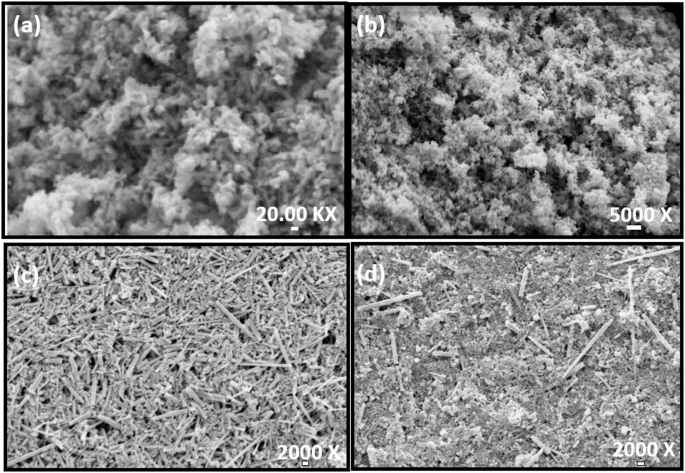
Structural analysis using scanning electron microscopy (SEM analysis)
Scanning Electron microscopy (SEM) study indicated the nano spherical morphology of the ZnO nanoparticles synthesized by the SSR method (Fig. 1.a, b.) with an average size of 50 nm. Typical nanorods morphology could be observed for ZnO nanoparticles synthesized by alkaline aqueous solution method (Fig. 1.c, d) with the average nanocrystal size in the range of 17 to 40 nm. The synthesized ZnO nanoparticles exhibited the typical wurtzite hexagonal structure43,44.

SEM image depicting spherical nanoparticle of ZnO (a) and ZnO: Mn (5%) (b) synthesized through solid state reaction method; while nanorod formation of ZnO nanoparticles could be visualized in both ZnO (c) and ZnO: Mn (2%) synthesized by alkaline aqueous method (d).
The change in the crystal parameters indicated that dopant electronegativity and ionic radii influence the growth of nanoparticles, as previously described by Field43,44. Theoretically, the rod shape of nanoparticles would have more available area for surface-adhesive interactions as compared to the spherical nanoparticles. Because of the high surface area, the nanorods of ZnO nanoparticles become more reactive; they perhaps can stay for longer periods of time and have high circulation time. Thus, they can be appropriately delivered to the deeper region of the tissue and can sustain for longer periods of time, enhancing their biological availability with relatively high clearance time57. As reported elsewhere, due to the higher aspect ratio of ZnO nanorods, compared to their spherical morphology, they effectively align with the cell membrane resulting in higher endocytosis58. As a result, we have preferred nanorod morphology of ZnO nanoparticles for penetration of the gut barrier including mucus layer and tight epithelial junctions especially to accomplish GI targeted drug delivery. For example, Sharma et al. have demonstrated that ZnO nanorods refereed higher drug absorption owing to their superior mucus penetrating ability due to their high transcellular uptake59. Thus, it could be deduced that rod shape morphology could effectively deliver the drugs as compared to the spherical morphology of nanoparticles60 by perhaps reducing the drug dosage and drug resistance.
Moreover, the nano size range of the synthesized nanoparticles (~ 50 nm) was found to be appropriate to deliver drugs in the gut region61. The magnetic properties of the as-synthesized nanoparticles of ZnO and ZnO: Mn were further studied to visualize the ferromagnetic behavior induced by the dopant.
Magnetic properties of the nanoparticles synthesized by solid state reaction method
In the case of the SSR method, the magnetic measurements of nanoparticles were conducted on the 2%, 5%, 10%, and 15% Mn-doped ZnO samples at room temperature.
In the present study, the result (Fig. 2a) characterized the diamagnetic behavior of ZnO nanoparticles as that of bulk ZnO. However, it has also been reported elsewhere that the change in electronic configuration also induces magnetism in ZnO nanoparticles without any doping62. However, here, the synthesized ZnO nanoparticles with an average size of 50 nm are relatively large and may not lie in the critical size range responsible for showing magnetic behavior; therefore, the undoped ZnO did not exhibit magnetic properties.

Magnetization curve of nanoparticles synthesized by solid state reaction (SSR) method (a to f), showing the ferromagnetic behavior of ZnO: Mn (5%) with typical hysteresis loop at room temperature (c) while diamagnetic behavior could be observed in ZnO nanoparticle both in capped (f) and bare ZnO (a); ferromagnetic behavior could not be observed in nanoparticles of ZnO: Mn (2%) (b), ZnO: Mn (10%) (d) and ZnO: Mn (15%) (e).
Magnetic properties were also not observed at 2% doping of Mn ions (Fig. 2b), indicating that by using high thermal treatment of the synthesis, magnetism cannot be induced in ZnO nanocrystals with 2% Mn doping. However, at the higher concentration, as Mn ions begin to be incorporated into the ZnO nanocrystals, the ferromagnetic behavior can be clearly visible with a typical S-shaped hysteresis loop in the case of 5% Mn doping (Fig. 2c). This magnetic behavior was not found due to magnetic impurities as no signal of that was observed in the XRD study44. The electronic structure of the ZnO semiconductor may be modified due to Mn doping and/or defects63. The ferromagnetism in the 5% Mn-doped ZnO (Fig. 2c) is possibly due to the different valence states of the transition element of the Mn ions attributing to the double exchange62,64,65. This provides compelling evidence, albeit indirect, that magnetization is not due to any precipitating secondary phase, as Mn-related secondary phases are antiferromagnetic.
Magnetization decreases rapidly with the further increase in Mn concentration (Fig. 2d, e), indicating that the magnetic moment of the transition metal ions is expected to show paramagnetic behavior at higher concentrations. The observed behavior arises due to spin alignment within a nanocrystal.
Although, it has been reported previously that the magnetic properties of the ZnO nanoparticles may not be only due to metal ions, but defects might also be responsible for imparting the magnetism62. Moreover, the type of capping agent was also found to be co-related with the type of defects and ferromagnetic properties at room temperature66,67. The capping material, especially thiol containing capping agent, may lead to the transfer of the charge to the ZnO nanoparticles which might induce magnetic properties68. However, in the present study, obtained magnetization curve was not found largely different for capped and uncapped ZnO nanoparticles (Fig. 2a and f). This may be due to the average size of nanoparticles (50 nm) which may not allow the change in electronic configuration without the presence of dopant.
Therefore, in our case, the undoped ZnO nanoparticles do not show any change in electronic structure even after capping with MSA; as a result, ferromagnetic behavior was absent. While magnetic measurements revealed that doped ZnO (5% Mn) showed typical ferromagnetic character, indicating that Mn doping induced the room temperature ferromagnetism to the ZnO nanoparticles.
Magnetic properties of the nanoparticles synthesized by alkaline aqueous solution method
In the case of the alkaline aqueous solution method, the magnetic measurements of nanoparticles were conducted on the 2% and 5% Mn-doped ZnO along with undoped ZnO samples at room temperature.
Similar to the SSR method, the undoped ZnO nanoparticles prepared by the alkaline aqueous solution method have shown diamagnetic behavior (Fig. 3a), while ferromagnetic behavior could be observed in 2% doping of Mn (Fig. 3b). Again, ferromagnetic behavior could not be observed at higher concentrations of Mn ions (Fig. 3c).

Magnetization curve of nanoparticles synthesized by alkaline aqueous method (a, b,c), showing the ferromagnetic behavior of ZnO: Mn (2%) with typical hysteresis loop at room temperature (b) while diamagnetic behavior could be seen in both bare ZnO (a) and ZnO: Mn(10%) (c).
In the case of the SSR method, the ferromagnetic behavior was observed at 5% Mn doping in ZnO nanoparticles, while in the case of the alkaline aqueous solution method, the magnetic behavior was observed at 2% doping of Mn at room temperature. Different thermal annealing environments may influence the induced magnetic properties63. Furthermore, inhomogeneity may be the result of the assembling of different Mn ions valences (Mn3+ and Mn4+) and differential magnetic interactions with nonmagnetic Zn2+ ions.64 The nanorod morphology of the ZnO nanoparticles has a relatively more active surface area and, thus, is more receptive for the interphase reaction. As a result, in the alkaline aqueous solution method, the thermal desorption of Zn + ions and defects promote greater diffusion of ions and therefore the magnetic behavior could be seen at lower Mn concentrations. These parameters provide control for better diffusion of Mn ions hence resulting in magnetic properties65. Interestingly, ferromagnetic behavior was not observed at extremely low or high Mn concentration in the ZnO nanoparticles.
Drug delivery agents could be effectively targeted to the desired region of the tissue if regulated by the external stimuli to release the drug’s payload69. Here, we extrapolate that the ZnO magnetic nanoparticles could be traced using imaging techniques, and bio conjugated MSA-capped ZnO nanoparticles could be targeted to the site to release drug payload, reducing the side effects and drug dosage11. Magnetic nanoparticles of iron oxide having super-paramagnetic behavior have been previously reported to be used for the diagnosis of metastasis and even used clinically for the surgical treatment of cancer-related to the esophagus and gastric area. However, in our case, the as-synthesized nanoparticles have diluted magnetic semiconductor behavior, so there would probably not be side effects as those of using stronger magnetic iron oxide nanoparticles70,71,72.
Room temperature ferromagnetic Mn doped ZnO nanoparticle exhibit biocompatibility and non-cytotoxic behavior as reported elsewhere73. Moreover, it has also been reported that Zn2+ ions and reactive oxygen species (ROS), if released in the application of targeted drug delivery, may have shown anticancer and antimicrobial activity due to electrostatic interactions occurring between the cell membrane and ZnO nanoparticles74. The ZnO nanoparticles with intrinsic magnetic and photoluminescence properties with the capacity to integrate multiple moieties possibly enable the precise delivery of the drug and possibly could be used as therapeutics. Therefore, synthesized nanoparticles could be potentially used as nano-theragnostic, which denotes both “Therapeutics” and “Diagnostics” using nanoparticles57,75.
The cytotoxicity of the nanoparticles could be further reduced by optimization of the nanoparticle dosage used in drug delivery vehicles. Their higher concentration could induce changes in the cell membrane properties as well as reduce the cellular adhesion capability. In addition, there may be unexpected interactions between biological molecules and nanoparticles that can be minimized by studying potential interactions in simulation analysis. Ferromagnetic Mn-doped ZnO nanoparticles exhibit biocompatibility76 and, thus, could be applied as magnetically guided delivery vehicles for therapeutic applications. The prospect of magnetic nanoparticles is enormous; however, it depends on the systematic synergistic study of pharmacokinetics and dosage to formulate non-toxic nanocarriers together with computational analysis for the prediction of biological interactions with nanoparticles76,77. We were not able to discern batch-to-batch variation in the nanoparticle synthesis. Another limitation of magnetism estimation is the possible diamagnetic background, which we were not able to subtract. There was no apparent toxicity of Mn-doped ZnO nanoparticles on Caco-2 cell lines in vitro. Future studies are required to assess toxicity in normal intestinal cells and other organ systems through in vivo and ex vivo studies.
Nanoparticle-AgomiR-200c-delivery causes an increase of miR-200 C expression and an increase in intestinal permeability
We next examined the use of these nanoparticles for delivery of Agomir 200c-3p in mouse intestinal permeability in vivo. We utilized these ZnO nanoparticles, to load the AgomiR-200c, and the size & shape of these nanoparticles were confirmed using Transmission electron microscopy (TEM. The miRNA-ZnO nanoparticle were coated with the intestinal epithelial cell membrane (Caco-2 cell membrane) fragments (Fig. 4). The nanoparticles were coated with Caco-2 cell membrane by sonication and breakage of Caco-2 cell membrane components and reconstitution of the cell membrane on the miRNA-ZnO nanoparticle. To validate the delivery of cell membrane-coated ZnO nanoparticles to the mouse intestinal mucosal surface, in these studies, we used high-resolution confocal microscopy to demonstrate the miRNA-ZnO nanoparticle delivery to the intestinal epithelial cells. The intestinal epithelial cell-coated, miRNA-loaded ZnO nanoparticles were localized in high concentration at the mouse intestinal cell surface compared to the ZnO nanoparticles without the bilipid cell membrane coating.

Schematic illustration of the construction of a Caco-2-cell membrane camouflaged nanoscale delivery system and its application in intestinal epithelial cells.
Additionally, the effect of oral-gastric gavage of cell membrane-coated ZnO nanoparticles loaded with AgomiR-200c on mouse intestinal permeability was determined by recycling perfusion of an isolated segment (6 cm) of small intestine in-vivo, using fluorescein isothiocyanate–labeled dextran (10 kilodaltons) as a paracellular marker. Oral gavaging of AgomiR-200c coated nanoparticle caused a 28-29-fold increase in miR-200c expression in the intestinal enterocytes’ cells (Fig. 5a) and a corresponding decrease in intestinal enterocytes cells occludin mRNA (Fig. 5b). AgomiR-200c also caused an increase in small intestinal permeability to fluorescein isothiocyanate– dextran (Fig. 5c). Because intestinal tissue consists of a variety of cell types that could disproportionately affect tissue miR-200c or occludin mRNA level, laser capture microdissection was used to isolate a pure population of intestinal epithelial cells from the intestinal mucosal surface. 28 The enterocytes were captured by a 7.5-m diameter laser beam (pulsed at a duration of 0.5 milliseconds). 28. These in vivo studies indicated that the Oral gavage administration of AgomiR-200-coated nanoparticles was associated with an increase in enterocyte miR-200c miRNA activity and a decrease in occluding mRNA level.

The effect of Nanoparticle-agomiR-200c on mouse small intestinal permeability in-vivo. AgomiR-200c induced increase in microRNA-200c expression (A), induced decrease in occludin mRNA expression (B), and induced increase in mouse intestinal permeability(C). Data represent mean values from n = 3 animals per group. Unpaired two-tailed Student’s t-tests were used to compare treatment and control groups. A p-value < 0.05 was considered statistically significant. Due to the small sample sizes typical of pilot in vivo studies, degrees of freedom were limited and are available upon request.
Nanoparticle-AgomiR-200c-delivery safety considerations
In our previous studies, we have administered a 200X concentration of antagomir, which we have now formulated. There was no toxicity, and we observed amelioration of colitis in a DSS injury mouse model29. The carrier system used is not new, and numerous studies have employed the same78. The limitation of this study is lack of complete safety profiling, metabolic fate and effect of this antagomir on other cell types. There is need of elaborate studies addressing the same.
Conclusions
The biologically active Agomir/Antagomir adsorbed on magnetic ZnO particles bound by a lipid bilayer membrane from Caco-2 cells (human colorectal adenocarcinoma) are present in our artificial extracellular vesicles-like nanoparticles, which we have tested for potential use as therapeutic nanoparticles or as delivery vehicles for therapeutic agents to the gut epithelia. Manganese doping was used in the solid-state reaction (SSR) and alkaline aqueous solution techniques to create magnetic ZnO nanoparticles. The alkaline solution method displayed morphology resembling nanorods, but the SSR method displayed ferromagnetic properties. Mercaptosuccinic acid was used to illustrate encapsulation. These biocompatible nanoparticles were utilized to assemble lipid membrane fragments from Caco-2 cells and adsorb miR200c antagomir/agomir molecules because of their increased surface area and nanorod-like shape. As a proof of concept for the therapeutic use of the Antagomir homologue, we conducted a number of in vivo efficacy tests of CNV (Caco-2NanoVesicles) or deleterious CNV (tCNV) loaded with miR200c Agomir that degrade occludin protein coding mRNA. Mice administered either CNVs or tCNVs showed no appreciable toxicity, death, or systemic inflammatory or immunological reactions. Mice with tCNV treatment had increased intestinal permeability. This lends credence to their application as biological nanotherapeutics for gut barrier restoration. As previously stated, our first hypothesis was to use an alternating current magnetic field (AcMF) to improve the tCNVs’ capacity to penetrate mucus. However, the most relevant thing to determine the efficacy of the nanoparticles for clinical relevance is to know their circulation time and half-life period. For future prospects of synthesized magnetic nanoparticles knowledge about their bio-distribution and, eventually, clearance through our biological system is critical11,79.
Data availability
The data that support the findings of this study are available from the corresponding author on reasonable request.
References
-
Vitulo, M., Gnodi, E., Meneveri, R. & Barisani, D. Interactions between Nanoparticles and Intestine. IJMS 23, 4339 (2022).
-
Zhao, Y. et al. A comparison between sphere and rod nanoparticles regarding their in vivo biological behavior and pharmacokinetics. Sci. Rep. 7, 4131 (2017).
-
Lee, J. H. et al. Rod-shaped iron oxide nanoparticles are more toxic than sphere-shaped nanoparticles to murine macrophage cells. Environ. Toxicol. Chem. 33, 2759–2766 (2014).
-
Lee, S. W. L. et al. MicroRNA delivery through nanoparticles. J. Controlled Release. 313, 80–95 (2019).
-
Stimuli-Responsive Nanocarriers for Drug Delivery. in. The ADME Encyclopedia 1–13 (Springer International Publishing, 2021). https://doi.org/10.1007/978-3-030-51519-5_177-1
-
Guo, L. et al. Research progress on antibacterial applications of metal-organic frameworks and their biomacromolecule composites. Int. J. Biol. Macromol. 261, 129799 (2024).
-
Suresh, S. & Karthikeyan, S. Optical, magnetic and photocatalytic properties of magnetically separable Fe3O4-doped ZnO and pristine ZnO nanospheres. J. Iran. CHEM. SOC. 13, 2049–2057 (2016).
-
Kianfar, E. Magnetic nanoparticles in targeted drug delivery: a review. J. Supercond Nov Magn. 34, 1709–1735 (2021).
-
Jain, T. K. et al. Magnetic nanoparticles with dual functional properties: drug delivery and magnetic resonance imaging. Biomaterials 29, 4012–4021 (2008).
-
Weizenecker, J., Gleich, B., Rahmer, J., Dahnke, H. & Borgert, J. Three-dimensional real-time in vivo magnetic particle imaging. Phys. Med. Biol. 54, L1–L10 (2009).
-
Próspero, A. G. et al. Real-time in vivo monitoring of magnetic nanoparticles in the bloodstream by AC biosusceptometry. J. Nanobiotechnol. 15, 22 (2017).
-
Miranda, J. R. A., Oliveira, R. B., Sousa, P. L. & Braga, F. J. H. Baffa, O. A novel biomagnetic method to study gastric antral contractions. Phys. Med. Biol. 42, 1791–1799 (1997).
-
Corá, L. A. et al. Magnetic images of the disintegration process of tablets in the human stomach by ac biosusceptometry. Phys. Med. Biol. 50, 5523–5534 (2005).
-
Andreis, U. et al. Gastric motility evaluated by electrogastrography and alternating current biosusceptometry in dogs. Physiol. Meas. 29, 1023–1031 (2008).
-
Quini, C. C. et al. Employment of a noninvasive magnetic method for evaluation of Gastrointestinal transit in rats. J. Biol. Eng. 6, 6 (2012).
-
Babayevska, N. et al. ZnO size and shape effect on antibacterial activity and cytotoxicity profile. Sci. Rep. 12, 8148 (2022).
-
Ezealisiji, K. M., Siwe-Noundou, X., Maduelosi, B., Nwachukwu, N. & Krause, R. W. M. Green synthesis of zinc oxide nanoparticles using solanum torvum (L) leaf extract and evaluation of the toxicological profile of the ZnO nanoparticles–hydrogel composite in Wistar albino rats. Int. Nano Lett. 9, 99–107 (2019).
-
Zöngür, A. & Er Zeybekler, S. Evaluation of the effects of zinc oxide (ZnO NPs) nanoparticles synthesized by green synthesis on caenorhabditis elegans. Biol. Futura. 75, 411–423 (2024).
-
Safari, P., Rahimabadi, E. Z., Vaezi, M. R., Behnamghader, A. & Tahergorabi, R. Development of ZnO-NPs reinforced Chitosan nanofiber Mats with improved antibacterial and biocompatibility properties. Sci Rep 15, 16567 (2025).
-
Al-Shehaby, N. et al. In vitro localization of modified zinc oxide nanoparticles showing selective anticancer effects against colorectal carcinoma using biophysical techniques. Sci. Rep. 15, 16811 (2025).
-
Rasmussen, J. W., Martinez, E., Louka, P. & Wingett, D. G. Zinc oxide nanoparticles for selective destruction of tumor cells and potential for drug delivery applications. Expert Opin. Drug Deliv. 7, 1063–1077 (2010).
-
Zhou, J., Xu, N. S. & Wang, Z. L. Dissolving behavior and stability of ZnO wires in biofluids: A study on biodegradability and biocompatibility of ZnO nanostructures. Adv. Mater. 18, 2432–2435 (2006).
-
Chauhan, R., Kumar, A., Tripathi, R. & Kumar, A. Advancing of Zinc Oxide Nanoparticles for Cosmetic Applications. in Handbook of Consumer Nanoproducts 1–16Springer Singapore, Singapore, (2021). https://doi.org/10.1007/978-981-15-6453-6_100-1
-
Ensign, L. M., Cone, R. & Hanes, J. Oral drug delivery with polymeric nanoparticles: the Gastrointestinal mucus barriers. Adv. Drug Deliv. Rev. 64, 557–570 (2012).
-
Lai, S. K., Wang, Y. Y., Wirtz, D. & Hanes, J. Micro-and macrorheology of mucus. Adv. Drug Deliv. Rev. 61, 86–100 (2009).
-
Pharmacokinetic and Pharmacodynamic Modulations of Therapeutically Active Constituents From Orally Administered Nanocarriers. Along with a glimpse of their advantages and limitations. in Nano- Microscale Drug Delivery Systems 357–375 (Elsevier, 2017). https://doi.org/10.1016/b978-0-323-52727-9.00019-4
-
Lu, L. et al. Nanoparticle-based oral delivery systems for colon targeting: principles and design strategies. Sci. Bull. 61, 670–681 (2016).
-
Li, B. et al. Magnetic natural lipid nanoparticles for oral treatment of colorectal cancer through potentiated antitumor immunity and microbiota metabolite regulation. Biomaterials 307, 122530 (2024).
-
Rawat, M. et al. IL1B increases intestinal tight junction permeability by Up-regulation of MIR200C-3p, which degrades occludin mRNA. Gastroenterology 159, 1375–1389 (2020).
-
Wang, D. et al. Nanocarriers transport across the Gastrointestinal barriers: the contribution to oral bioavailability via blood circulation and lymphatic pathway. Adv. Drug Deliv. Rev. 203, 115130 (2023).
-
Zhang, Y., Wang, Y., Lu, Y. & Guo, H. Advanced oral drug delivery systems for Gastrointestinal targeted delivery: the design principles and foundations. J. Nanobiotechnol. 23, 400 (2025).
-
Zou, H. et al. Synthetic mucus barrier arrays as a nanoparticle formulation screening platform. RSC Pharm. 1, 218–226 (2024).
-
Yao, S. et al. pH-activated DNA nanomachine for miRNA-21 imaging to accurately identify cancer cell. Microchim Acta 189, 266 (2022).
-
Yang, J. et al. Construction and application of star polycation nanocarrier-based MicroRNA delivery system in Arabidopsis and maize. J. Nanobiotechnol. 20, 219 (2022).
-
Meng, W. et al. Prospects and challenges of extracellular vesicle-based drug delivery system: considering cell source. Drug Deliv. 27, 585–598 (2020).
-
Marquez, C. A. et al. Synergistic vesicle-vector systems for targeted delivery. J Nanobiotechnol 22, 6 (2024).
-
Maruf, A., Milewska, M., Lalik, A., Student, S. & Wandzik, I. A simple synthesis of Reduction-Responsive Acrylamide-Type nanogels for MiRNA delivery. Molecules 28, 761 (2023).
-
Cui, Z., Lan, X., He, J. & Liu, L. Potential therapeutic effects of milk-derived exosomes on intestinal diseases. J. Nanobiotechnol. 21, 496 (2023).
-
Yang, R. et al. Laminarin-mediated oral delivery of miRNA-223 for targeted macrophage polarization in inflammatory bowel disease. Int. J. Biol. Macromol. 311, 143052 (2025).
-
Tavares, G. A. et al. Oral Delivery of miR-320-3p with Lipidic Aminoglycoside Derivatives at Mid-Lactation Alters miR-320-3p Endogenous Levels in the Gut and Brain of Adult Rats According to Early or Regular Weaning. IJMS 24, 191 (2022).
-
Xu, Q. et al. osa-miR168a, a plant MiRNA that survives the process of in vivo food digestion, attenuates dextran sulfate Sodium-Induced colitis in mice by oral administration. J. Agric. Food Chem. 72, 25146–25160 (2024).
-
Liu, H., Su, Y. Y., Jiang, X. C. & Gao, J. Q. Cell membrane-coated nanoparticles: a novel multifunctional biomimetic drug delivery system. Drug Deliv Transl Res. 13, 716–737 (2023).
-
Sharda, Jayanthi, K. & Chawla, S. Synthesis of Mn doped ZnO nanoparticles with biocompatible capping. Appl. Surf. Sci. 256, 2630–2635 (2010).
-
Chawla, S., Jayanthi, K. & Sharda & Fabrication of zno:mn nanoparticles with organic shell in a highly alkaline aqueous environment. Appl. Surf. Sci. 257, 2935–2939 (2011).
-
Al-Sadi, R. Mechanism of cytokine modulation of epithelial tight junction barrier. Front. Biosci. 14, 2765–2778 (2009).
-
Ma, T. Y. et al. TNF-α-induced increase in intestinal epithelial tight junction permeability requires NF-κB activation. Am. J. Physiology-Gastrointestinal Liver Physiol. 286, G367–G376 (2004).
-
Gaiser, B. K. et al. Effects of silver nanoparticles on the liver and hepatocytes in vitro. Toxicol. Sci. 131, 537–547 (2013).
-
Chopra, M., Dharmarajan, A. M., Meiss, G. & Schrenk, D. Inhibition of UV-C Light–Induced apoptosis in liver cells by 2,3,7,8-Tetrachlorodibenzo-p-Dioxin. Toxicol. Sci. 111, 49–63 (2009).
-
Ye, D., Guo, S., Al–Sadi, R. & Ma, T. Y. MicroRNA regulation of intestinal epithelial tight junction permeability. Gastroenterology 141, 1323–1333 (2011).
-
Shatnawi, M. et al. Influence of Mn doping on the magnetic and optical properties of ZnO nanocrystalline particles. Results Phys. 6, 1064–1071 (2016).
-
Song, Y. et al. In vitro cytotoxicity of silver nanoparticles and zinc oxide nanoparticles to human epithelial colorectal adenocarcinoma (Caco-2) cells. Mutat. Research/Fundamental Mol. Mech. Mutagen. 769, 113–118 (2014).
-
Fan, S. et al. Oral colon-targeted pH-responsive polymeric nanoparticles loading naringin for enhanced ulcerative colitis therapy. J. Translational Med. 22, 878 (2024).
-
Egwuche, B. et al. Manganese doped zinc oxide nanoparticles capped with chitosan, cetyltrimethylammonium bromide and Gongronema latifolium for hyperthermia applications. J. Macromolecular Sci. Part. B 0, 1–23. https://doi.org/10.1080/00222348.2024.2425562 (2024).
-
Sun, C., Lee, J. S. & Zhang, M. Magnetic nanoparticles in MR imaging and drug delivery. Adv. Drug Deliv. Rev. 60, 1252–1265 (2008).
-
Haque, S., Tripathy, S. & Patra, C. R. Manganese-based advanced nanoparticles for biomedical applications: future opportunity and challenges. Nanoscale 13, 16405–16426 (2021).
-
Khan, S. A., Shahid, S., Bashir, W., Kanwal, S. & Iqbal, A. Synthesis, characterization and evaluation of biological activities of manganese-doped zinc oxide nanoparticles. Trop. J. Pharm. Res. 16, 2331–2339 (2017).
-
Angelakeris, M. Magnetic nanoparticles: A multifunctional vehicle for modern theranostics. Biochim. Et Biophys. Acta (BBA) – Gen. Subj. 1861, 1642–1651 (2017).
-
Gratton, S. E. A. et al. The effect of particle design on cellular internalization pathways. Proc. Natl. Acad. Sci. U.S.A. 105, 11613–11618 (2008).
-
Sharma, V. et al. DNA damaging potential of zinc oxide nanoparticles in human epidermal cells. Toxicol. Lett. 185, 211–218 (2009).
-
Cooley, M. et al. Influence of particle size and shape on their margination and wall-adhesion: implications in drug delivery vehicle design across nano-to-micro scale. Nanoscale 10, 15350–15364 (2018).
-
An, S. S. A. et al. Tissue distribution and excretion kinetics of orally administered silica nanoparticles in rats. IJN 251 https://doi.org/10.2147/IJN.S57939 (2014).
-
Garcia, M. A. et al. Magnetic properties of ZnO nanoparticles. Nano Lett. 7, 1489–1494 (2007).
-
Rubi, D. et al. Reversible ferromagnetic switching in ZnO:(Co, Mn) powders. Phys. Rev. B. 75, 155322 (2007).
-
Typek, J. et al. Magnetic study of znmno₃ in zno/mno nanocomposites. IEEE Trans. Magn. 57, 1–12 (2021).
-
Rubio-Marcos, F. et al. Some clues about the interphase reaction between ZnO and MnO2 oxides. J. Solid State Chem. 182, 1211–1216 (2009).
-
Kittilstved, K. R., Norberg, N. S. & Gamelin, D. R. Chemical manipulation of High- T C ferromagnetism in ZnO diluted magnetic semiconductors. Phys. Rev. Lett. 94, 147209 (2005).
-
Kittilstved, K. R. & Gamelin, D. R. Activation of High- TC ferromagnetism in Mn2+ -Doped ZnO using amines. J. Am. Chem. Soc. 127, 5292–5293 (2005).
-
Lima, R. D. et al. Iron oxide nanoparticles show no toxicity in the comet assay in lymphocytes: A promising vehicle as a nitric oxide releasing nanocarrier in biomedical applications. J. Phys. : Conf. Ser. 429, 012021 (2013).
-
Sadighian, S., Rostamizadeh, K., Hosseini-Monfared, H. & Hamidi, M. Doxorubicin-conjugated core–shell magnetite nanoparticles as dual-targeting carriers for anticancer drug delivery. Colloids Surf., B. 117, 406–413 (2014).
-
Ishiyama, K. et al. Visualization of lymphatic basin from the tumor using magnetic resonance lymphography with superparamagnetic iron oxide in patients with thoracic esophageal cancer. J. Comput. Assist. Tomogr. 30, 270–275 (2006).
-
Pultrum, B. B. Detection of lymph node metastases with ultrasmall superparamagnetic iron oxide (USPIO)-enhanced magnetic resonance imaging in oesophageal cancer: a feasibility study. Cancer Imaging. 9, 19–28 (2009).
-
Tokuhara, T. et al. Evaluation of lymph node metastases in gastric cancer using magnetic resonance imaging with ultrasmall superparamagnetic iron oxide (USPIO): diagnostic performance in post-contrast images using new diagnostic criteria. Gastric Cancer. 11, 194–200 (2008).
-
Muthusamy, C., Ashokkumar, M., Boopathyraja, A. & Priya, V. V. Enhanced ferro magnetism of (Cu, Fe/Mn) dual doped ZnO nanoparticles and assessment of in-vitro cytotoxicity and antimicrobial activity for magnetically guided immunotherapy and hyperthermia applications. Vacuum 205, 111400 (2022).
-
Bisht, G. & Rayamajhi, S. ZnO nanoparticles: A promising anticancer agent. Nanobiomedicine 3, 9 (2016).
-
De Crozals, G., Bonnet, R., Farre, C. & Chaix, C. Nanoparticles with multiple properties for biomedical applications: A strategic guide. Nano Today. 11, 435–463 (2016).
-
Coenegrachts, K. et al. Evaluation of peri-tumoral vessels surrounding colorectal liver metastases after intravenous injection of extruded magnetoliposomes in rats: correlation with 3t mri and histopathology. J. Belg. Soc. Radiol. 93, 87 (2005).
-
Liang, M., Li, L. D., Li, L. & Li, S. Nanotechnology in diagnosis and therapy of Gastrointestinal cancer. WJCC 10, 5146–5155 (2022).
-
Khan, M., Ullah, R., Shah, S. M., Farooq, U. & Li, J. Manganese-Based nanotherapeutics for targeted treatment of breast cancer. ACS Appl. Bio Mater. 8, 3571–3600 (2025).
-
Sun, C., Lee, J. S. H. & Zhang, M. Magnetic nanoparticles in MR imaging and drug delivery. Adv. Drug Deliv. Rev. 60, 1252–1265 (2008).
Acknowledgements
We are thankful to Mr. Aadi Chaudhary for his excellent technical inputs and editing help.
Funding
This research has been supported in part by the startup Grant-120000003048 from Penn State-COM (to M. Rawat).
Ethics declarations
Competing interests
The authors declare no competing interests.
Additional information
Publisher’s note
Springer Nature remains neutral with regard to jurisdictional claims in published maps and institutional affiliations.
Rights and permissions
Open Access This article is licensed under a Creative Commons Attribution-NonCommercial-NoDerivatives 4.0 International License, which permits any non-commercial use, sharing, distribution and reproduction in any medium or format, as long as you give appropriate credit to the original author(s) and the source, provide a link to the Creative Commons licence, and indicate if you modified the licensed material. You do not have permission under this licence to share adapted material derived from this article or parts of it. The images or other third party material in this article are included in the article’s Creative Commons licence, unless indicated otherwise in a credit line to the material. If material is not included in the article’s Creative Commons licence and your intended use is not permitted by statutory regulation or exceeds the permitted use, you will need to obtain permission directly from the copyright holder. To view a copy of this licence, visit http://creativecommons.org/licenses/by-nc-nd/4.0/.
About this article

Cite this article
Rawat, M., Nara, S., Gupta, Y. et al. Lipid membrane-camouflaged biomimetic nanoparticle for MicroRNA based therapeutic delivery to intestinal epithelial cells. Sci Rep 15, 31363 (2025). https://doi.org/10.1038/s41598-025-17382-7
-
Received:
-
Accepted:
-
Published:
-
DOI: https://doi.org/10.1038/s41598-025-17382-7