
Introduction
Cultured meat, also referred to as cultivated or cell-based meat, is an emerging biotechnology designed to produce authentic animal muscle tissue in vitro without the need for conventional livestock rearing or slaughter, thereby offering a sustainable and ethical alternative to traditional meat production1,2. Given that global meat demand is projected to increase by approximately 70% by 2050, cultured meat has the potential to significantly reduce environmental burdens such as land and water use by up to 90%, while also lowering greenhouse gas emissions and improving animal welfare and food safety3,4. A key component of cultured meat bioprocessing is the cell culture media. Traditionally, FBS has been widely used due to its complex mixture of growth factors that promote cell adhesion, proliferation, and differentiation. However, reliance on FBS presents substantial ethical, economic, and regulatory challenges. Economically, FBS is a major cost driver accounting for more than 60% of media costs and the majority of total production expenses5. Furthermore, its supply is inherently tied to the livestock industry, rendering it unsustainable for large-scale production. Consequently, the development of serum-free media has become a critical objective in advancing cultured meat biotechnology6. Researchers are developing SFM formulations that exclude animal serum, formulating either chemically defined media (CDM) composed of purified nutrients and growth factors, or chemically undefined media (CUM) containing alternatives such as plant or insect protein extracts to support cell growth7,8,9,10,11,12. Kolkmann et al. (2022) formulated the first fully defined medium specifically for primary bovine myoblasts, incorporating different factors to replace serum in media13. Stout et al. (2023) introduces Beefy-R, a chemically undefined media for bovine satellite cells that replaces costly recombinant albumin with rapeseed protein isolate. Beefy-R not only reduces production costs significantly but also supports faster cell growth and maintains key muscle cell functions like proliferation and differentiation14.
An important consideration for developing SFM is that optimal media composition can be cell-type and species specific. Cells from different species (bovine, ovine, fish, etc.) may have differing nutrient requirements and responses to growth factors and their combinations15. Therefore, researchers in the cultivated meat field have utilized a variety of optimization strategies ranging from traditional one-factor-at-a-time (OFAT) approaches to advanced statistical and computational methods such as design of experiments (DOE), response surface methodology (RSM), a statistical technique that uses mathematical modeling to explore relationships between multiple variables and optimize responses through designed experiments, and various machine learning (ML) techniques to develop serum-free or serum-reduced media formulations16,17,18. Skrivergaard et al. (2023) developed a serum-free proliferation media for muscle cell expansion using DOE and RSM. They statistically optimized and simplified a complex media ingredient to several key supplements, and their resulting media supported long-term proliferation of bovine and porcine satellite cells as well as murine C2C12 myoblasts, even outperforming 10% FBS in growth rate. This demonstrates how factorial RSM optimization can yield a robust serum-free formulation across multiple species19. Recent efforts to develop cost-effective, serum-free media for cultured meat have increasingly turned to advanced computational and statistical approaches. Cosenza et al. (2021) demonstrated the effectiveness of nonlinear DOE in reducing the number of trials needed to optimize muscle cell proliferation media, balancing cell growth and media cost using a hybrid optimization algorithm20. Building on this, a multi-objective Bayesian optimization framework was introduced to explore trade-offs between cost and long-term cell proliferation. This approach successfully identified serum-free formulations that achieved higher cell growth at reduced costs by leveraging low- and high-fidelity experimental data in an active learning loop21. Further refinement using a multi-information source model enabled efficient navigation of the complex media design space, underscoring the utility of machine learning-guided experimentation for serum-free media optimization in cellular agriculture17. For lamb muscle cells, which have received less research attention, dedicated optimization of SFM is crucial. This study presents a systematic development of serum-free media formulations specifically optimized for immortalized lamb muscle cells. Recognizing the species-specific nature of media requirements, we employed a dual approach to formulate both chemically defined media (LM7) and chemically undefined media (LM8), integrating cost modeling, and biological performance metrics. Media compositions were optimized based on a multi-objective framework that jointly maximized cell proliferation and morphological viability while minimizing cost. Notably, we developed a rapid, image-based analysis pipeline using a custom MATLAB script to classify cell health from microscopic images, enabling morphology-driven optimization. Among the resulting formulations, several LM8 achieved proliferation rates statistically comparable to those supported by 20% FBS a benchmark not previously reached in muscle cell culture. Moreover, the LM7 formulations demonstrated defined and reproducible performance that was comparable to, and in some cases outperformed, most commercially available serum-free media. Furthermore, we validated the scalability of these optimized media in three-dimensional microcarrier-based systems, demonstrating their applicability for biomanufacturing. This work represents a novel and critical advancement in species-specific media engineering and establishes a scalable, serum-free foundation to produce cultivated lamb meat.
Materials and methods
Isolation, culture and maintenance of immortalized lamb muscle cells (ILMCs)
Primary satellite cells were isolated from the longissimus dorsi muscle of a 10-month-old Suffolk lamb obtained post-mortem from the Rosenthal Meat Science Center, Department of Animal Science, Texas A&M University. The animal was part of the Center’s routine meat processing program and was not sacrificed specifically for this research. Accordingly, no live animal experiments were performed, and additional Institutional Animal Care and Use Committee (IACUC) approval was not required. All procedures were carried out in accordance with institutional and national guidelines and regulations for the ethical use of animals in research. This study is reported in accordance with the ARRIVE guidelines (Animal Research: Reporting of In Vivo Experiments; https://arriveguidelines.org), and all relevant details of animal sourcing and tissue handling are transparently described. Lamb primary satellite cells were immortalized by co-transduction with lentiviral vectors encoding human telomerase reverse transcriptase (hTERT) and cyclin-dependent kinase 4 (hCDK4). Following transduction at a multiplicity of infection (MOI) of 5, cells were selected with puromycin and expanded. Successful integration of the transgenes was confirmed by PCR (data not shown), and the cells were cultured under optimized conditions. ILMCs were maintained in Dulbecco’s Modified Eagle Medium (DMEM, Thermo Fisher, #10566024), supplemented with 20% fetal bovine serum (FBS, Thermo Fisher, #26140079), 1 ng/mL human Fibroblast Growth Factor-2 (FGF-2, Thermo Fisher, #100-18B), and 1% antibiotic-antimycotic solution (Thermo Fisher, #15240062). The culture flasks were pre-coated with 0.1% (W/V) gelatin (VWR, 97062-618) to facilitate cell attachment. Upon reaching approximately 80% confluency, the cells were detached using 0.25% trypsin-EDTA solution (Thermo Fisher, #25200056) and passaged, maintained at 37 °C with 5% CO₂ incubation conditions.
Mung bean protein isolation
Mung bean (Tootsi Impex Inc.) protein isolate was prepared using an acid-base precipitation method, modified from our previous work22. Initially, 100 g of mung bean were finely ground into a powder. The ground material was then treated with 1 L of 0.1 M NaOH, and the pH was adjusted to 9 to facilitate protein solubilization. The mixture was stirred at 50 °C for 2 h. Following this, the solution was centrifuged at 4000g (AccuSpin MAX + R 4 L Refrigerated Centrifuge) for 15 min to separate the supernatant containing the solubilized proteins. The pH of the supernatant was then adjusted to 4.5 using HCl to induce protein precipitation. A second centrifugation step (4000g for 20 min) was conducted to collect the protein-enriched pellet, which was freeze-dried and stored at -20 °C.
Experimental design for serum-free media (SFM) components
To develop SFM, two approaches were considered: (I) chemically undefined media (LM8) and (II) chemically defined media (LM7). The experimental design was structured using a central composite design (CCD) and implemented through Design-Expert® software (version 13, Stat-Ease Inc., USA; https://www.statease.com/software/design-expert/). Seven chemically defined components were considered for the LM7, and these components, along with mung bean protein isolate, were considered for the LM8. The components considered in both media types included insulin (Millipore Sigma, #I9278), ascorbic acid-2-phosphate (Millipore Sigma, #49752-10G), transferrin (Millipore Sigma, #T8158-100MG), sodium selenite (Millipore Sigma, #S9133-1MG), TGF-β3 (Thermo Fisher, #AF10036E10), NRG1 (Cell Guidance Systems, #GFH46-10), and thermostable FGF2/bFGF (Thermo Fisher, #100-18B). Table S1 shows the media components and their corresponding concentration ranges. DMEM/F-12 (Thermo Fisher, #10565018) basal media was used for both serum- free media.
The experimental design for the LM8 utilized a ½ fraction factorial design. This fraction design was implemented to minimize the number of experimental runs, as higher-order factor interactions (e.g., 4th or 5th order) were unlikely to have significant effects. According to the ½ fraction factorial design, 144 non-centered points and 10 center points were designed, resulting in a total of 154 experimental runs, as detailed in Table S2.
For the LM7, a mini-run with resolution V was performed, which considered all two-factor interactions. The rationale behind this design was that the results from the chemically undefined media indicated minimal higher-order (e.g., 3rd or 4th order) interactions. In this approach, 44 non-centered points and 6 center points were designed, resulting in a total of 50 experimental runs, as outlined in Table S3.
The responses for both media were evaluated based on the cell proliferation assay through a short-term, three-day study, microscopic image, and media cost assessment. The total cost for each formulation was calculated for both the LM8 (Table S4) and LM7 (Table S5) media. Additionally, the percentage contribution of each individual component to the overall cost was determined for both approaches, as shown in Figure S1a. The cost was calculated based on unit prices, corresponding to the largest available packaging size offered by the supplier. Multiple linear regression (MLR) in stepwise mode and analysis of variance (ANOVA) were employed to develop the model, which was identified as the top-performing model. Ultimately, the optimum response that maximizing cell proliferation, ideal morphology, and minimizing media cost was determined using the Nelder-Mead simplex optimization method.
Short-term growth study
The evaluation of both LM7 and LM8 formulations was conducted through a short-term (3-day) experiment, as previously described in our work22. In brief, ILMCs (passage #25) were seeded in 96-well plates (VWR, #734–2327) at a density of 1,000 cells/well. The wells were precoated with 0.1% (w/v) gelatin (VWR, #97062-618), and the cells were seeded in media supplemented with serum. After an overnight incubation to allow for cell attachment, the culture media was removed, and the cells were gently washed with Dulbecco’s Phosphate-Buffered Saline (DPBS) (Millipore Sigma, #D8537). Subsequently, the LM7 and LM8 formulations were added to the respective wells, and the plates were incubated for 3 days at 37 °C with 5% CO₂. Each formulation was tested in triplicate per plate to ensure consistency and reliability of the results. Following the 3-day treatment period, the cells were imaged (CKX53 Olympus) prior to performing the CyQUANT™ NF (Thermo Fisher, #C35006) cell proliferation assay.
Cell microscopic image analysis and cell viability classification
A custom MATLAB script (version[R2024a], MathWorks, USA; https://www.mathworks.com/products/matlab.html) was developed to process microscopic images from both SFM formulations, enabling morphological classification as one of the response variables for optimization. Microscopic images from 20%FBS-containing media (GM), along with formulations showing highly similar morphology to GM, were used as references to establish threshold criteria based on cell viability. Images were first subjected to contrast enhancement to improve visibility of cellular features. Following enhancement, Gaussian filtering was applied to reduce noise, and adaptive thresholding was used to segment individual cells. Live cells were identified by high eccentricity, representing elongated, spindle-like morphology. In contrast, dead cells were classified based on high circularity and area thresholds, indicative of rounded morphology. The number of pixels associated with each category was used to compute the percentage of green area (live cells) and red area (dead cells) per image. Each image was then assigned a viability class (1–4) based on defined thresholds for live and dead cell proportions. Class 1 represented healthy cultures with high viability, whereas class 4 indicated poor viability, characterized by a low percentage of live cells and/or a high presence of dead cells. We then assigned scores as follows: 20 for class 1, 15 for class 2, 10 for class 3, and 5 for class 4.
ILMCs characterization and differentiation in LM8 and LM7 optimized media
A short-term study was conducted to evaluate the stemness and differentiation potential of ILMCs cultured in both LM8 and LM7 optimized formulations. Cells were stained for paired box protein 7 (Pax7) and Phalloidin to assess stemness and cytoskeletal organization, respectively. ILMCs were fixed with 4% paraformaldehyde (Thermo Fisher, #J61899.AP) for 20 min at room temperature, followed by permeabilization with 0.5% Triton X-100 (Millipore Sigma, #T8787) for 20 min. After three washes with DPBS, cells were blocked using DPBS containing 5% (v/v) goat serum (Thermo Fisher, #16210064). Cells were washed with DPBS + 0.1% Tween-20 and incubated overnight at 4 °C with a primary antibody Pax7 (1:250 dilution; Thermo Fisher, #PA5-68506) and Phalloidin 488 (1:1000 dilution; Thermo Fisher, #A12379) in blocking buffer. After washing with DPBS containing Tween-20, cells were blocked again for 30 min and incubated with a secondary antibody (anti-rabbit, 1:500 dilution; Thermo Fisher, #A-11072) for 1 h at room temperature in the dark. Nuclei were stained with DAPI (1:250 dilution; Abcam, #ab104139) for 15 min. Finally, the cells were washed, and fluorescence images were acquired using a fluorescence microscope (CKX53 Olympus).
Following a 3-day incubation of ILMCs with LM7 and LM8, the culture media were replaced with differentiation media composed of Neurobasal (Invitrogen, #21103049) and L15 (Invitrogen, #11415064) at a 1:1 ratio, 100 ng/mL epidermal growth factor (EGF) (Sigma, #100-26-500UG), 10 ng/mL insulin-like growth factor 1 (IGF-1) (Sigma, #100-34AF-100UG), and 1% antibiotic-antimycotic. The cells were maintained in the differentiation media for a period of 21 days with media changing every 2 days. Following the differentiation period, cells were stained to assess early and late stages of differentiation. Primary antibody was stained using myosin heavy chain (MHC 1:100; Developmental: Studies Hybridoma Bank, #MF-20) and desmin (1:200 dilution; Abcam, #ab15200) in blocking solution. Following primary antibody incubation, secondary antibodies specific for MHC (1:200 dilution; Thermo Fisher, #A32723) and desmin (1:500 dilution; Thermo Fisher, #A11072;) were applied, along with DAPI (1:200 dilution; Thermo Fisher) for nuclear labeling, all in blocking buffer.
Comparison of optimized formulations with commercial samples
The optimized LM8 and LM7 formulations were compared with four commercial media (C1–C4) to evaluate their ability to support cell growth. C1 is a chemically defined, xeno-free medium for pluripotent stem cell expansion; C2 is a bovine platelet lysate supplement; and C3 and C4 are animal-component-free FBS replacements designed for mammalian cell proliferation. Except for C1, all commercial media were prepared at two concentrations of 10% and 20% of supplement in basal media. All commercial media were prepared according to manufacturer’s instructions and used at recommended concentrations. The performance of these media was evaluated in a short-term study using the CyQUANT assay, providing a preliminary comparison of their ability to promote optimal cell growth.
Preparation of microcarriers (MCs) for 3D cell culture
To test the capability of the optimized media formulations in scaling up, a thorough experiment was conducted to evaluate different microcarriers. Microcarriers, including CellBIND® (Corning, 4620), Cytodex-1® (Cytiva, #17044801), Synthemax II (Corning, #3535), and SoloHill® Star Plus (Sartorius, SP-221-020), were prepared following their respective manufacturers’ protocols. Briefly, each MC type was initially hydrated in Ca²⁺ and Mg²⁺ free PBS (50 mL/g of MC) for a minimum of 3 h at room temperature. Following hydration, the supernatant was carefully decanted, and the MCs were washed with fresh PBS for 2–3 min. MCs were sterilized by autoclaving at 115 °C and 15 psi for 15 min. After autoclaving, MC stock suspensions were stored at 4 °C until use in the experiments. Prior to use in cell culture experiments, MCs were allowed to settle, and the supernatant was carefully removed using a serological pipette. For conditioning, MCs were reconstituted in 5 to 10 mL of chemically defined media and stored for at least 1 h at 37 °C and 5% CO₂ prior to inoculation.
Well plate cultures for MCs screening
Microcarrier screening experiments were conducted in 24-well ultra-low attachment plates (Thermo Fisher, #174930). MCs were conditioned with LM8 and LM7 media, and cells were seeded onto microcarriers in separate plates containing LM8 or LM7 at a density of 2,000 cells/cm² with a microcarrier concentration of 10 cm²/mL. Plates were placed on a digital orbital shaker (OHAUS™, #02-106-1013) at 100 rpm to ensure uniform suspension of microcarriers. The comparative performance of different microcarriers was evaluated by assessing cell attachment efficiency via microscopy images and cell proliferation using the CyQUANT™ NF assay on day 3 under both chemically defined and undefined culture media conditions.
Scaling up assay of LM7 and LM8
Following the identification of optimal microcarriers (CellBIND microcarrier) based on the Sect. Well plate cultures for MCs screening, ILMCs cultures were established in three-dimensional (3D) shaking flask systems (MTC Bio, #F4060-F). Three distinct culture media were evaluated in parallel: serum-containing medium (20% FBS) or growth media (GM), LM7, LM8. For each media condition, cells were seeded using two experimental approaches: one flask containing MCs and one flask without MCs, to serve as suspension culture controls. All seeding and MCs preconditioning steps were conducted in chemically defined media (LM7) to ensure consistency across experimental conditions. A CellBIND microcarrier stock suspension was prepared at a surface area density of 36 cm²/mL. 4 mL of this suspension were transferred to a 15 mL conical tube and allowed to settle by gravity for approximately 2 min. The supernatant was carefully aspirated using a 5 mL pipette without disturbing the microcarrier pellet. The pellet was then gently resuspended in 10 mL of pre-warmed LM7 and maintained at 37 °C until cell seeding. For cell inoculation, 5 mL of cell suspension in LM7, at concentrations of 3 × 10⁵ cells/mL, was added to the prepared microcarrier suspension, resulting in final seeding densities of approximately 10,000 cells/cm². Flasks were placed in a incubator maintained at 37 °C and 5% CO₂ and initially agitated at a low speed to ensure uniform distribution of cells and microcarriers. Cells were allowed to adhere to the microcarriers under static conditions overnight. Following this adhesion period, the culture volume was increased to a final volume of 45 mL using fresh media of each condition (GM, LM7, and LM8), and continuous agitation was resumed at 115 rpm. Cultures were maintained under agitation for a total of 6 days. On day 3, a partial medium exchange was performed. The flask contents were allowed to settle for 5 min, after which 50% of the culture media (22.5 mL) was removed using a 25 mL pipette and replaced with an equal volume of fresh, pre-warmed medium. On day 6, LIMCs cultures were assessed for total yield, and viability using cell counting (Thermo Fisher, Countess 3 FL automated cell counter) and the PrestoBlue™ (Thermo Fisher, #A13261) viability assay.
Cell harvesting from microcarrier and suspension cultures
Cells were harvested from both microcarrier-based and suspension cultures on Day 6 for quantification and analysis. Cell-MCs suspensions were mixed several times using a 25 mL serological pipette to ensure homogeneity. A 10 mL aliquot of the suspension was then transferred to a 15 mL conical tube and allowed to settle for 5 min to pellet the microcarriers. Following removal of the supernatant, the pellet was washed twice with DPBS, allowing complete sedimentation of the microcarriers between washes. For enzymatic detachment, cultures maintained in GM were treated with 5 mL of 0.25% trypsin-EDTA and incubated at 37 °C for 5 min. For serum-free cultures, 5 mL of TrypLE Express (Thermo Fisher, #12604021) was used, with a 10-minute incubation at 37 °C. Following enzymatic treatment, the entire cell-MCs suspension was passed through a 70 μm cell strainer (Millipore Sigma, #CLS431751) into tubes containing either serum-containing medium (to neutralize trypsin) or directly into LM7 or LM8 (for TrypLE) depending on the culture condition. Then, the cells were centrifuged (300 × g for 5 min) and counted. Cultured cells in suspension were harvested by centrifugation at 300 × g for 5 min. The resulting cell pellets were resuspended and analyzed using the Presto Blue, according to the manufacturer’s protocol.
Statistical analyses
All statistical analyses were conducted using GraphPad Prism version 10.0 (GraphPad Software, San Diego, CA, USA; https://www.graphpad.com). For experiments involving comparisons across multiple groups, a one-way analysis of variance (ANOVA) was performed to evaluate overall differences among groups, followed by Tukey’s post hoc multiple comparison test to assess pairwise differences between group means (n = 3). Statistical significance was inferred when groups did not share the same superscript letter above the bars (a, b, c, d, e), with distinct letters denoting significant differences at p < 0.05. For direct pairwise comparisons, an unpaired two-tailed Student’s t-test was applied. A threshold of p < 0.05 was considered statistically significant.
Results and discussion
Multi-response optimization of the LM8 and LM7
Designing the LM8 and LM7 experiments using CCD, a stepwise MLR model was developed to optimize three key responses in ILMCs: cell proliferation, image morphology, and media cost. Cell proliferation was quantified on day three using the CyQUANT, and fluorescence intensity values for each formulation are presented in Table S2 and Table S3. For LM8, as evidenced by the data, formulations containing high concentrations of mung bean protein consistently exhibited the lowest proliferation rates, accompanied by a higher prevalence of round-shaped cells, a morphological marker of cell death. This finding suggests that, regardless of other components, higher concentration of mung bean protein exerts the most dominant influence on cell viability. At low to moderate mung bean concentrations, a broader range of fluorescent intensity was observed, reflecting a more nuanced interaction between mung bean and other media components. These findings align with previous results from our recent work22, where high protein concentrations were shown to induce apoptosis in muscle satellite cells.
Regarding media cost, analysis reveals that basal media (40.81%), insulin (18.72%), and FGF2/bFGF (16.91%) are the top three contributors to the overall cost of our serum-free media formulation. While basal media accounts for the largest share, all components could benefit from cost-efficient sourcing, bulk purchasing, or scalable production methods to improve the economic viability of serum-free systems for large-scale applications such as cultivated meat.
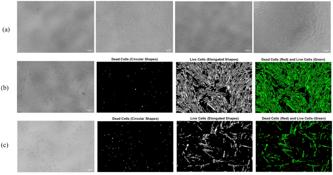
As the third response, microscopic image analysis was conducted to evaluate cell viability across different media formulations using the custom MATLAB script described in the material and methods section. Control images Fig. 1a displaying normal and healthy cell morphology were used to establish the threshold for identifying live cells and dead cells. Figure 1b illustrates a representative example of a high-viability culture media. The live cells (highlighted in green) display elongated, spindle-like morphologies consistent with healthy cells, occupying a substantial portion of the image area. Quantitative assessment (green area calculation) placed this sample within class 1 as an indicative of optimal culture conditions. In contrast, Fig. 1c shows a sample cultured in a media formulation with poor performance exhibiting significantly reduced viability. In this image, the green-highlighted live cells are noticeably fewer and less uniformly distributed, while the red-labeled dead cells are more prevalent throughout. Based on green and red area calculation, the image is categorized as class 4.

Microscopic image analysis for viability classification of cells cultured in different media formulations. (a) Images used to establish threshold criteria for identifying live and dead cells. (b) Representative example of a high-viability condition, and (c) example of a low-viability condition. For (b) and (c), binary segmentation was performed using a custom MATLAB script to distinguish dead cells (circular shapes) and live cells (elongated shapes). The proportions of green and red areas were used for classification, with (b) categorized as class 1 (high viability) and (c) as class 4 (low viability). Scale bars: 50 μm.
To optimize both LM8 and LM7 formulations, a multi-response optimization using Derringer desirability function was employed, aiming to maximize cell proliferation and image-based viability scores while minimizing media cost. Prior to modeling the data using MLR, various transformations were applied to the cell proliferation response to improve model fit. The optimal transformation was selected based on diagnostic criteria including adjusted R², residual distribution, and predictive accuracy, ensuring robust and reliable modeling of the experimental data. An Inverse transformation (1/response) was applied to both LM8 and LM7 datasets as the best transformation to achieve a better model fit. Moreover, outlier detection was performed using Cook’s distance, as well as externally and internally studentized residuals, to reduce noise and improve the model’s accuracy.
Biological roles and interactions of key media components in LM8 and LM7
Table 1 presents the ANOVA results for both media types. The LM8 model demonstrated high statistical significance (F = 66.99, p < 0.0001), confirming that the chosen variables and their interactions significantly influenced ILMC responses. Efficient serum-free media formulations require a comprehensive understanding of the individual and interaction roles played by essential growth factors and supplements in supporting cell proliferation, viability, and differentiation. Insulin plays a fundamental role in promoting the proliferation and myogenic differentiation of skeletal muscle satellite cells under serum-free conditions. As demonstrated by Lee et al., insulin enhances muscle cell expansion by activating the PI3K/AKT and MAPK/ERK signaling pathways, which regulate cell cycle progression, protein synthesis, and glucose metabolism. Their study on bovine satellite cells showed that insulin supplementation significantly increased both proliferation rates and expression of myogenic markers such as MyoD and Myogenin, indicating its dual role in early growth and differentiation. Moreover, insulin’s mitogenic effect was shown to be dose-dependent and most effective when used in synergy with other media components such as transferrin and selenium (interaction terms), confirming its essential role in serum-free media formulations for cultured meat production23. Ascorbic Acid-2-Phosphate (Asc-2P) mitigates oxidative stress through reactive oxygen species (ROS) scavenging and supports extracellular matrix formation by enhancing collagen synthesis, factors essential for maintaining viability, proliferation, and differentiation of muscle cells in the absence of serum24,25. Our findings demonstrate a significant interaction between insulin and ascorbic acid in promoting cell proliferation. This is consistent with prior in vitro study by Okita et al., which showed that ascorbic acid enhances insulin receptor signaling by upregulating insulin receptor substrates and activating the PI3K/Akt pathway. The antioxidant properties of ascorbic acid may also preserve insulin sensitivity by protecting signaling components from oxidative stress26. Transferrin is an essential iron-transport protein widely used in serum-free media formulations to regulate iron availability for skeletal muscle cell growth and mitochondrial function. Transferrin prevents iron-induced cytotoxicity by tightly binding free iron and delivering it safely into cells, supporting processes such as DNA synthesis, oxidative metabolism, and proliferation. According to Skrivergaard et al. (2023), the inclusion of transferrin in a chemically defined medium significantly improved the proliferation of porcine muscle cells, underscoring its critical role in supporting satellite cell expansion in the absence of serum19. Sodium selenite emerged as one of the most potent enhancers of cell growth and proliferation in LM8. Sodium selenite acts as a critical trace element in serum-free media, enhancing lamb muscle cell proliferation by contributing to the biosynthesis of antioxidant selenoproteins such as glutathione peroxidase and thioredoxin reductase. These enzymes support redox balance, protect against oxidative stress, and promote cellular survival and differentiation. Findings in bovine satellite cells demonstrate that sodium selenite inclusion is essential for muscle cell expansion under defined conditions13. TGF-β3 plays a regulatory role in skeletal muscle cell culture by controlling the timing and extent of myoblast fusion during differentiation. It signals through the SMAD2/3 pathway to act as a molecular brake, preventing excessive or premature fusion and allowing proper tissue maturation. As demonstrated by Melendez et al. (2021), constitutive activation of TGF-β signaling inhibits fusion, while its suppression leads to over-fusion, highlighting its essential role in maintaining balance during myogenesis. In serum-free systems, where controlled differentiation is critical, TGF-β3 supports ordered myotube formation and structural alignment, making it a valuable supplement in engineered muscle media27. NRG1 promotes skeletal muscle regeneration by activating ErbB receptors, which stimulate PI3K/AKT and MAPK/ERK pathways. This supports satellite cell proliferation, differentiation, and survival. As shown by Kubo et al. (2007), NRG1 is upregulated in both satellite cells and motoneurons during regeneration, indicating its dual role in muscle growth and neuromuscular signaling, making it a valuable component in advanced serum-free or co-culture media systems28. FGF2 plays a crucial role in promoting muscle satellite cell proliferation under serum-free conditions by activating MAPK/ERK and PI3K/AKT signaling pathways. It maintains the expression of Pax7, a key marker of satellite cell identity, and preserves the self-renewing capacity of myogenic precursors while preventing premature differentiation. This function supports sustained cell expansion and is essential in serum-free media formulations for cultured meat production29. Mung bean protein significantly enhances muscle cell growth and proliferation in serum-free media, as shown by its high F-value (1055.86, p < 0.0001) in the LM8 model. According to Amirvaresi et al. (2025), its rich amino acid composition supports protein synthesis, ECM formation, and metabolic efficiency. Moreover, mung bean showed strong synergistic interactions with sodium selenite (DH), NRG1 (FH), and TGF-β3 (EH), suggesting that its benefits are amplified when combined with antioxidants and growth factors. These interactions likely enhance redox balance, signaling, making mung bean a potent serum alternative in muscle cell culture systems.
In contrast, the LM7 model, though also significant (F = 29.21, p < 0.0001), highlighted a different set of influential factors, including A-Insulin (F = 92.07), CE (F = 158.44), and EF (F = 79.34). Interestingly, D-Sodium Selenite, a highly significant factor in LM8, was not significant in LM7 (F = 0.0272, p = 0.8705), indicating a difference in nutrient utilization or metabolic requirements/interaction between the chemically defined and undefined conditions. Figure S1 b and c provides a visual representation of the effects of both main and interaction terms on the responses in the LM8 and LM7 models. In addition, both models showed non-significant lack-of-fit results (LM8 p = 0.5217; LM7 p = 0.7880), validating the robustness of the MLR approach. These findings underscore the necessity of tailored optimization strategies depending on media composition to enhance ILMC performance effectively. Table S6 represents the coded equation for both LM8 and LM7. Furthermore, Fig. 2c provides a representative example for both media formulations and offers a clearer visual understanding of the interactions among key factors.

Model diagnostics and response surface visualization for cell proliferation response. (a) Predicted vs. actual plots showing high correlation and good model fit for both media. (b) Normal probability plots of residuals indicate that the residuals follow a random behavior. (c) 3D response surface plots depicting the interactive effects of Sodium Selenite (SS) and Mung Bean extract (MB) for LM8, as well as TGF-β3 and NRG1 for LM7.
In addition, fit statistics and diagnostic evaluations of both models demonstrate their robustness and suitability for describing cell responses. The LM8 model exhibited strong performance, with an R² of 0.93, adjusted R² of 0.91, and predicted R² of 0.90 (Fig. 2a, LM8), Similarly, the LM7 model showed an R² of 0.96, an adjusted R² of 0.93, and a predicted R² of 0.87 (Fig. 2a, LM7), confirming its validity. The normal probability plots of residuals for both models show that residuals closely follow a normal distribution (Fig. 2b).
Optimized LM8 and LM7 media formulations
Finally, using the Nelder-Mead nonlinear optimization method, the obtained equation was optimized with the goals of maximizing proliferation and image score while minimizing media cost, giving higher importance and weight to cell proliferation. As a result, we derived several formulations and selected multiple candidates for both LM8 and LM7 (Table S7). Figure 3a shows the CyQUANT assay results on day three, demonstrating that among the LM8 formulations, formulation 9 achieved the highest proliferation, significantly exceeding the 10% FBS GM and matching the 20% FBS GM control without a statistically significant difference. This underscores formulation 9’s ability to support ILMC proliferation at levels equivalent to high-serum conditions. Formulation 5 also demonstrated a strong proliferative capacity comparable to 20% FBS, while offering a substantially lower total media cost, making it the most practical choice for cost-sensitive applications. The other LM8 formulations displayed proliferation similar to the 10% FBS control, with formulation 8 standing out as the most economical option, effectively balancing cost with satisfactory growth performance. The choice between these formulations depends on whether minimizing cost or maximizing proliferation is the primary goal, with formulation 9 recommended for maximal proliferation and formulation 5 offering a cost-effective alternative without significant compromise in performance. The LM7 formulations (10–13), representing optimized defined media for ILMCs proliferation, are shown in Fig. 3b. Among these, Formulation 11 demonstrated the highest proliferation, surpassing other formulations and most commercial serum-free media tested. Importantly, all LM7 formulations exhibited better performance compared to the B8 media, which served as the baseline for component selection for our serum-free media development. Figure 3c further compares formulation 8 ($65) from the LM8 group with the B8 control, showing that this formulation significantly enhances ILMC growth compared to B8. However, it should be noted that the B8 media was specifically optimized for the culture and maintenance of human iPSCs, underscoring the need for an ILMC-specific formulation to achieve optimal performance11. Among the nine LM8 media formulations tested, formulation 9, which had the highest cell proliferation, was designated as the optimized LM8 medium, and for the LM7 formulations, formulation 11 was designated as the optimized LM7 medium. All subsequent references to LM8 and LM7 refer specifically to these top-performing formulations. Figures 3d–f present comparisons of top-performing LM8 and LM7 formulations with each other and against commercial media. Notably, none of the commercial media or LM7 formulations achieve proliferation levels comparable to LM8 media (Fig. 3d). Figure 3e highlights formulation 5’s marked improvement in ILMC proliferation compared to all market samples. Furthermore, Fig. 3f demonstrates that formulation 5 outperforms all LM7 and B8 media, representing its dual advantages of efficacy and cost-efficiency.

Comparative analysis of ILMC proliferation across LM8, LM7, commercial media, control and market samples. (a) CyQUANT assay measuring ILMC proliferation on day 3 across LM8 formulations 1–9, compared to 10% and 20% FBS controls. Formulation 9 exhibits the highest proliferation, statistically comparable to 20% FBS. (b) Proliferation of ILMCs in LM7 formulations 10–13 relative to B8. (c) Growth comparison between formulation 8 and B8 medium analyzed by paired t-test, highlighting significant proliferation enhancement despite lower cost. (d) Overall comparison of top-performing LM8 (formulation 9) and LM7 (formulation 11) formulations with commercial media demonstrating superior proliferation in LM8. (e) Comparison of formulation 5 with commercial media shows significant improvement in ILMC proliferation. (f) Proliferation of ILMCs in formulation 5 relative to all LM7 and B8 media showing superior growth support and cost-effectiveness. Results are based on n = 3 distinct samples. Statistical significance among multiple groups was determined using one-way ANOVA followed by Tukey’s multiple comparison test, while pairwise differences were assessed using unpaired t-tests. Groups not sharing the same superscript letter differ significantly (p < 0.05).
Validation of stemness and differentiation potential of ILMCs in LM7 and LM8
To further validate the optimized media, we evaluated their ability to support both stemness maintenance and differentiation potential of ILMCs. The stemness and cytoskeletal organization capacity of ILMCs cultured in the optimized formulations were assessed via Pax7 and Phalloidin immunostaining after three days of culture. As shown in Fig. 4a and b, positive Pax7 staining indicates that ILMCs maintained in both LM8 and LM7 retained their stem-like phenotype, while actin staining confirms cytoskeletal integrity. In addition, after three days of culture in the optimized LM8 or LM7 formulations, the growth media were replaced with differentiation media to allow ILMCs to adapt and initiate differentiation. Cells were maintained under these conditions for 21 days, with media changes every other day. Myogenic differentiation was assessed by immunostaining for Desmin (Fig. 4c), an early differentiation marker, and MHC, a marker of late-stage differentiation (Figure d). The immunostaining results are in accordance with previous reports that evaluated the maintenance of stemness and myogenic potential in bovine satellite cells under serum-free or optimized media conditions. Similar to our observations, other studies have demonstrated the capability of alternative media formulation for maintaining Pax7 expression in early culture stages3. Consistent with Phalloidin staining to assess cytoskeletal organization, Dolgin et al. and Stout et al. confirmed healthy morphology in serum-free conditions3,30. Furthermore, the progression from early to late myogenic differentiation in our study (Desmin and MHC), aligns with findings reported by Amirvaresi et al. and others, who observed the formation of multinucleated myotubes and expression of terminal differentiation markers under optimized protein-supplemented conditions3,22,30.

Assessment of stemness, cytoskeletal integrity, and differentiation potential of ILMCs in optimized culture conditions. (a) Immunofluorescence staining of ILMCs cultured in GM, LM8 and LM7 shows positive expression of (a) Pax7 (red) indicating maintenance of stem-like phenotype. (b) F-actin, highlighting cytoskeletal organization and cell morphology, nuclei were counterstained with DAPI (blue). (c) After 21 days of culture in differentiation media, ILMCs were stained for Desmin (green), an early myogenic differentiation marker. Robust Desmin expression indicates the initiation of the differentiation. (d) Late-stage myogenic differentiation was evaluated via immunostaining for myosin heavy chain (MHC) (red), confirming advanced muscle fiber development. Merged images show co-localization of markers with nuclear staining. Scale bars: 50 μm.
MCs screening and 3D culture of ILMCs in LM7 and LM8
To identify the most suitable MCs for supporting the attachment and proliferation ILMCs, a high-throughput screening assay was conducted. Each MC has unique physicochemical properties such as surface charge, material composition, and functional coatings that can influence cell adhesion and interaction with specific media formulations. ILMCs were seeded at a density of 2,000 cells/cm² onto four distinct MCs, each added at a concentration equivalent to 10 cm²/mL. Plates were agitated on an orbital shaker at 100 rpm for 72 h under standard culture conditions. After three days, cell proliferation and attachment efficiency on each microcarrier type were quantitatively assessed using the CyQUANT cell proliferation assay. In LM8, ILMCs exhibited comparable levels of attachment and proliferation across CellBIND, Cytodex-1, and Star-Plus microcarriers (Fig. 5a). However, in LM7, the CellBIND microcarriers demonstrated significantly higher cell growth compared to the other carriers (Fig. 5b). The screening of MCs for ILMCs revealed significant variation in attachment and proliferation depending on the physicochemical properties of each MC and the type of media used. This observation aligns with previous studies that emphasized the interplay between MC surface characteristics and medium composition.

Microcarrier screening and 3D culture of ILMCs in LM7 and LM8 media. Cell attachment and proliferation on different microcarriers (CellBIND, Cytodex-1, Star Plus, and Synthemax II) in (a) LM8 and (b) in LM7 media. (c) Cell number in 45 mL media after 6 days of culture in GM, LM8, and LM7 media and (d) cell viability normalized to control in FBS20%, LM8, and LM7 media formulations. Results show that cell numbers and viability in LM8 and LM7 media are significantly reduced compared to FBS20%.
Positively charged MCs like Cytodex-1 and Star Plus can enhance cell attachment due to the electrostatic attraction between negatively charged cell membranes and positively charged surfaces31. In our study, however, CellBIND, a negatively charged MC, showed superior performance. This suggests that additional factors such as protein adsorption or integrin-mediated binding may override surface charge effects under serum-free conditions. This finding is in accordance with Hoshiba et al. (2018), who reported that electrostatic charge alone does not determine cell adhesion; instead, protein adsorption and surface stiffness also play critical roles32. Our observation that Star Plus, despite being positively charged, failed to support ILMC attachment, reinforces this point. As also reported by Bodiou et al. (2024), microcarrier stiffness can impede cell attachment, potentially explaining Star-Plus’s poor performance due to its cross-linked polystyrene matrix. Based on these findings, CellBIND microcarriers were selected for subsequent 3D culture study using shaking flask systems.
To evaluate the scalability and performance of ILMCs under our optimized media, a 3D culture assay was conducted using CellBIND microcarriers in shaking flasks. For each condition, cells were cultured with and without MCs to compare attachment-dependent and suspension growth. Cultures were maintained for 6 days with partial medium exchange on day 3. Cell counting and proliferation for MCs flasks, as well as the viability assay for cell suspension flasks, were performed on day 6 using total cell counts and the PrestoBlue viability assay.
Figures 5c depict the cell counts in MCs flasks for GM, LM8, and LM7 conditions. We observed significantly higher cell numbers in GM media (with 20% FBS) compared to LM8 and LM7 media conditions in 3D flask cultures. This discrepancy between 2D and 3D results can likely be attributed to nutrient limitations in LM8 and LM7 media. As cells proliferate over time, nutrient consumption increases, and the media’s ability to support cell growth becomes more critical. Consequently, further optimization of these media formulations might be necessary to better support cell growth over extended culture periods. This observation is consistent with the findings of Dai et al. (2024), who reported that serum-containing media promote strong myoblast expansion, whereas serum-free formulations exhibited reduced initial attachment and higher susceptibility to detachment during dynamic culture33. Furthermore, Tsai et al. (2020) emphasized that microcarrier culture systems introduce unique microenvironmental challenges, including nutrient diffusion constraints, shear stress, and particle aggregation, all of which can alter cell behavior compared to planar systems. The lower cell growth observed in LM8 and LM7 media under 3D conditions may thus reflect compounded effects of suboptimal nutrient availability and the dynamic shear environment34. It is important to note that the media used for comparison in this study contained 20% FBS, whereas many studies typically use 10% FBS as a baseline for media comparison. Based on our previous experiments, usually cells in 10% FBS media grew at approximately half the rate of those in 20% FBS, which suggests that the observed differences between LM8, LM7, and GM media could be significantly lower if a 10% FBS formulation were used, and these formulations could be considered as cost-effective and ethically favorable alternatives. Additionally, although a comprehensive analysis was conducted to select the most suitable MC for these cultures, variations in experimental parameters such as cell seeding density, shaking conditions, and overall setup could affect the outcomes. These factors should be considered when optimizing for this specific step in the 3D flask culture process. In suspension cultures, where cells form a biomass (Figure S2), we encounter challenges in accurately quantifying cell numbers via conventional cell counting methods. The enzymatic treatment required for dissociation of cells in suspension failed to yield single-cell suspensions, therefore the PrestoBlue assay was used to assess cell viability instead. The results from the PrestoBlue assay aligned (Fig. 5d) with the cell count data, providing a reliable estimate of cell health and proliferation. Notably, visual inspection of the biomass after centrifugation revealed a substantially higher cell number in the MCs-containing flasks compared to the suspension cultures, confirming the superior cell growth supported by the microcarriers. In summary, while our findings support the effectiveness of MCs for enhancing cell growth in optimized media, further refinements in both media formulations and experimental protocols are required to achieve consistent and scalable growth in 3D cultures.
Cell-specific productivity and economic analysis
To comprehensively assess media performance for cultivated meat applications, we evaluated cell-specific productivity and cost-effectiveness metrics for our optimized formulations relative to conventional FBS-containing media in both 2D and 3D culture systems.
In 2D culture, LM8 achieved the highest cell density (6.47 × 10⁴ cells/cm²), exceeding FBS (20%, 5.88 × 10⁴ cells/cm²). It also demonstrated exceptional cost efficiency at $3.42 per million cells, representing a 25% cost reduction compared to FBS ($4.62 per million cells). LM7 showed moderate performance (3.24 × 10⁴ cells/cm²) but incurred higher costs ($6.53 per million cells) due to reduced cell yield despite its lower media price.
In 3D microcarrier culture, FBS maintained superior absolute cell yields (1.74 × 10⁵ cells/cm²) and the best cost efficiency ($0.066 per million cells) due to high total cell production (282 million cells from 45 mL culture). LM8 reached 3.33 × 10⁴ cells/cm² at $0.297 per million cells, while LM7 achieved 2.54 × 10⁴ cells/cm² at $0.372 per million cells. These represent 4.5-fold and 5.6-fold higher costs relative to FBS, respectively.
These productivity and cost analyses demonstrate that LM8 achieved superior economic performance in 2D culture, outperforming FBS in both cell density and cost efficiency. However, the substantial performance gap observed in 3D microcarrier systems highlights the critical challenge of translating serum-free media optimization from 2D to production-relevant 3D environments.
Conclusion
In conclusion, the development of LM7 and LM8 serum-free media formulations represents a critical step forward in the scalable, ethical, and economically viable cultivation of lamb muscle cells. Through a robust, statistically driven optimization framework incorporating RSM and DOE, we established two novel media platforms, LM7 (chemically defined) and LM8 (chemically undefined) that sustain high levels of ILMC proliferation while preserving both stemness and myogenic differentiation potential. Notably, LM8 achieved the highest proliferation rate, matching FBS performance. Furthermore, the scalability of these media in 3D microcarrier-based cultures illustrates their potential for large-scale biomanufacturing of cultivated lamb meat. In addition to their biological performance, both LM7 and LM8 demonstrated significant cost-effectiveness, particularly LM8-5, which offers a robust alternative to more expensive FBS-containing media, thereby reducing overall production costs. Moving forward, these formulations could play a crucial role in reducing dependency on animal-derived products, aligning with the growing demand for sustainable and ethical alternatives to traditional meat production. The findings from this research establish a solid foundation for the continued development of optimized, serum-free culture media for large-scale, economically viable, and environmentally sustainable cultivated meat production. Future directions for this work should focus on the long-term stability and batch-to-batch consistency of these formulations under bioreactor conditions. Additionally, metabolomic profiling and systems biology approaches could also be applied to refine nutrient utilization and waste byproduct management in culture.