Introduction
Bone regeneration is a cornerstone of implant-based dental rehabilitation, especially in cases of advanced alveolar bone resorption due to premature tooth loss, periodontal disease, odontogenic cysts, trauma, or sinus pneumatization. These clinical scenarios often necessitate bone grafting procedures such as socket preservation, ridge augmentation, and sinus floor elevation to re-establish an anatomically and functionally favorable site for implant placement1,2.
While autogenous bone grafts are considered the gold standard due to their osteogenic, osteoconductive, and osteoinductive capabilities, their use is frequently constrained by donor site morbidity, surgical complexity, and inconsistent long-term success rates3,4. Consequently, clinicians increasingly rely on allogeneic, xenogeneic, and synthetic alternatives for reconstructing critical-size defects (CSDs). However, these materials lack intrinsic biological activity, offering passive osteoconduction with limited ability to promote cell recruitment, differentiation, or matrix mineralization5.
To overcome these limitations, next-generation biomaterials are being developed to mimic the extracellular matrix (ECM) at the nanoscale and deliver instructive cues for bone regeneration. Self-assembling peptide amphiphile (PA) nanofibers have emerged as a powerful platform in this regard. These molecules consist of a hydrophobic alkyl tail conjugated to a functional peptide sequence, allowing them to form ECM-like nanofibrous networks under physiological conditions6,7,8. Their modular design enables the rational incorporation of multiple biologically active peptide sequences within a single supramolecular framework, allowing for the concurrent presentation of distinct regenerative functions. In the present study, four peptide amphiphiles were co-assembled into a multifunctional nanofiber interface, each selected to address a critical biological process involved in bone healing. The DGEA motif, derived from type I collagen, facilitates integrin-mediated osteogenic differentiation by engaging α2β1 integrins, thereby supporting early commitment of mesenchymal stem cells to the osteoblastic lineage9. The EEE sequence, composed of a negatively charged glutamic acid triplet, mimics calcium-binding domains of native bone matrix proteins and enhances mineralization by promoting hydroxyapatite nucleation on the scaffold surface10. To ensure robust surface adhesion under physiological and wet conditions, DOPA—a catechol-containing residue inspired by mussel adhesive proteins—was incorporated, providing strong binding to the irregular and moist topography of graft materials through hydrogen bonding and metal coordination11,12. Finally, GL13K, a synthetic cationic peptide derived from salivary proteins, was included for its broad-spectrum antimicrobial activity, which has been shown to inhibit bacterial adhesion and biofilm formation, thereby reducing the risk of infection-driven graft failure13,14,15.
While surface modification approaches in dentistry have traditionally relied on single-function peptides or have been tested on non-clinical materials, few have been evaluated on clinically approved xenograft, allograft, or synthetic substrates widely used in oral surgery16. Moreover, these commercial grafts—although effective in space maintenance—often exhibit limited regenerative performance, including slow resorption (xenografts), biological variability (allografts), or low osteogenic capacity (synthetics), particularly in compromised clinical scenarios17,18,19,20. Our multifunctional nanofiber interface directly addresses these limitations by providing a tunable, bioactive coating compatible with real-world grafts, aiming to enhance biological performance across multiple regenerative domains. The multifunctional peptide nanofiber platform used herein has previously shown both osteoinductive and antimicrobial effects, as reported in our earlier study21.
Materials and methods
Materials
All Fmoc-protected amino acids, rink amide resin (0.65 mmol/g), and coupling reagents including HBTU, HOBt, and DIPEA used for peptide synthesis were obtained from Iris Biotech GmbH (Marktredwitz, Germany). Lauric acid (≥ 98%) was purchased from Sigma-Aldrich (St. Louis, MO, USA). Analytical grade solvents such as N, N-dimethylformamide (DMF), dichloromethane (DCM), and diethyl ether were sourced from Merck (Darmstadt, Germany). Trifluoroacetic acid (TFA) and triisopropylsilane (TIS) were also obtained from Merck.
Peptide purification was carried out using a preparative reverse-phase high-performance liquid chromatography (HPLC) system (Shimadzu, Japan) equipped with a C18 column (Grace Vydac, USA). The identity and purity of synthesized peptides were confirmed by electrospray ionization mass spectrometry (ESI-MS) using a Bruker amaZon SL ion trap mass spectrometer. Peptide self-assembly behavior and secondary structure were analyzed using a Jasco J-1500 circular dichroism (CD) spectropolarimeter (Tokyo, Japan). Fourier-transform infrared (FTIR) spectra were recorded with a Bruker Tensor II ATR-FTIR spectrometer, and static contact angles were measured using a Dataphysics OCA goniometer (Filderstadt, Germany).
Graft materials included bovine-derived xenografts, human-derived allografts, and synthetic hydroxyapatite particles, all obtained from Bioland (Istanbul, Türkiye). Human dental pulp stem cells (DPSCs), pre-characterized for mesenchymal markers, were supplied by Genkök Biotechnology (Ankara, Türkiye). For cell culture, Dulbecco’s Modified Eagle Medium (DMEM, high glucose), fetal bovine serum (FBS), penicillin-streptomycin, and trypsin-EDTA were procured from Gibco, Thermo Fisher Scientific (Waltham, MA, USA).
Fluorescent staining of viable cells was performed using Calcein-AM (Thermo Fisher Scientific), and metabolic activity was evaluated using the MTT assay reagent (Sigma-Aldrich). Alkaline phosphatase activity was measured using p-nitrophenyl phosphate (pNPP; Sigma-Aldrich), while extracellular matrix mineralization was assessed with Alizarin Red S and cetylpyridinium chloride, both purchased from Sigma-Aldrich. Total RNA was isolated using TRIzol reagent (Invitrogen, Thermo Fisher), and cDNA synthesis was carried out with the RevertAid First Strand cDNA Synthesis Kit (Thermo Fisher Scientific). Gene expression was analyzed using SYBR Green-based qPCR reagents from Applied Biosystems (Foster City, CA, USA).
Micro-computed tomography was conducted with a SkyScan 1176 system (Bruker, Kontich, Belgium), and morphometric analysis was performed using CTAn and CTVol software (Bruker). For histological analysis, decalcification was performed with EDTA (Merck), and sections were stained using 0.1% toluidine blue (Sigma-Aldrich). All other general laboratory reagents and solutions were obtained from Sigma-Aldrich or Merck unless otherwise specified.
Peptide amphiphile synthesis and characterization
Peptide amphiphile (PA) molecules were synthesized via standard solid-phase peptide synthesis (SPPS) using Fmoc (9-fluorenylmethyloxycarbonyl) chemistry22. The syntheses were performed on a rink amide resin (0.65 mmol/g loading capacity) using an automated peptide synthesizer (e.g., Liberty Blue, CEM Corporation). Fmoc-protected amino acids were sequentially coupled using HBTU/HOBt activation in the presence of DIPEA. After completion of chain assembly, lauric acid (C12:0) was conjugated to the N-terminus to confer amphiphilic properties, followed by cleavage and side chain deprotection using a TFA: TIS: water (95:2.5:2.5, v/v) cocktail for 2 h at room temperature. Crude products were precipitated with cold diethyl ether and lyophilized.
Four distinct peptide amphiphile sequences were synthesized:
-
DGEA-PA (Lauryl–Val–Val–Ala–Gly–Lys–Lys–Gly–Asp–Gly–Glu–Ala–amide) – collagen I mimetic
-
EEE-PA (Lauryl–Val–Val–Ala–Gly–Lys–Lys–Gly–Glu–Glu–Glu–Ala–amide) – hydroxyapatite mineralization promoting
-
DOPA-PA (Lauryl–Val–Val–Ala–Gly–Lys–Lys–Gly–DOPA–Gly–Glu–Ala–amide) – inspired by mussel adhesive protein
-
GL13K-PA (Lauryl–Val–Val–Ala–Gly–Lys–Lys–Gly–Gly–Leu–Lys–Lys–amide) – antimicrobial sequence
Purification was performed via reverse-phase HPLC using a C18 preparative column under a linear water–acetonitrile gradient with 0.1% TFA. Molecular identity and purity (> 95%) were confirmed by electrospray ionization mass spectrometry (ESI-MS). To investigate secondary structures and self-assembly behavior, peptide amphiphiles were dissolved in deionized water or phosphate buffer (10 mM, pH 7.4) at a final concentration of 1 wt%. Solutions were sonicated and incubated at room temperature for 24 h to promote nanofiber formation under physiological ionic conditions (150 mM NaCl, pH 7.4). Circular dichroism (CD) spectra were acquired between 190 and 260 nm to evaluate β-sheet content. The zeta potential of individual peptide amphiphile solutions and the assembled nanofiber system was measured to evaluate surface charge characteristics. Measurements were performed at 25 °C using a Malvern Zetasizer Nano ZS (Malvern Instruments Ltd., UK) equipped with a 633 nm He-Ne laser and a detection angle of 173°. Peptide solutions were prepared at a final concentration of 0.1% (w/v) in 10 mM phosphate-buffered saline (PBS, pH 7.4) and filtered through a 0.22 μm syringe filter prior to analysis. Each sample was measured in triplicate, and average zeta potential values were reported with standard deviation.
Graft materials and surface functionalization with peptide nanofibers
Commercially available xenografts (Xeno; bovine-derived), allografts (Allo; human-derived), and synthetic hydroxyapatite-based grafts (Synth) were used as base scaffold materials. Prior to functionalization, all grafts were sterilized by UV irradiation for 30 min and stored under sterile conditions. Peptide amphiphile (PA) powders—DGEA-PA, EEE-PA, DOPA-PA, and GL13K-PA—were mixed in equimolar ratios to form a bioactive cocktail. The combined mixture was dissolved in sterile deionized water or phosphate-buffered saline (PBS, pH 7.4) at a total concentration of 1% (w/v) and incubated for 24 h at room temperature under physiological salt conditions (150 mM NaCl) to promote spontaneous nanofiber self-assembly.
Each graft type (100 mg) was immersed in 1 mL of the assembled peptide solution and incubated overnight at 4 °C under gentle agitation to ensure homogeneous coating. Excess solution was removed by centrifugation (500 × g, 5 min), and grafts were washed once with sterile PBS and air-dried under aseptic conditions.
Experimental groups were defined as follows:
-
Xeno, Allo, Synth: uncoated grafts
-
Xeno-P, Allo-P, Synth-P: grafts coated with the DGEA-PA + EEE-PA + DOPA-PA + GL13K-PA nanofiber mixture
Successful peptide immobilization was confirmed via Fourier-transform infrared (FTIR) spectroscopy by detecting characteristic amide I and II absorption bands, indicating the presence of peptide secondary structures on graft surfaces.
Morphological and chemical characterization of functionalized grafts
Surface morphology of functionalized and unmodified graft materials was examined using scanning electron microscopy (SEM). Samples were mounted on aluminum stubs using carbon adhesive tape, sputter-coated with a 10 nm layer of gold, and imaged using a scanning electron microscope (e.g., Zeiss EVO LS10) operated at 5–15 kV. Multiple fields and magnifications were recorded to evaluate nanofiber coverage and surface architecture. Chemical characterization of the peptide coatings was performed using attenuated total reflectance Fourier-transform infrared (ATR-FTIR) spectroscopy. Spectra were acquired in the range of 4000–600 cm⁻¹ using a spectrometer (e.g., Bruker Tensor II) with 32 scans per sample at 4 cm⁻¹ resolution. Particular attention was given to amide I and II bands, which are indicative of peptide backbone structure. In addition, phosphate- and carbonate-associated absorption bands were monitored to assess interactions with the mineral components of the grafts. Optional analysis of surface wettability was performed using contact angle goniometry (e.g., Dataphysics OCA system). A sessile drop of distilled water (5 µL) was placed on the surface of each sample, and static contact angles were measured at room temperature. Measurements were repeated in triplicate for each group, and values were recorded for comparative assessment of surface hydrophilicity following peptide coating.
In vitro cell culture and experimental design
Human dental pulp stem cells (DPSCs) were utilized for in vitro evaluation of graft biocompatibility and osteogenic potential. DPSCs were obtained from a commercial cell supplier (Genkök Biotechnology, Ankara, Türkiye), pre-characterized for mesenchymal stem cell surface markers (CD73⁺, CD90⁺, CD105⁺, CD34⁻, CD45⁻) and confirmed for multipotent differentiation potential24.
Cells were cultured in standard growth medium composed of Dulbecco’s Modified Eagle Medium (DMEM, high glucose; Gibco) supplemented with 10% fetal bovine serum (FBS; Biowest), 1% penicillin-streptomycin (Gibco), and maintained in a humidified incubator at 37 °C with 5% CO₂. Media was refreshed every 2–3 days.
For experimental studies, DPSCs at passages 3–5 was detached using 0.25% trypsin-EDTA, counted, and seeded onto the graft samples at a density of 1 × 10⁵ cells per well in 24-well tissue culture plates. Each well contained one graft sample (either unmodified or peptide-functionalized).
The experimental groups were organized as follows:
Unmodified graft groups (negative controls)
Xeno (uncoated xenograft), Allo (uncoated allograft), Synth (uncoated synthetic scaffold).
Peptide-coated graft groups (test groups)
Xeno-P, Allo-P, Synth-P — each coated with a multifunctional peptide nanofiber blend (DGEA-PA + EEE-PA + DOPA-PA + GL13K-PA).
In this setup, the unmodified grafts served as negative controls to assess baseline cell behavior, while the peptide-coated groups were used to evaluate the bioactivity and osteoinductive potential of the nanofiber system.
All groups were cultured in standard growth medium for initial adhesion and viability studies. For osteogenic differentiation studies, cells were cultured in osteogenic induction medium (DMEM supplemented with 10% FBS, 10 mM β-glycerophosphate, 50 µg/mL ascorbic acid, and 100 nM dexamethasone) for up to 14 days, with medium changes every 3 days.
Cell adhesion assay
Cell adhesion was evaluated 24 h after seeding using a live-cell fluorescence staining method. DPSCs (1 × 10⁵ cells/well) were seeded onto each graft sample in 24-well plates and incubated under standard culture conditions (37 °C, 5% CO₂). After 24 h, non-adherent cells were gently removed by washing the samples twice with phosphate-buffered saline (PBS, pH 7.4). Adherent viable cells were stained using the Calcein-AM live-cell labeling dye (Thermo Fisher Scientific). A working solution of Calcein-AM (2 µM in PBS) was added to each well and incubated for 30 min at 37 °C in the dark. After incubation, excess dye was removed by PBS washing, and the samples were immediately imaged using an inverted fluorescence microscope (e.g., Zeiss Axio Observer) with FITC filter settings. Fluorescence images were captured from three predefined regions per sample. Quantitative analysis of adherent cell density was performed using ImageJ software (National Institutes of Health, USA)25. by thresholding and counting fluorescent cell areas per field. Cell adhesion efficiency was expressed as mean fluorescence area per field (n = 3 per group), providing a relative estimate of the number of viable adherent cells.
Cell viability and proliferation assay
To evaluate the metabolic activity and proliferation of DPSCs cultured on graft surfaces, the MTT [3-(4,5-dimethylthiazol-2-yl)−2,5-diphenyltetrazolium bromide] assay was employed at 24-, 48-, and 72-hours post-seeding. Cells were seeded at a density of 1 × 10⁵ cells per well in 24-well plates containing either uncoated or peptide-functionalized grafts and incubated under standard conditions. At each time point, culture medium was aspirated and replaced with 500 µL of serum-free DMEM containing 0.5 mg/mL MTT (Sigma-Aldrich). Samples were incubated for 4 h at 37 °C in the dark to allow for the formation of purple formazan crystals by metabolically active cells. Following incubation, the MTT solution was carefully removed, and the formed formazan crystals were solubilized in 500 µL of dimethyl sulfoxide (DMSO) per well. The resulting solution was mixed thoroughly and transferred to a 96-well plate for absorbance measurement. Optical density was recorded at 570 nm using a microplate reader (e.g., BioTek Epoch 2), with a reference wavelength of 630 nm. All experiments were conducted in triplicate (n = 3 per group per time point). Cell viability was expressed as mean absorbance values, which are directly proportional to the number of viable cells.
The antimicrobial activity of peptide nanofiber components (DOPA, GL3K, DGEA, and EEE) was evaluated using the MTT-based viability assay, adapted for bacterial cells. Escherichia coli (ATCC 25922) and Staphylococcus aureus (ATCC 29213) were used as representative Gram-negative and Gram-positive strains, respectively. Bacterial suspensions were adjusted to 1 × 10⁸ CFU/mL using the McFarland standard. Peptide nanofiber solutions were prepared at final concentrations of 50, 100, and 200 µg/mL in sterile PBS. For each condition, 500 µL of the bacterial suspension was mixed with 500 µL of the corresponding peptide solution in sterile microtubes and incubated at 37 °C for 4 h under shaking. After incubation, 100 µL from each mixture was transferred to a 96-well plate, followed by the addition of 10 µL of MTT reagent (5 mg/mL in PBS). The plate was incubated for 2 h at 37 °C in the dark. Formazan crystals were solubilized using 100 µL of DMSO, and absorbance was measured at 570 nm using a microplate reader. Untreated bacteria were used as the viability control. All experiments were performed in triplicate, and data are presented as mean ± standard deviation (Table S2-3).
Osteogenic differentiation and gene expression analysis
To assess osteogenic commitment at the molecular level, real-time quantitative polymerase chain reaction (qRT-PCR) was used to analyze the expression of key osteogenesis-associated genes. DPSCs were cultured on unmodified and peptide-functionalized grafts in osteogenic induction medium (DMEM supplemented with 10% FBS, 10 mM β-glycerophosphate, 50 µg/mL ascorbic acid, and 100 nM dexamethasone) for up to 14 days. Medium was refreshed every 3 days. At day 3 and day 14, total RNA was extracted from the cell-graft complexes using a phenol-chloroform-based method (e.g., TRIzol reagent, Invitrogen), according to the manufacturer’s protocol. RNA concentration and purity were determined spectrophotometrically (NanoDrop™ 2000, Thermo Scientific). Reverse transcription was performed using a cDNA synthesis kit (e.g., RevertAid First Strand cDNA Synthesis Kit, Thermo Fisher), employing 1 µg of total RNA per reaction. Gene expression analysis was carried out using SYBR Green-based qRT-PCR (e.g., Applied Biosystems™ StepOnePlus™ system). Specific primers were designed for the following target genes: Runt-related transcription factor 2 (RUNX2), Osteopontin (OPN), Type I collagen alpha 1 (COL1A1). GAPDH was used as the internal reference gene. The relative expression of each gene was calculated using the ΔΔCt method26, and results were expressed as fold change relative to control samples at each time point. All qPCR reactions were run in triplicate, and negative controls (no template) were included to confirm specificity. Primer sequences and annealing temperatures are provided in Supplementary Table 1.
Alkaline phosphatase (ALP) activity and mineralization assay
To evaluate functional osteogenic differentiation, alkaline phosphatase (ALP) activity and extracellular matrix mineralization were assessed at day 14 of induction.
Alkaline Phosphatase (ALP) Activity: DPSCs cultured on grafts were lysed using ALP assay lysis buffer (e.g., 0.1% Triton X-100 in Tris-HCl, pH 7.6). Cell lysates were collected and incubated with p-nitrophenyl phosphate (pNPP; Sigma-Aldrich) substrate solution at 37 °C for 30 min. The enzymatic reaction was terminated by adding 3 N NaOH, and the absorbance was measured at 405 nm using a microplate reader (BioTek Epoch 2). Protein concentrations in lysates were determined by BCA protein assay (Thermo Scientific), and ALP activity was normalized to total protein content (U/mg protein) (Figure S2). All measurements were performed in triplicate.
Calcium Deposition by Alizarin Red S (ARS) Staining: To assess matrix mineralization, samples were fixed with 4% paraformaldehyde for 15 min and rinsed with distilled water. Fixed samples were stained with 2% (w/v) Alizarin Red S (ARS; Sigma-Aldrich) solution (pH 4.2) for 20 min at room temperature. Excess dye was removed by repeated washing with distilled water. Stained calcium deposits were visualized under a stereomicroscope and imaged. For quantification, the bound ARS dye was extracted using 10% (w/v) cetylpyridinium chloride in 10 mM sodium phosphate buffer (pH 7.0) for 30 min, and the absorbance was measured at 550 nm. Quantitative values were reported as mean ± standard deviation (n = 3 per group). (Standard curve can be found in Supplemantary Information). To quantify calcium deposition following osteogenic differentiation, Alizarin Red S-stained samples were imaged under identical exposure settings using a brightfield microscope. For each sample, representative images were acquired from at least three independent fields per group. Red channel intensity was then quantified using ImageJ (NIH) software. Briefly, images were converted to RGB stack, and the red channel was isolated. Mean red intensity values were measured using standardized ROI selections, ensuring consistent area and thresholding across all groups. The resulting values were averaged for each group and presented as mean ± standard deviation (n = 3). This method provides a semi-quantitative assessment of mineralized matrix content, complementing the visual ARS staining results.
In vivo calvarial bone defect model and treatment groups
To evaluate the bone regenerative capacity of peptide-functionalized grafts in vivo, a critical-sized calvarial defect model was utilized in adult male New Zealand White rabbits (Oryctolagus cuniculus). A bilateral critical-sized calvarial defect (8 mm in diameter) was selected, as such defects do not heal spontaneously and are widely accepted for assessing bone regeneration strategies in preclinical studies5,27.
All animals used were adult male New Zealand White rabbits, weighing between 2.5 and 3.0 kg and approximately 5–6 months old at the time of surgery. Animals were obtained from Kobay Deney Hayvanlari Laboratuvari (Ankara, Turkey). All animals were clinically healthy with no known immunodeficiencies or genetic modifications, and no previous surgical or experimental procedures had been performed prior to this study.
Upon arrival, animals were individually housed in standard cages under controlled ambient temperature conditions with a 12-hour light/dark cycle. They were fed a standard commercial rabbit chow and had ad libitum access to fresh water throughout the study. Animals were acclimatized to the housing environment for seven days prior to surgical procedures to minimize stress-induced variability.
All experimental procedures were approved by the Local Animal Ethics Committee of Kobay Deney Hayvanlari Laboratuvari (Protocol No: 709, Approval Date: 02/2024) and conducted in accordance with Turkish national regulations, the European Directive 2010/63/EU on the protection of animals used for scientific purposes and reported in compliance with the ARRIVE guidelines (Animal Research: Reporting of In Vivo Experiments).
Inclusion criteria required that all animals be healthy, adult male New Zealand White rabbits (2.5–3.0 kg) without any clinical signs of systemic disease prior to surgery. Furthermore, only male rabbits were used in this study to avoid the potential confounding effects of hormonal fluctuations associated with the estrous cycle in females. Estrogen and other sex hormones are known to influence bone turnover, inflammatory responses, and soft tissue remodeling, which could introduce variability into regenerative outcomes. By limiting the cohort to skeletally mature males, we aimed to ensure a more uniform biological background, enhancing the reproducibility and interpretability of the observed effects. Exclusion criteria included intraoperative complications, postoperative infections, significant surgical site morbidity, or systemic illness that could impact bone healing. No animals or defects met exclusion criteria; therefore, all defects were included in the final analysis. Each experimental group (Blank, Xeno, Xeno-P, PA-NF) consisted of 6 defects (n = 6). The sample size was determined based on standard practice in preclinical rabbit calvarial defect models. Six animals were used, each contributing four defects, resulting in 24 defects total. No formal a priori sample size calculation was performed; the selected sample size is consistent with previous studies that reported sufficient statistical power for detecting significant differences in bone regeneration outcomes28.
Under general anesthesia induced by intramuscular injection of xylazine (5 mg/kg) and ketamine (35 mg/kg), the calvarial region was shaved, disinfected, and a midline sagittal incision was made to expose the parietal bone. Bilateral full-thickness flaps were reflected, and circular, critical-sized defects (8 mm in diameter) were created on both sides of the parietal bone using a trephine bur under sterile saline irrigation27,29. The following experimental conditions were randomly assigned to the defects:
-
Blank: untreated critical-sized defect (empty control)
-
Xeno: defect filled with unmodified xenograft particles
-
Xeno-P: defect filled with peptide-functionalized xenograft (coated with DGEA-PA, EEE-PA, DOPA-PA, and GL13K-PA nanofibers)
-
PA-NF: defect filled with self-assembled peptide nanofiber hydrogel alone (no graft material)
The experimental unit for all analyses was the individual defect. To minimize potential confounding effects, defects were created bilaterally, and treatment allocations were randomized using a computer-generated random number. Randomization of defects to treatment groups was performed using a computer-generated random number table; the individual performing the allocation was aware of group assignments. The surgical procedures were conducted by personnel who were aware of the treatment groups due to the nature of the interventions. However, all outcome assessments, including micro-CT imaging and histological evaluations, were performed by investigators blinded to the group allocations. Data analysis was conducted using anonymized datasets to maintain blinding during statistical evaluation. Following implantation, surgical sites were closed in anatomical layers using resorbable sutures. Postoperative analgesia (meloxicam, 0.2 mg/kg) and prophylactic antibiotic therapy (enrofloxacin, 10 mg/kg) were administered subcutaneously once daily for three consecutive days. At six weeks post-surgery, animals were euthanized with an overdose of anesthesia, and the calvarial bones were harvested for micro-computed tomography (micro-CT) and histological analyses. The primary outcome measure was bone volume fraction (BV/TV) assessed via micro-CT imaging. Secondary outcome measures included trabecular thickness (Tb.Th), trabecular spacing (Tb.Sp), and histological evaluation of new bone formation and tissue integration.
Micro-CT imaging and bone morphometric analysis
Quantitative evaluation of newly formed bone within calvarial defects was performed using high-resolution micro-computed tomography (micro-CT)27. Harvested calvarial specimens were fixed in 10% neutral-buffered formalin for 48 h, then scanned using a micro-CT imaging system (e.g., SkyScan 1176, Bruker) at 9 μm isotropic voxel resolution, with 50 kV voltage, 500 µA current, and a 0.5 mm aluminum filter. Regions of interest (ROIs) were defined based on the full defect diameter. All groups—including peptide-functionalized grafts, unmodified grafts, and the blank group (defect without graft material)—were analyzed under identical scanning and processing conditions.Thresholding was applied to distinguish mineralized tissue from soft tissue and background.
Quantitative morphometric parameters included:
-
Bone volume fraction (BV/TV, %)
-
Trabecular thickness (Tb.Th, µm)
-
Trabecular number (Tb.N, 1/mm)
-
Trabecular separation (Tb.Sp, µm)
-
Bone surface to volume ratio (BS/TV, 1/mm)
For each experimental group, a minimum of three independent biological replicates (n = 3) were analyzed, and group-level comparisons were performed based on the mean values derived from these replicates. Outcome assessors were blinded to group allocation during analysis. Three-dimensional renderings were generated to visualize the extent and spatial pattern of bone regeneration within the defect area.
Histological evaluation
Following micro-CT analysis, calvarial bone specimens were decalcified in 10% ethylenediaminetetraacetic acid (EDTA, pH 7.4) at room temperature for 3 weeks with regular solution changes. After complete decalcification, samples were dehydrated through a graded ethanol series, cleared in xylene, and embedded in paraffin. Serial Sect. (5 μm thickness) were obtained in the coronal plane using a microtome (e.g., Leica RM2235). Sections were stained with toluidine blue (0.1% w/v, pH 2.5) to visualize cellular and extracellular components, including newly formed bone tissue and residual graft material. Slides were examined under a light microscope (e.g., Olympus BX51), and digital images were captured for qualitative assessment29. Regions of interest (ROI) were delineated manually around the original defect margins (Figure S7). ImageJ software (NIH, USA) was used to quantify three key parameters: graft retention (% area occupied by residual graft material), new bone formation (% area occupied by newly formed trabecular bone), and void/soft tissue area (% area lacking mineralized tissue or graft). For each sample, three non-overlapping fields were analyzed, and values were averaged per animal (n = 3 per group). Statistical comparisons were conducted using one-way ANOVA followed by Tukey’s post hoc test. Results were reported as mean ± standard deviation (SD), and significance thresholds were set at p < 0.05.
Statistical analysis
All experimental data are presented as mean ± standard deviation (SD). Statistical analyses were performed using GraphPad Prism software (version 9.0, GraphPad Software Inc., San Diego, CA, USA). The Shapiro–Wilk test was used to assess data normality. For comparisons involving more than two groups, one-way analysis of variance (ANOVA) followed by Tukey’s post hoc test was applied to determine intergroup differences.For gene expression analysis (ΔΔCt values), log-transformation was applied before statistical testing where necessary to meet parametric assumptions. A significance level of p < 0.05 was considered statistically significant. All experiments were conducted in triplicate (n = 3 for in vitro assays) or as indicated in figure legends for in vivo analyses.
Results
Design and integration strategy of peptide nanofiber-coated graft interfaces
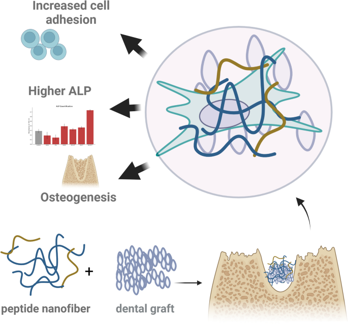
To develop a bioactive interface capable of enhancing osseointegration and osteogenic differentiation, synthetic peptide amphiphile nanofibers were designed and integrated with conventional dental graft materials (xenograft, allograft, and synthetic scaffolds). As schematically illustrated in Fig. 1, the functionalization strategy aimed to create an extracellular matrix-mimicking environment at the graft surface, promoting cell adhesion, early osteogenic signaling, and mineralized tissue formation. Peptide sequences were tailored to deliver osteoinductive cues, while maintaining injectability and nanoscale architecture suitable for complex defect geometries.

Schematic illustration of peptide nanofiber-enhanced dental grafts for bone regeneration. The graphical abstract represents the combination of peptide nanofiber structures with conventional dental graft materials to enhance their biological performance. Upon integration, the composite promotes enhanced cell adhesion, higher alkaline phosphatase (ALP) activity, and accelerated osteogenesis, as demonstrated by the increased osteoblast differentiation and bone-forming capacity. The nanofiber system mimics extracellular matrix-like environments, supporting cellular activity at the graft interface.
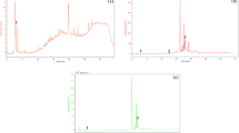
To elucidate the compositional and surface properties of the multifunctional nanofiber system and its interaction with clinically used bone grafts, a series of physicochemical and morphological analyses were performed (Fig. 2). Figure 2A shows the chemical structures of the four distinct peptide amphiphiles (PAs) used in this study—DOPA-PA, DGEA-PA, EEE-PA, and GL13K-PA—each bearing a hydrophobic lauric acid tail to drive self-assembly. Their respective net charges and mixing ratios were optimized to achieve a synergistic balance of adhesion, osteogenic, mineralization, and antimicrobial functions in the final nanofiber formulation. Figure 2B presents a scanning electron microscopy (SEM) image of the self-assembled nanofiber network, displaying an interconnected fibrillar architecture at the submicron scale, consistent with the formation of an extracellular matrix-like microenvironment. The secondary structure of individual PAs and the final assembled nanofiber system was assessed using circular dichroism (CD) spectroscopy (Fig. 2C). the final assembled nanofiber system displayed spectra characteristic of β-sheet-rich structures, while DOPA-PA and GL13K-PA exhibited more disordered conformations. The composite nanofiber system demonstrated a complex ellipticity profile, indicating the coexistence of multiple secondary structures within the hybrid interface.

Peptide-based nanofiber system characterization and interface formation on synthetic, xenogeneic, and allogeneic grafts. (A) Chemical structures of individual peptide amphiphiles (DOPA-PA, DGEA-PA, EEE-PA, GL13K-PA) with their corresponding net charges and mixing ratios used for nanofiber formulation. (B) Scanning electron microscopy (SEM) image showing the entangled nanofiber morphology of the self-assembled peptide amphiphile matrix. (C) Circular dichroism (CD) spectra of individual peptide amphiphiles and the composite nanofiber system, demonstrating secondary structure formation, including β-sheet and random coil features. (D) Zeta potential measurements indicating surface charge profiles of each PA component and the assembled nanofiber system. (E) SEM images of peptide-coated synthetic, xenograft, and allograft surfaces, demonstrating nanofiber coverage. FTIR spectra below confirm the presence of characteristic amide I/II bands and additional surface peaks after peptide functionalization.
To evaluate the surface charge characteristics of individual components and the resulting nanofiber network, zeta potential measurements were conducted (Fig. 2D). DOPA-PA and GL13K-PA exhibited positive charges, while DGEA-PA and EEE-PA were highly negative. The assembled nanofiber system showed a near-neutral but slightly positive charge, reflecting its compositional balance and anticipated biocompatibility.
Finally, Fig. 2E illustrates SEM images of peptide-functionalized synthetic (Synth-P), xenogeneic (Xeno-P), and allogeneic (Allo-P) graft surfaces, each showing uniform nanofiber coverage and integration. The peptide nanofibers were immobilized primarily via physical adsorption mechanisms (Figure S1). In particular, the DOPA-functionalized PA plays a crucial role in the adhesion process. Inspired by mussel adhesive proteins, DOPA residues form multiple non-covalent interactions—such as hydrogen bonding, π–π stacking, and coordination with metal ions present in mineralized graft surfaces—thereby enhancing the stability and retention of the peptide coating under physiological conditions30.
Representative SEM images were intentionally acquired from regions of the graft surfaces where nanofiber adhesion was visually evident, in order to illustrate the interaction between the peptide assemblies and the underlying scaffold microstructure. Corresponding FTIR spectra confirm successful peptide immobilization via the appearance of characteristic amide I (~ 1650 cm⁻¹) and amide II (~ 1540 cm⁻¹) peaks on coated surfaces, which were absent or weaker in unmodified grafts31.
Initial cell adhesion and viability assessment on graft surfaces
To investigate early cellular interactions with graft surfaces, adhesion and viability analyses were conducted following short-term incubation periods. As shown in Fig. 3, after 24 h of seeding, the number of adherent cells was substantially reduced on unmodified xenograft, allograft, and synthetic graft surfaces compared to the control. This suggests that these scaffolds alone may not sufficiently support initial attachment. In contrast, peptide-functionalized surfaces markedly improved early cell adhesion, with Allo-P and Xeno-P groups exhibiting the highest attachment levels. The enhancement observed in these groups highlights a potential synergistic effect between graft material and peptide amphiphile integration.

In vitro assessment of cell adhesion and proliferation on peptide-functionalized grafts. A) Representative fluorescence images of adherent cells on different graft surfaces at 24 h. Peptide-functionalized groups (Xeno-P, Allo-P, Synth-P) show a higher density of adhered cells compared to non-functionalized counterparts (Xeno, Allo, Synth). The TCP surface serves as the control condition. (B) Quantification of adherent cell numbers per field confirms significantly enhanced adhesion on peptide-treated grafts, especially Allo-P and Xeno-P groups. (C) MTT assay results over 72 h indicate increased cell viability and proliferation on peptide-modified grafts, notably Xeno-P and Allo-P, in comparison to controls.
In parallel, cell viability was assessed via MTT assay at 24, 48, and 72 h to evaluate metabolic activity over time. All groups demonstrated a time-dependent increase in mitochondrial activity; however, cells cultured on peptide-modified scaffolds exhibited significantly higher absorbance values, particularly at 72 h. Among these, Xeno-P and Allo-P showed the most prominent increase, suggesting enhanced proliferation and metabolic maintenance.
To assess the antimicrobial potential of the peptide nanofiber components, MTT assays were performed against Escherichia coli (Gram-negative) and Staphylococcus aureus (Gram-positive) at concentrations of 50, 100, and 200 µg/mL (Figure S3 A–B). Among the tested sequences (DOPA, GL3K, DGEA, and EEE), GL3K consistently exhibited the most potent antibacterial activity, as evidenced by significantly reduced MTT absorbance values in both bacterial strains (p < 0.001). This effect was observed in a dose-independent manner, suggesting strong inherent antimicrobial properties of the GL3K-containing nanofibers. In contrast, DGEA and EEE groups demonstrated moderate activity, while the DOPA group showed limited antimicrobial effect.
Peptide-functionalized grafts enhance osteogenic differentiation at the molecular and functional levels
To assess the osteogenic differentiation potential of cells cultured on various graft substrates, expression levels of key osteogenic genes—osteopontin (OPN), Runt-related transcription factor 2 (RUNX2), and type I collagen (COL1A1)—were evaluated by qRT-PCR on days 3 and 14. As shown in Fig. 4A–C, all three genes exhibited a time-dependent increase, with significantly higher expression in the peptide-functionalized groups (Xeno-P, Allo-P, and Synth-P) compared to their unmodified counterparts. The most notable upregulation of RUNX2, a master transcription factor for osteoblast differentiation, was observed in the Allo-P and Synth-P groups at day 14, whereas COL1A1, an essential extracellular matrix protein, demonstrated a robust and sustained increase particularly in the Synth-P group. These results suggest that peptide incorporation into the graft interface augments both early commitment and matrix synthesis during osteogenic differentiation. To further explore the intrinsic osteoinductive potential of the peptide nanofibers independent of graft substrates, qRT-PCR analysis was performed under basal (non-osteogenic) conditions. As shown in Figure S5, peptide treatment alone led to a time-dependent upregulation of OPN, COL1A1, and RUNX2 gene expression, indicating that the peptide nanofibers are capable of directly promoting osteogenic commitment even in the absence of external differentiation cues.

Osteogenic gene expression, ALP activity, and mineralization on peptide-functionalized grafts. (A–C) Gene expression analysis of osteogenic markers: osteopontin (OPN), RUNX2, and collagen type I alpha 1 (COL1A1) at days 3 and 14. Peptide-functionalized groups (Xeno-P, Allo-P, Synth-P) consistently exhibit higher expression levels, indicating upregulated osteogenic differentiation. (D) Alizarin Red staining at day 14 reveals increased calcium deposition in PA-modified groups, particularly in Allo-P and Synth-P. (E) Quantitative analysis of ALP activity further confirms the osteogenic potential of peptide-enhanced grafts, with Synth-P showing the highest ALP levels (p < 0.01).
To corroborate the molecular findings with functional outcomes, Alizarin Red S (ARS) staining and alkaline phosphatase (ALP) activity quantification were performed on day 14 (Fig. 4D–E). Minimal calcium deposition was detected in cells cultured on unmodified xenograft, allograft, and synthetic scaffolds, whereas the peptide-functionalized variants displayed extensive mineralization. Among all groups, Synth-P exhibited the highest degree of calcium-rich nodule formation. To further support the mineralization findings, red channel intensity analysis of Alizarin Red-stained images was performed to provide a quantitative evaluation of calcium deposition across groups. As shown in Figure S4, peptide-functionalized grafts demonstrated significantly higher red signal intensity compared to their unmodified counterparts, corroborating the enhanced mineral content observed qualitatively. In alignment with the mineralization results, ALP activity—a hallmark of early osteogenic activity—was also significantly elevated in the Synth-P group, followed by Allo-P and Xeno-P, confirming the superior osteoinductive capacity of peptide-modified surfaces.
Bone regeneration in vivo: micro-CT and histological evaluation of peptide nanofiber-augmented grafts
To evaluate the regenerative efficacy of peptide-functionalized grafts in vivo, critical-sized calvarial bone defects were treated with unmodified xenografts (Xeno), peptide-coated xenografts (Xeno-P), or standalone peptide nanofiber hydrogels. While in vitro assays were performed across xenograft (Xeno), allograft (Allo), and synthetic (Synth) scaffolds with and without peptide nanofiber coatings, in vivo evaluation was limited to the xenograft-based groups (Xeno, Xeno-P) and peptide nanofiber-only treatment. This decision was informed by our in vitro findings, where xenograft scaffolds exhibited the most pronounced improvement in cell adhesion and viability upon peptide modification, compared to allograft and synthetic substrates (see Fig. 3). Moreover, peptide-enhanced xenografts showed substantial upregulation of osteogenic markers such as RUNX2 and COL1A1 and increased mineral deposition. These findings suggested a strong biological response when combining xenograft material with peptide nanofibers, justifying their prioritization for in vivo validation.
From a clinical standpoint, xenografts remain among the most widely used graft materials in oral surgery due to their availability and structural similarity to native bone.
Micro-computed tomography (micro-CT) analysis was performed after 6 weeks to assess bone volume and microarchitecture (Fig. 5A–B). To establish a baseline for comparison, empty defects (blank group) were also analyzed and included in the statistical evaluation. The blank group exhibited minimal new bone formation, as expected, and served as a negative control to assess the relative efficacy of each graft formulation (Table S5). Quantitative morphometric parameters—including bone volume to total volume ratio (BV/TV), trabecular thickness (Tb.Th), trabecular spacing (Tb.Sp), bone surface-to-volume ratio (BS/TV), and trabecular number (Tb.N)—were significantly improved in the peptide nanofiber group compared to both Xeno and Xeno-P. Specifically, BV/TV and Tb.Th were markedly increased (p < 0.001), while Tb.Sp was substantially reduced, indicating denser and more mature bone formation.

In vivo evaluation of bone regeneration using peptide nanofiber-enhanced grafts. (A) Micro-CT-based quantification of bone regeneration parameters: bone volume fraction (BV/TV), trabecular thickness (Tb.Th), trabecular spacing (Tb.Sp), bone surface-to-volume ratio (BS/TV), and trabecular number (Tb.N). Peptide nanofiber-treated defects demonstrate significantly superior outcomes across all metrics compared to Xeno and Xeno-P. The blank group (defect without graft material) is included as a negative control. (**p < 0.05 to *p < 0.001). (B) Representative 3D reconstructions and explant images of calvarial defects confirm enhanced bone fill in the peptide nanofiber group. (C) Histological analysis of harvested bone sections reveals robust new bone formation and integration in the peptide nanofiber group, compared to minimal regeneration in the Blank and Xeno groups.
Histological assessment via toluidine blue staining corroborated the micro-CT findings (Fig. 5C). The peptide nanofiber-treated group exhibited extensive new bone formation with evidence of active osteogenesis and seamless integration with host tissue. In contrast, the Xeno and Blank groups showed sparse bone formation and fibrous tissue infiltration, while the Xeno-P group displayed moderate regeneration. Importantly, the peptide-only group demonstrated bone ingrowth not only at the defect margin but also centrally, suggesting that the nanofiber scaffold alone can support robust osteogenesis. These histological observations were further validated by quantitative histomorphometric analysis (Table S4, Figure S6). Compared to the unmodified xenograft group (Xeno), the Xeno-P and Peptide nanofiber (Peptide n.) groups demonstrated significantly greater new bone formation (p < 0.01 and p < 0.001, respectively). In parallel, graft retention was also enhanced in Xeno-P (36 ± 4%) compared to Xeno (32 ± 3%), while void/soft tissue areas were markedly reduced in both peptide-modified groups. Interestingly, the Peptid n. group yielded the highest percentage of new bone among all, despite having lower initial graft content.
Discussion
The present study demonstrates that functionalization of clinically available xenograft, allograft, and synthetic bone substitutes with a bioactive peptide amphiphile (PA) nanofiber system markedly improves cellular and osteogenic responses both in vitro and in vivo. These improvements are attributed to the synergistic integration of osteoinductive (DGEA-PA), mineralization-enhancing (EEE-PA), adhesive (DOPA-PA), and antimicrobial (GL13K-PA) peptide sequences, which collectively mimic key components of the native bone extracellular matrix. Among these, the DOPA-functionalized PA contributed to a robust and durable surface interaction. The catechol group of DOPA is known to form strong non-covalent bonds—including hydrogen bonding and metal coordination—with mineralized substrates, thereby supporting stable nanofiber retention. This binding mechanism enhances coating persistence under physiological conditions, which is crucial for maintaining biological function over time30.This binding mechanism enhances coating persistence under physiological conditions, which is crucial for maintaining biological function over time. Furthermore, the stability and degradability of the nanofiber coating are critical for its in vivo performance. Although not presented in this manuscript, we have previously confirmed that the peptide amphiphile-based nanofiber structure undergoes time-dependent enzymatic degradation when exposed to Proteinase K, as monitored by Rhodamine B-labelled absorbance tracking (Yaylaci et al., unpublished data). These observations support the hypothesis that the nanofibers maintain sufficient stability under physiological conditions to promote early osteogenesis, while also ensuring eventual biodegradation compatible with tissue remodeling. Although localized SEM imaging revealed heterogeneous surface patterns in some scaffolds, particularly Synth-P and Allo-P, this appearance is primarily attributed to the native roughness of the materials and the presence of submicron debris rather than a lack of peptide coating. Importantly, macroscopic evaluation confirmed that the peptide nanofiber network formed a continuous and stable coverage across the entire graft surface. This distinction between local surface morphology and global coating behavior underscores the robust adhesion capacity of the nanofibers, which is further supported by the observed improvements in biological performance.
In vitro analyses showed that peptide-functionalized grafts significantly enhanced early cell adhesion and viability compared to their unmodified counterparts. This finding is consistent with previous reports demonstrating that peptide-based nanostructures provide integrin-binding motifs and nanotopographical cues that promote stem cell attachment and spreading32. The observed improvement in cellular proliferation further supports the notion that nanofiber coatings create a more hospitable microenvironment, potentially by modulating surface energy and hydrophilicity.
qRT-PCR results confirmed the upregulation of osteogenic markers such as RUNX2, COL1A1, and OPN, indicating that PA-coated grafts not only support cellular viability but actively guide lineage-specific differentiation. RUNX2, a master regulator of osteoblast differentiation, was significantly elevated in the Allo-P and Synth-P groups, while COL1A1 expression was notably high on synthetic scaffolds—highlighting the context-dependent interplay between material type and bioactive signaling. Although peptide-coated groups were evaluated under osteogenic conditions in the present study, the intrinsic osteoinductive potential of the peptide nanofiber system has been previously demonstrated by our group under basal (non-osteogenic) culture conditions, where significant ALP activity and mineral deposition were observed on PA-NF-coated surfaces without exogenous osteoinductive supplements33. While these findings indicate transcriptional activation of osteogenic pathways, future work incorporating protein-level validation through immunostaining or Western blotting will be valuable to confirm translational effects at the cellular level. Likewise, although Alizarin Red S staining was used to quantitatively assess calcium deposition, future studies may benefit from complementary methods such as Von Kossa staining to provide additional insight into phosphate-based mineral accumulation and matrix maturation.
These findings align with studies emphasizing the role of nanoscale matrix mimicry in accelerating osteogenesis34.
Importantly, in vivo calvarial defect experiments validated the functional relevance of these molecular effects. Micro-CT analyses demonstrated increased bone volume, trabecular thickness, and number in PA-coated groups, particularly in the peptide-only and Xeno-P conditions. Histological evaluation further revealed seamless integration with host bone and robust new bone formation, in contrast to fibrous tissue infiltration observed in the control groups. These data suggest that the multifunctional peptide nanofiber interface acts as both a structural scaffold and an instructive biochemical environment.
The superior regenerative performance of the Xeno-P group, observed in both in vitro and in vivo analyses, may result from a synergistic interplay between the peptide nanofiber coating and the physicochemical properties of the xenograft material. Cerabone®, the xenograft used in this study, exhibits relatively high porosity, roughness, and surface area, which can enhance peptide adsorption and retention, improve cellular infiltration, and support better nutrient and oxygen exchange. These features may have enabled a more uniform and bioavailable presentation of the osteoinductive (DGEA), mineralization-promoting (EEE), adhesive (DOPA), and antimicrobial (GL13K) sequences, thus maximizing the regenerative response. In contrast, the more variable structure of allografts or the limited surface functionality of synthetic materials may have attenuated the bioactivity of the same peptide formulation when applied to those substrates.
Our findings align with previous studies demonstrating that bioactive peptide-based nanostructures can significantly enhance osteogenic differentiation. For instance, Lee et al. (2013) reported that heparin-binding peptide amphiphile nanofibers, when integrated into collagen scaffolds with low-dose BMP-2, significantly enhanced bone regeneration in a rat femoral defect model. This approach amplified BMP-2 signaling, leading to improved bone formation compared to conventional collagen scaffolds35. Similarly, Gaharwar et al. (2020) highlighted the potential of engineered biomaterials in in situ tissue regeneration, emphasizing the role of bioactive cues in directing endogenous progenitor or stem cells to the injury site, thereby facilitating tissue healing and regeneration36.
In our study, the use of a multicomponent PA system (DGEA, EEE, DOPA, and GL13K) yielded synergistic effects not only on osteogenic gene expression but also on in vivo bone volume and microarchitecture. This multifunctional approach addresses multiple aspects of bone healing, including osteoinduction, mineralization, adhesion, and antimicrobial activity, potentially offering enhanced performance in complex clinical scenarios. While the RGD peptide is widely employed to enhance cell-material interactions via integrin binding37, its primary role does not address the core challenge of anchoring a coating to the graft material itself, particularly under wet conditions. The selection of DOPA in our study was therefore a strategic choice driven by the specific demands of modifying granular bone grafts. DOPA’s key advantage lies in its superior ‘wet adhesion’ performance, a characteristic inspired by mussel adhesive proteins that is essential for this application38. Its catechol side chains form robust, non-covalent bonds with inorganic, mineralized surfaces like hydroxyapatite, ensuring the physical stability of the entire nanofiber platform. In our multifunctional system, DOPA serves as the foundational anchor for the coating, whereas the DGEA peptide already provides a specific integrin-binding signal for osteogenic differentiation9. Therefore, DOPA was chosen to fill the critical, non-redundant role of ensuring platform integrity—a prerequisite for the bioactivity of all other components.
Our study uniquely evaluates the efficacy of a multifunctional peptide amphiphile (PA) nanofiber system across various clinically relevant graft materials—xenograft, allograft, and synthetic scaffolds. The consistent enhancement of cellular activity and bone regeneration across these diverse substrates underscores the broad applicability of our PA system. Notably, the significant bone formation observed in the peptide nanofiber-only group (PA-NF), devoid of any graft particles, highlights the instructive potential of the nanofiber microenvironment. This finding aligns with previous studies demonstrating the osteoinductive capabilities of self-assembling peptide hydrogels in bone regeneration models39..
In terms of clinical translatability, the injectable and conformable nature of our PA system offers practical advantages for irregular defect coverage. This characteristic is consistent with the properties of shear-thinning hydrogels, which have been shown to effectively fill irregular bone defects and support bone regeneration. However, our formulation distinguishes itself by integrating multiple functional domains—osteogenic (DGEA), mineralization-promoting (EEE), adhesive (DOPA), and antimicrobial (GL13K)—within a single nanofiber scaffold. This multifunctionality addresses various aspects of bone healing, potentially offering enhanced performance in contaminated or high-risk implantation sites, a challenge not often addressed by prior single-function systems40.
Interestingly, despite the conventional understanding that allografts offer superior biological performance compared to xenografts, our in vitro findings revealed enhanced cell adhesion and metabolic activity in the Xeno-P group relative to Allo-P. This outcome may be attributed to structural and physicochemical differences between the substrates: the bovine-derived xenograft used (Cerabone®) exhibits higher porosity and surface area than the human allograft (Maxgraft®), potentially allowing for greater peptide nanofiber immobilization and subsequent cell–material interaction. Furthermore, donor variability and residual biological content in allografts may introduce inconsistencies that affect coating uniformity and cellular response. It should also be noted that the in vivo portion of the study did not include the Allo-P group, which represents a limitation. Future studies directly comparing peptide-coated allografts and xenografts in vivo are warranted to elucidate these observations more thoroughly.
Nonetheless, this study has some limitations. The in vivo evaluation was limited to a 6-week endpoint, and longer-term analyses are warranted to assess remodeling dynamics and biodegradation. Additionally, future work should investigate peptide release kinetics and immune response in more clinically relevant load-bearing models.
Conclusion
This study demonstrates that multifunctional peptide amphiphile nanofiber coatings significantly enhance the biological performance of clinically available bone grafts. By integrating osteoinductive (DGEA), mineralization-promoting (EEE), adhesive (DOPA), and antimicrobial (GL13K) peptide sequences into a self-assembled nanofiber interface, we developed a versatile platform capable of improving cell adhesion, viability, and osteogenic differentiation in vitro. Moreover, the system promoted robust bone regeneration in vivo, as evidenced by increased bone volume and quality in a rabbit calvarial defect model.
Importantly, the peptide nanofiber platform was effective across multiple graft types, indicating broad applicability and translational potential. Its injectable and bioadaptive nature further supports its use in minimally invasive surgical contexts. By mimicking the multifunctionality of the native extracellular matrix, this strategy provides an innovative and clinically relevant solution to overcome the biological limitations of conventional grafts.
Future work should focus on long-term in vivo studies, peptide release dynamics, and immunomodulatory effects to fully validate the clinical feasibility of this biomimetic approach in bone tissue engineering.
Data availability
The datasets generated and analyzed during the current study are available from the corresponding author upon reasonable request.
References
-
Nkenke, E., Schultze-Mosgau, S., Kloss, F., Neukam, F. W. & Radespiel‐Tröger, M. Morbidity of harvesting of chin grafts: a prospective study. Clin oral implants res, 12(5), 495–502 (2001). (2001).
-
Misch, C. M. Autogenous bone: Is it still the gold standard? Implant Dent.[accessed 2025 Apr 24];19(5):361 (2010).
-
Schmitt, C. M., Schlegel, K. A., Gammel, L. & Moest, T. Gingiva thickening with a Porcine collagen matrix in a preclinical dog model: histological outcomes. J. Clin. Periodontol. 46 (12), 1273–1281 (2019).
-
Albrektsson, T., Zarb, G., Worthington, P. & Eriksson, A. R. The long-term efficacy of currently used dental implants: a review and proposed criteria of success. Int. J. Oral Maxillofac. Implants. 1 (1), 11–25 (1986).
-
Naito, Y. et al. The effect of simvastatin-loaded polymeric microspheres in a critical size bone defect in the rabbit calvaria. Int. J. Pharm. 461 (1–2), 157–162 (2014).
-
Hartgerink, J. D., Beniash, E. & Stupp, S. I. Peptide-amphiphile nanofibers: A versatile scaffold for the preparation of self-assembling materials. Proceedings of the National Academy of Sciences of the United States of America. 2002;99(8):5133–5138. (2002).
-
Hartgerink, J. D., Beniash, E. & Stupp, S. I. Self-assembly and mineralization of peptide-amphiphile nanofibers. Science (New York, N.Y.). 2001;294(5547):1684–8. (2001).
-
Hartgerink, J. D., Beniash, E. & Stupp, S. I. Self-assembly and mineralization of peptide-amphiphile nanofibers. Science (New York, N.Y.). 2001;294(5547):1684–8. (2001).
-
Mehta, M., Madl, C. M., Lee, S., Duda, G. N. & Mooney, D. J. The collagen I mimetic peptide DGEA enhances an osteogenic phenotype in mesenchymal stem cells when presented from cell-encapsulating hydrogels. J. Biomedl Mat. Part. A. 103 (11), 3516–3525 (2015).
-
Ceylan, H. et al. Bone-like mineral nucleating peptide nanofibers induce differentiation of human mesenchymal stem cells into mature osteoblasts. Biomacromolecules 15 (7), 2407–2418 (2014).
-
Guvendiren, M., Brass, D. A., Messersmith, P. B. & Shull, K. R. Adhesion of DOPA-functionalized model membranes to hard and soft surfaces. J. Adhes. 85 (9), 631–645 (2009).
-
Moulay, S. Dopa/catechol-tethered polymers: dioadhesives and biomimetic adhesive materials. Polym. Rev. 54 (3), 436–513 (2014).
-
Hirt, H. & Gorr, S. U. Antimicrobial peptide GL13K is effective in reducing biofilms of Pseudomonas aeruginosa. Antimicrob. Agents Chemother. 57 (10), 4903 (2013).
-
Li, T. et al. Antibacterial activity and cytocompatibility of an implant coating consisting of TiO2 nanotubes combined with a GL13K antimicrobial peptide. Int J. Nanomedicine 12, 2995–3007 (2017).
-
Mutreja, I., Lan, C., Li, Q. & Aparicio, C. Chemoselective coatings of GL13K antimicrobial peptides for dental implants. Pharmaceutics 15 (10), 2418 (2023).
-
Bermúdez, M. et al. Bioactive synthetic peptides for oral tissues regeneration. Front. Mater. 8, 655495 (2021).
-
Ceccarelli, G., Presta, R., Benedetti, L., De Angelis, C. & Lupi, M. G. Rodriguez y baena, R. Emerging perspectives in scaffold for tissue engineering in oral surgery. Stem Cells Int. 2017 (1), 4585401 (2017).
-
Ciszyński, M., Dominiak, S., Dominiak, M., Gedrange, T. & Hadzik, J. Allogenic bone graft in dentistry: a review of current trends and developments. Int. J. Mol. Sci. 24 (23), 16598 (2023).
-
Feroz, S., Cathro, P., Ivanovski, S. & Muhammad, N. Biomimetic bone grafts and substitutes: A review of recent advancements and applications. Biomed. Eng. Adv. 6, 100107 (2023).
-
Miron, R. J. Optimized bone grafting. Periodontol 2000. 94 (1), 143–160 (2024).
-
Yaylacı, S., Eberliköse, H. & Ceylan, H. Translational advances in regenerative dentistry: functional biomaterials and emerging technologies. Curr. Oral Health Rep. 12 (1), 20 (2025).
-
Mäde, V., Els-Heindl, S. & Beck-Sickinger, A. G. Automated solid-phase peptide synthesis to obtain therapeutic peptides. Beilstein J. Org. Chem. 10 (1), 1197–1212 (2014).
-
Trajkovski, B. et al. Hydrophilicity, viscoelastic, and physicochemical properties variations in dental bone grafting substitutes. Materials 11 (2), 215 (2018).
-
Ustun, S., Kocabey, S., Guler, M. O. & Tekinay, A. B. Peptide nanofiber scaffolds for multipotent stromal cell culturing. Editor: Kursat Turksen; In Stem Cell Nanotechnology: Methods and Protocols 61–76 (Humana, 2013).
-
Schneider, C. A., Rasband, W. S. & Eliceiri, K. W. NIH image to imageJ: 25 years of image analysis. Nat. Meth. 9 (7), 671–675 (2012).
-
Jolla, L. Quantification strategies in real-time PCR Michael W. Pfaffl. :87–112 (2004). (2004).
-
Liu, W. et al. Vertical guided bone regeneration in the rabbit calvarium using porous nanohydroxyapatite block grafts coated with rhVEGF165 and cortical perforation. Intl J. Nanomedicine 15, 10059–10073 (2020).
-
Erdoğmuş, Z. & Gülsün, B. The effect of mesenchymal stem cells, demineralized bone graft and platelet-rich plasma on osteogenesis in rat tibia defects. Int Dent Res. 11(Suppl. 1):47–55 (2021). (2021).
-
Shiu, S. T. et al. Feng S.W. Effect of different bone grafting materials and mesenchymal stem cells on bone regeneration: A micro-computed tomography and histomorphometric study in a rabbit calvarial defect model. Int. J. Mol. Sci. 22 (15), 8101 (2021).
-
Li, Y. et al. Molecular design principles of Lysine-DOPA wet adhesion. Nat. Commun. 11 (1), 3895 (2020).
-
Bakshi, K., Liyanage, M. R., Volkin, D. B. & Middaugh, C. R. Fourier transform infrared spectroscopy of peptides. Editor: Andrew A. Nixon; In Therapeutic Peptides: Methods and Protocols 255–269 (Humana, 2014).
-
Acar, H. et al. Self-assembling peptide-based Building blocks in medical applications. Adv. Drug Deliv Rev. 110, 65–79 (2017).
-
Gao, X. et al. Osteoinductive peptide-functionalized nanofibers with highly ordered structure as biomimetic scaffolds for bone tissue engineering. Int J. Nanomed 10, 7109–7128 (2015).
-
Singh, A. K. & Khademhosseini, I. Engineered biomaterials for in situ tissue regeneration. Nat. Rev. Mat. 5 (9), 686–705 (2020).
-
Lee, S. S. et al. Stupp S.I. Bone regeneration with low dose BMP-2 amplified by biomimetic supramolecular nanofibers within collagen scaffolds. Biomaterials 34 (2), 452–459 (2013).
-
Gaharwar, A. K., Singh, I. & Khademhosseini, A. Engineered biomaterials for in situ tissue regeneration. Nat. Rev. Mater. 5 (9), 686–705 (2020).
-
Hersel, U., Dahmen, C. & Kessler, H. RGD modified polymers: biomaterials for stimulated cell adhesion and beyond. Biomaterials 24 (24), 4385–4415 (2003).
-
Lee, H., Lee, B. P. & Messersmith, P. B. A reversible wet/dry adhesive inspired by mussels and geckos. Nature 448 (7151), 338–341 (2007).
-
He, B. et al. Functionalized d-form self-assembling peptide hydrogels for bone regeneration. Drug Des. Devel Thera 10, 1379–1388 (2016).
-
Alarçin, E. et al. Injectable shear-thinning hydrogels for delivering osteogenic and angiogenic cells and growth factors. Biomat Sci. 6 (6), 1604–1615 (2018).
Acknowledgements
The authors gratefully acknowledge the support of Genkök Biotechnology for providing pre-characterized dental pulp stem cells and Bioland for supplying the graft materials. The authors also thank Zeynep Ergül Ülger for her valuable technical assistance during the experimental procedures.
Funding
This research was supported by Türkiye Sağlık Enstitüleri Başkanlığı, with the Project ID: 39033.
Ethics declarations
Competing interests
The authors declare no competing interests.
Conflict of interest
The authors declare that they have no competing interests.
Ethics approval
All in vivo experimental procedures were approved by the the Local Animal Ethics Committee of Kobay Deney Hayvanları Laboratuarı (Protocol No: 709, Approval Date: 2/2024), and conducted in accordance with national regulations and the European Directive 2010/63/EU.
Additional information
Publisher’s note
Springer Nature remains neutral with regard to jurisdictional claims in published maps and institutional affiliations.
Supplementary Information
Rights and permissions
Open Access This article is licensed under a Creative Commons Attribution-NonCommercial-NoDerivatives 4.0 International License, which permits any non-commercial use, sharing, distribution and reproduction in any medium or format, as long as you give appropriate credit to the original author(s) and the source, provide a link to the Creative Commons licence, and indicate if you modified the licensed material. You do not have permission under this licence to share adapted material derived from this article or parts of it. The images or other third party material in this article are included in the article’s Creative Commons licence, unless indicated otherwise in a credit line to the material. If material is not included in the article’s Creative Commons licence and your intended use is not permitted by statutory regulation or exceeds the permitted use, you will need to obtain permission directly from the copyright holder. To view a copy of this licence, visit http://creativecommons.org/licenses/by-nc-nd/4.0/.
About this article

Cite this article
Eberliköse, H., Yaylacı, S., Kaçaroğlu, D. et al. Multifunctional peptide nanofiber coatings enhance bone regeneration on xenograft materials. Sci Rep 15, 31541 (2025). https://doi.org/10.1038/s41598-025-15743-w
-
Received:
-
Accepted:
-
Published:
-
DOI: https://doi.org/10.1038/s41598-025-15743-w