
Main
Snakebite envenoming is a neglected tropical disease that disproportionately affects populations in rural tropical regions, with sub-Saharan Africa bearing a substantial burden. It is estimated that more than 300,000 snakebites occur annually in this region, leading to over 7,000 deaths and 10,000 amputations, although the actual incidence and mortality may be up to 5 times higher1,4 (Fig. 1a). Currently, the only specific treatment for snakebite envenoming is antivenoms derived from the plasma of hyperimmunized large animals, such as horses. Although they are life-saving, these antivenoms suffer from batch-to-batch variation, high production costs, sometimes restricted efficacy across species (that is, limited polyvalency), and the risk of causing immunological adverse reactions2,5,6. In addition, existing antivenoms may contain as little as 10% active ingredient—that is, antibodies specifically neutralizing venom toxins7. Consequently, large doses need to be administered, increasing treatment costs. Furthermore, current antivenoms are often ineffective at mitigating local tissue damage caused by envenoming, such as dermonecrosis, which contributes to the high number of amputations and tissue debridement procedures among snakebite victims8,9,10.

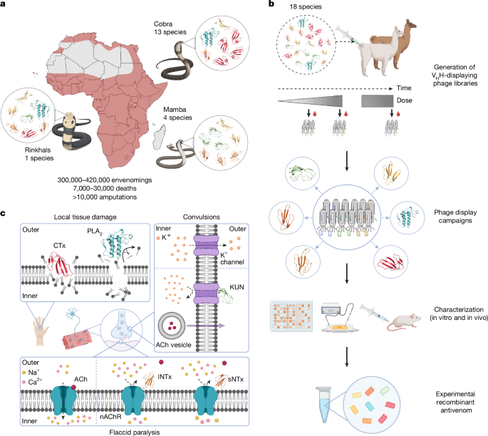
a, Distribution of the 18 most medically relevant elapid snakes from sub-Saharan Africa with the estimated snakebite envenoming incidence, fatality rate and morbidity1,61. b, Schematic overview of the pipeline used for the development of the experimental recombinant antivenom. An alpaca and a llama were immunized with a mixture of whole venoms from 18 elapid snakes to generate immune VHH-displaying phage libraries for discovery of VHHs with broad cross-reactivity and high affinity. The VHHs were characterized in vitro and in vivo, and eight VHHs were chosen and combined into an experimental recombinant antivenom. c, Visual representation of the pathology of the included major toxin families. A type IA cytotoxin62 (CTx, dark red, Protein Data Bank (PDB) ID: 9BK6) disrupts lipid bilayers, and a PLA2 (cyan, PDB ID: 1A3D)63 catalyses the hydrolysis of fatty acids from phospholipids, both of which cause local tissue damage37,64,65. An sNTx66 (dark orange, PDB ID: 1VB0) and an lNTx62 (light orange, PDB ID: 9BK5) both block acetylcholine (ACh) from binding to nAChRs, preventing ion flux and leading to flaccid paralysis31. A KUN67 (light green, PDB ID: 1DEM) blocks voltage-gated K+ channels, causing enhanced acetylcholine release and subsequent convulsions68. Created in BioRender. Burlet, N. (2025) https://BioRender.com/1lypaqd.
To overcome the limitations of current antivenoms, the development of alternative broad-spectrum antivenoms, with a high content of therapeutically active antibodies and improved efficacy against both lethality and local tissue damage, is critically warranted. To this end, recombinant monoclonal broadly neutralizing antibodies have shown great promise11. Specifically, it has been demonstrated that a single broadly neutralizing antibody targeting a specific toxin (sub)family can prevent toxicity and/or lethality in mice injected with whole venoms predominantly composed of toxins from that same (sub)family12,13,14,15,16,17. In addition, mixtures of a few recombinant monoclonal antibodies or antigen-binding fragments of camelid heavy-chain-only antibodies, VHHs (nanobodies), have been shown to neutralize at least one or two venoms18,19,20,21. However, to truly qualify as an alternative to existing therapy, a recombinant antivenom must effectively neutralize venoms from all medically relevant snake species within a region, across different genera. This is particularly challenging to achieve owing to the complex compositions of snake venoms, where the venom from a single species can contain up to a hundred toxins from multiple different protein families22, which can vary extensively both inter- and intraspecifically3. This complexity has created a prevailing view that neutralizing all medically relevant toxins will require an impractically large number of antibodies.
Here we demonstrate that neutralization of 17 out of the 18 most medically relevant elapid snakes in sub-Saharan Africa can be achieved with a surprisingly low number of broadly neutralizing toxin-targeting VHHs. After identifying the medically relevant toxins in the venoms of the 18 elapid snakes (mambas, cobras and rinkhals), we used phage display technology23,24 to identify VHHs that were evaluated for their ability to bind and neutralize their target toxins (Fig. 1b). We then designed an experimental recombinant antivenom based on an oligoclonal mixture of the top 8 VHHs and demonstrated that this VHH cocktail can preclinically prevent venom-induced lethality against all of the 18 most medically relevant elapid snakes in sub-Saharan Africa, except Dendroaspis angusticeps, in pre-incubation experiments. We further validated the efficacy of the antivenom using a representative panel of 11 venoms in a more stringent rescue model that better simulates a real-world snakebite scenario. We also demonstrated that the recombinant antivenom can inhibit or reduce morbidity-causing venom-induced dermonecrosis, a previously unexplored therapeutic area for VHHs. Collectively, this study demonstrates the feasibility of developing a polyvalent recombinant antivenom with continent-wide species coverage, offering hope for advancing such snakebite envenoming therapeutics to future clinical application.
Discovery of VHHs against elapid toxins
Africa is inhabited by many venomous snake species, primarily belonging to the viperid or elapid families; however, many are of no or little medical concern. As outlined by the World Health Organization (WHO), within sub-Saharan Africa there are a total of 18 elapid snakes that can be considered the most medically important25,26 (Fig. 1a and Supplementary Table 1). They belong to 3 genera: Dendroaspis (mambas; 4 species), Hemachatus (rinkhals; 1 species) and Naja (cobras; 13 species); with the genus Naja being further divided into 3 subgenera: Uraeus (cape cobras; 5 species), Boulengerina (forest/water cobras; 1 species) and Afronaja (African spitting cobras; 7 species). The clinical manifestations of envenoming vary markedly among these five (sub)genera. Species belonging to Dendroaspis, Uraeus and Boulengerina primarily induce neurotoxic symptoms, whereas species belonging to Hemachatus and Afronaja mostly cause severe local tissue damage27 (Fig. 1c). Neutralizing the wide range of toxic effects that underlie these manifestations is thus a complex endeavour, which complicates the development of effective therapies with broad coverage of syndromes and snake species28.
To identify VHHs targeting toxins from these venoms, we first generated immune VHH-displaying phage libraries from an alpaca and a llama injected with a mixture of venoms from the 18 medically important elapid snakes (Fig. 1b). Injection doses were increased across bi-weekly intervals over 8 time points, and 3 additional booster injections were administered 52, 54 and 60 weeks after the first immunization20. We collected blood samples at different time points during the immunization time course to generate three unique VHH-displaying phage libraries (Supplementary Table 2).
We next identified the most medically relevant toxins present in the 18 elapid venoms, on the basis of our previous work29. These toxins belong to three distinct protein families: three-finger toxins (3FTx), phospholipase A2 (PLA2) and Kunitz-type serine protease inhibitors (KUN). We purified the toxins using reversed-phase high-performance liquid chromatography (RP-HPLC) and performed proteomic analysis of the 47 fractions in which the key toxins were expected to be present (Extended Data Fig. 1 and Supplementary Table 1). The most abundant 3FTx found in these fractions belonged to five different subfamilies, namely type I α-neurotoxin (sNTx), type II α-neurotoxin (lNTx), type IA/IB cytotoxin (CTx), orphan group XI (Og XI) and aminergic toxin (AgTx)30,31,32. In this study, fractions and purified toxins are denoted by the name of the main medically relevant toxin (sub)family present in the fraction, followed by a number (for example, lNTx-1, lNTx-2, and so on) for ease of reference (Supplementary Table 1).
Overall, 16 fractions (3 lNTx, 3 sNTx, 2 KUN, 2 Og XI, 1 AgTx, 1 PLA2 and 4 CTx) were selected to be used as targets in subsequent phage display selections, on the basis of their abundance in the corresponding venom and their purity (Supplementary Table 3, Supplementary Table 1, Extended Data Fig. 2). Throughout the phage display campaigns, we utilized cross-panning strategies33,34, including for example, exposure to sNTx fractions from different snake species and/or decreasing antigen concentrations in consecutive rounds to enrich for VHHs with broad cross-reactivity and/or high affinity (Supplementary Table 3).
VHHs show broad neutralization in vitro
Following the phage display campaigns, we subcloned 15 phage pool outputs, expressed more than 3,000 monoclonal VHHs in Escherichia coli, and screened them for binding to their cognate target toxins using an expression-normalized capture dissociation-enhanced lanthanide fluorescence immunoassay (DELFIA). Approximately 60% of the VHHs bound to their target toxins (Fig. 2a), and 25% of the clones with varying signal intensity were tested for cross-reactivity in a secondary DELFIA-based screening. Here we observed that more than 50% of the VHHs bound multiple toxins from the same toxin (sub)family, which after sequencing revealed more than 100 unique VHH clones. Sequences of the lead VHHs and complementarity-determining region 3 of hit VHHs are provided in Supplementary Table 4. We speculate that the identification of multiple high-affinity, cross-reactive VHHs may have been facilitated by our immunization strategy, which involved multiple venoms with a broad range of similar and dissimilar toxins (antigens). After our screening campaign, we further evaluated the top 21 unique cross-reactive VHHs against their corresponding toxin families in a dose–response DELFIA, yielding half-maximal effective concentrations (EC50s) ranging from 1 nM to 15 nM (Fig. 2b). Subsequently, we selected the top 15 VHHs on the basis of broad cross-reactivity and low EC50s, and evaluated their binding kinetics using biolayer interferometry (BLI). All of the 15 tested cross-reactive VHHs displayed low nanomolar affinity (Kd) with slow dissociation rates (koff < 5.5 × 10⁻⁴ s−1) for most of their target toxins (Fig. 2c, Extended Data Fig. 3 and Supplementary Table 5).

a, Screening of more than 3,000 VHH clones for binding to their cognate toxin in an expression-normalized capture DELFIA. Only VHHs with a signal intensity ten times above the background level are shown, and the number of clones exceeding this threshold is displayed on the x axis. RFU, relative fluorescence units. b, Dose-response binding curves for 21 VHHs against a venom fraction or a toxin representing each targeted toxin (sub)family, measured using expression-normalized capture DELFIA. c, Isoaffinity plot of binding of 15 cross-reactive VHHs to venom fractions and toxins from various snake (sub)genera measured with BLI. Owing to the instrument’s detection limit for koff of 10−7 s−1, four points below this cutoff are plotted at ≤10−7 s−1. The red diagonal lines represent specific Kd values, which are labelled above each line. kon, association rate.
Next, we evaluated the ability of these VHHs to neutralize their target toxins in vitro. lNTx and sNTx exert their function by binding to the nicotinic acetylcholine receptors (nAChRs) at neuromuscular junctions, preventing acetylcholine binding and ion influx, which disrupts nerve–muscle communication and often results in paralysis35 (Fig. 1c). To address the neutralization of lNTx and sNTx, we measured nAChR currents in a human rhabdomyosarcoma cell line in patch clamp assays36 and observed that pre-incubation of anti-lNTx (a-lNTx) VHHs with lNTx-3, lNTx-5 and lNTx-7 or anti-sNTx VHHs with sNTx-1, sNTx-3 and sNTx-6 before addition to the cells protected the nAChR-mediated current—that is, they showed complete inhibition of neurotoxicity down to a 1:1 molar ratio between VHH and toxin (Fig. 3a).

a, Neutralization of sNTx- and lNTx-mediated blocking of the muscle type nAChR current in whole-cell patch clamp. Dose-response curves are shown with increasing concentrations of VHH to prevent the blocking of nAChRs by sNTx-1, sNTx-3, sNTx-6, lNTx-3, lNTx-5 and lNTx-7. Experiments were performed using eight technical replicates, and results are expressed as mean ± s.d. Toxin concentrations are shown in parentheses, and the dotted line represents a 1:1 molar ratio of toxin:VHH. b, Neutralization of venom-induced cytotoxicity using a cell viability assay with a N/TERT keratinocyte cell line. The positive control (PC) includes medium supplemented with PBS and was set to 100% cell viability. For the negative control (NC), medium supplemented with Triton X-100 was added, resulting in complete cell death. As an additional control, VHH without any venom was included (VHH). For all venoms, − indicates 2 × half-maximal inhibitory concentration (IC50) of the venom without VHH addition, and + indicates venom incubated with a VHH at a 1:5 molar ratio of CTx or PLA2 to VHH. Experiments were conducted in triplicate, and results are expressed as mean ± s.d. c, Neutralization of PLA2 enzymatic activity in the whole venoms of H. haemachatus and all Afronaja species by VHH20 a-PLA2 using a colorimetric enzymatic activity assay with the chromogenic substrate 4-nitro-3-(octanoyloxy)benzoic acid (NOBA). Experiments were conducted in duplicate, and results are expressed as mean ± s.d.
Besides neurotoxicity, envenoming from 8 of the 18 elapid snakes included in this study—7 Afronaja and 1 Hemachatus species—can cause severe local tissue damage. This is primarily owing to the toxic actions of CTx and PLA2 present in their venoms, acting either alone or synergistically37,38 (Fig. 1c). We therefore assessed the ability of the anti-CTx VHHs and an anti-PLA2 VHH to neutralize venom-induced cytotoxicity in cell viability assays. Specifically, we pre-incubated venoms from the eight snake species with three different VHHs individually—VHH1 a-CTx, VHH4 a-CTx and VHH20 a-PLA2—before addition to a keratinocyte cell line. Both anti-CTx VHHs demonstrated broad neutralization, providing 75–95% and 50–85% protection, respectively, against the cytotoxic effects from the venoms of all seven Afronaja species, whereas VHH20 a-PLA2 showed almost no neutralization of any of the tested venoms (Fig. 3b). We observed no neutralization of Hemachatus haemachatus venom, which was in accordance with the binding data, which showed that neither VHH1 a-CTx nor VHH4 a-CTx bound to CTx from this venom. Among the 18 elapid snakes studied, PLA2 exhibit the highest enzymatic activity in Afronaja, moderate activity in Boulengerina and Hemachatus, and negligible activity in Uraeus and Dendroaspis29. Thus, we also assessed the neutralizing effect of VHH20 a-PLA2 in an enzymatic assay and observed complete inhibition of PLA2 activity in the Afronaja venoms, but no neutralization of H. haemachatus venom (Fig. 3c).
Design of a VHH cocktail
After demonstrating in vitro neutralization of several medically important toxin subfamilies by multiple VHHs, we performed a series of WHO-recommended mouse pre-incubation experiments39,40 to examine whether these neutralizing effects would translate to in vivo protection. We first determined the median lethal dose (LD50) for venom fractions, toxins and whole venoms when administered intravenously (Supplementary Table 6), after which venoms were pre-incubated with either single or multiple VHHs and administered to mice, with survival monitored for 24 h.
With the aim of having as few VHHs as possible in the recombinant antivenom, we evaluated the most broadly neutralizing VHHs against each toxin (sub)family. We started with neutralization of individual pure toxins or toxin fractions, then moved to simple whole venoms with a few different toxin subfamilies present, and finally evaluated the neutralization of more complex venoms. First, we evaluated the neutralization of sNTx-3 and lNTx-7 using VHH5 a-sNTx and VHH9 a-lNTx, respectively (Extended Data Fig. 4a,b and Supplementary Table 7); this resulted in the survival of all mice. Thereafter, we combined these two VHHs and tested them on the venoms from Naja haje and Naja melanoleuca, species that are known to produce venoms rich in sNTx and lNTx. Pre-incubation of the venoms with these two VHHs prevented lethality in all mice (Extended Data Fig. 4c,d and Supplementary Table 7). We then moved to the more complex venom of Dendroaspis viridis, which contains sNTx and lNTx as well as AgTx and Og XI25. Pre-incubation of venom with a mixture of VHH5 a-sNTx, VHH9 a-lNTx, VHH15 a-Og XI and VHH13 a-AgTx protected all mice (Extended Data Fig. 4e and Supplementary Table 7), qualifying these four VHHs for inclusion in the recombinant antivenom. Next, we also wanted to ensure the neutralization of black mamba venom (Dendroaspis polylepis), which is rich in dendrotoxins from the KUN family but also contains sNTx and lNTx. We observed that adding VHH17 a-KUN to the four VHHs resulted in the survival of all mice when challenged with D. polylepis venom (Extended Data Fig. 4f and Supplementary Table 7). To evaluate the necessity of VHH15 a-Og XI and VHH13 a-AgTx, a mixture of VHH5 a-sNTx, VHH9 a-lNTx and VHH17 a-KUN was tested against D. viridis and Dendroaspis jamesoni venom. Although mice challenged with D. viridis venom all survived, they showed signs of lethargy, and only two mice survived after being challenged with D. jamesoni venom, illustrating that full neutralization is not achieved without VHH15 a-Og XI and VHH13 a-AgTx VHHs (Extended Data Fig. 4g and Supplementary Table 7). Finally, we evaluated the neutralization of PLA2 and CTx using Naja nigricollis venom and observed that a combination of VHH20 a-PLA2 and two anti-CTx VHHs (VHH1 a-CTx and VHH4 a-CTx), which show different binding patterns to CTxs and therefore might provide broader neutralization than each of the anti-CTx VHHs alone, protected all mice from lethal venom effects (Extended Data Fig. 4h and Supplementary Table 7). In total, we selected eight lead VHHs targeting seven medically important toxin subfamilies and combined these into an experimental polyvalent recombinant antivenom.
To gain insights into the molecular basis of the broad neutralization of the eight lead VHHs, co-crystallization, cryo-EM and in silico structural modelling predictions of the VHHs in complex with one of their target toxins were used to determine their binding interactions. These experiments indicated that the VHHs interact with residues that are predominantly conserved across the target toxins that they neutralize (Fig. 4 and Extended Data Figs. 5a–c, 6a–c and 7) and that the VHH–sNTx and VHH–lNTX interactions are similar to those previously reported14,17,41. Of note, we observed that VHH1 a-CTx was biparatopic, which may explain why this VHH was better at neutralizing cell cytotoxicity than VHH4 a-CTx (Fig. 3b). To our knowledge, such biparatopic behaviour has not previously been reported and is worthy of further investigation.

a,b, The biparatopic VHH1 a-CTx (light blue) binding two different epitopes of cardiotoxin (UniProt: P01468) (salmon) (a) and VHH5 a-sNTx (light blue) in complex with short neurotoxin 1 (P01426) (purple) (b). Magnified views illustrate hydrogen bonds (black dotted lines), π–π and π–CH interactions (orange dotted lines) and van der Waals interactions (red dotted lines). Sequence alignments of the verified target toxins of each VHH are shown under their structures. The epitope residues of the target toxin present in the co-crystal structure are highlighted in salmon and pink, respectively.
The VHH cocktail prevents lethality
To assess whether the recombinant antivenom (that is, the defined cocktail of 8 selected VHHs) could neutralize venom-induced lethality caused by all of the 18 most medically relevant snakes in sub-Saharan Africa, we performed a series of pre-incubation experiments in mice39. In this set of experiments, we first determined the LD50s for the whole venoms injected intravenously (Supplementary Table 6). Then, three times the LD50s of the venoms were pre-incubated with the same dose of recombinant antivenom and administered intravenously to groups of five mice. The composition and dose of the recombinant antivenom were kept consistent across all venoms to simulate a real product being deployed in Africa, with the dose corresponding to at least a 1:10 molar ratio between each targeted toxin (sub)family and the respective VHH (Supplementary Table 8). Survival and signs of envenoming were monitored over a 24 h period post-injection. We observed that the recombinant antivenom prevented venom-induced lethality for all included species except D. angusticeps (Fig. 5a). No evident signs of envenoming were observed in mice that were administered recombinant antivenom pre-incubated with venoms from D. jamesoni, H. haemachatus, Naja ashei, Naja katiensis, Naja mossambica, Naja nigricincta, N. nigricollis, Naja nubiae, Naja pallida, Naja anchietae, N. haje, Naja nivea and Naja senegalensis. In mice injected with the recombinant antivenom pre-incubated with the venoms from N. melanoleuca and D. viridis, only minor signs of envenoming, including closed eyes and periods of lethargy interspersed with episodes of excessive grooming, were observed. For mice injected with recombinant antivenom pre-incubated with Naja annulifera venom, similar signs of envenoming appeared after 15 h. The venom from D. polylepis pre-incubated with the recombinant antivenom caused an initial state of severe lethargy, which disappeared after 1–2 min. We speculate that this rapid transient effect could be caused by small molecules in the venom such as adenosine or acetylcholine, which the recombinant antivenom is not designed to neutralize42. For D. angusticeps venom, we observed that the recombinant antivenom prolonged the survival of mice to between 3 and 6 h. This venom is rich in muscarinic toxins, fasciculins and synergistically acting toxins43, and we speculate that the synergy between these toxins, which are not targeted by the recombinant antivenom, could be responsible for the observed delayed lethality.

a, Neutralization efficacy of the recombinant antivenom (AV) against venom-induced lethality caused by 18 elapid venoms in a pre-incubation setup. Venom and the recombinant antivenom were pre-incubated before intravenous injection. b, Neutralization efficacy of the recombinant antivenom against venom-induced lethality caused by 11 snake venoms in a rescue setup. Venom was injected subcutaneously followed by intravenous injection of the recombinant antivenom 5 min later. In both setups, Inoserp PAN-AFRICA antivenom was included for comparison. c, Neutralization of venom-induced dermonecrosis with a mixture of three VHHs (VHH1 a-CTx, VHH4 a-CTx and VHH20 a-PLA2) against three representative cytotoxic venoms from H. haemachatus, N. mossambica and N. nigricollis, in both pre-incubation and rescue setups. In the pre-incubation setup, the VHHs and venom were pre-incubated before intradermal (i.d.) injection, and in the rescue setup, the VHH mixture was injected into the same region as the venom injection 15 min after venom injection. The lesion size was measured 72 h post-injection and results are shown as mean ± s.d. d, The neutralization efficacy of the recombinant antivenom against dermonecrosis caused by the same three venoms in a second rescue setup. In this rescue setup, venom was injected intradermally and the recombinant antivenom was injected intravenously (i.v.) 15 min later. The lesion size was measured 48 h post-injection and results are shown as mean ± s.d. For N. nigricollis venom, in addition to the recombinant antivenom, Inoserp PAN-AFRICA antivenom was included for comparison. c,d, Lesion sizes were compared to PBS controls using Welch’s t-test. Normality (Shapiro–Wilk) and outliers (ROUT) were assessed. Significance was set at α = 0.05. *P ≤ 0.05, **P < 0.01, ***P < 0.001; NS, not significant. n = 5 mice. Created in BioRender. Burlet, N. (2025) https://BioRender.com/rkd7bzp.
To better evaluate the efficacy of the recombinant antivenom in a scenario that mimics real snakebite envenoming, we further performed rescue experiments using 11 venoms from 4 different (sub)genera12,44. To perform these experiments, we first determined the subcutaneous venom LD50s. However, when attempting to determine subcutaneous LD50s for Afronaja venoms, we observed severe muscle and skin damage at the injection sites, and mice had to be euthanized owing to ethical considerations. Given that local tissue damage is the most clinically relevant effect of Afronaja envenomings, we deemed rescue-from-lethality experiments for these species to be ethically unjustifiable. For the remaining species, we administered three times the LD50 of the venom subcutaneously (Supplementary Table 6), followed by intravenous administration of the recombinant antivenom 5 min later, and monitored survival and signs of envenoming over a 24 h period post-injection (Supplementary Table 9). In these rescue experiments, the dose of the recombinant antivenom was increased compared with the pre-incubation experiments relative to the increase in LD50s that was observed when switching from intravenous to subcutaneous administration of venom (Supplementary Table 9). For comparison, the commercial Inoserp PAN-AFRICA antivenom was included at a dose recommended by the manufacturer to neutralize three times the LD50 of the venom. We observed that the recombinant antivenom completely prevented lethality induced by the venoms from N. haje, N. annulifera, N. nivea, N. senegalensis, N. nubiae and H. haemachatus (Fig. 5b and Supplementary Table 9), and the mice showed no signs of envenoming. Furthermore, the recombinant antivenom also prevented lethality induced by the venom of D. viridis (Fig. 5b and Supplementary Table 9), although we observed signs of envenoming, including limited movement, approximately 4 h after venom injection and continuing throughout the experiment. In addition, after 20 h, four out of five mice developed swelling and haemorrhage of the eyeballs, and one mouse developed an increased abdominal volume. For N. melanoleuca venom, partial neutralization was observed, with three out of five mice surviving, and the time of death being substantially extended for the other two mice (Fig. 5b and Supplementary Table 9). The surviving mice presented similar signs of envenoming as observed for D. viridis, but did not show an increased abdominal volume. Finally, for D. polylepis venom, the recombinant antivenom delayed the time of death from approximately 0.5 h in the venom-only control mice to 2 h; for D. jamesoni venom, the time of death was delayed from approximately 0.5 h to 1 h; and no protective effect was seen with the venom from D. angusticeps (Fig. 5b and Supplementary Table 9). On the basis of the difference between pre-incubation and rescue experiments for D. polylepis and D. jamesoni, we hypothesize that this is owing to the complex interplay between venom and antivenom pharmacokinetics. It has been observed that the ability of antivenoms to redistribute and neutralize the toxins once they have reached their target strongly depends on the affinity of the antibodies present in the antivenom for the toxin as well as the affinity of the toxin for its respective target within the tissue45. Furthermore, a delayed release of the toxins from the subcutaneous injection site, due to the depot effect, could occur after the VHHs have been cleared from the circulation, which could have resulted in a recurrence of envenoming signs in the absence of repeated therapeutic dosing46,47. Notably, in the rescue setting, the Inoserp PAN-AFRICA antivenom showed only partial neutralization of all of the tested venoms and an extension of time of death for the venom from N. melanoleuca, demonstrating that, except for D. polylepis, the recombinant antivenom performed better than the commercial antivenom on all included venoms at the tested doses (Fig. 5b and Supplementary Table 9).
The VHH cocktail prevents dermonecrosis
Current plasma-derived antivenoms are typically poor at preventing local tissue damage, resulting in a high morbidity rate, including limbs lost, in snakebite victims8,9,10. In elapid snakes in sub-Saharan Africa, local tissue damage is primarily associated with spitting snake species (Afronaja and Hemachatus spp.), and CTx and PLA2 have the most important role38. We therefore examined the ability of our two anti-CTx VHHs (VHH1 a-CTx and VHH4 a-CTx) and anti-PLA2 VHH (VHH20 a-PLA2) to prevent venom-induced dermonecrosis caused by N. mossambica, N. nigricollis and H. haemachatus using an in vivo pre-incubation setup. We mixed the three VHHs and pre-incubated this mixture with each of the venoms before intradermal injection and measured the lesion size after 72 h. The VHH mixture significantly reduced the dermonecrotic lesion areas, and all but one mouse per treatment group showed a complete absence of lesion for the two Naja venoms (Fig. 5c). We then moved to a rescue setup, where the VHH mixture was delivered intradermally to the same injection region 15 min after the venom. Despite observations of rapid discoloration at the injection site within the 15 min treatment window, the VHH mixture significantly reduced the size of the dermonecrotic lesions caused by each of the three venoms (Fig. 5c and Extended Data Fig. 8a).
After demonstrating the efficacy of the mixture of VHH1 a-CTx, VHH4 a-CTx and VHH20 a-PLA2 in both pre-incubation and rescue setups, we evaluated the recombinant antivenom (containing the eight VHHs) in another rescue assay, where the recombinant antivenom was delivered intravenously 15 min after intradermal injection of the venom. Inoserp PAN-AFRICA antivenom was included for comparison with N. nigricollis venom at a dose recommended by the manufacturer to neutralize three times the LD50 of the venom. The recombinant antivenom reduced the size of the lesions caused by each of the three venoms at the 48 h experimental endpoint, although these results were statistically significant only for N. nigricollis (Fig. 5d and Extended Data Fig. 8b). In comparison, we observed less, and statistically insignificant, reduction of lesion size for Inoserp PAN-AFRICA antivenom on the tested venom from N. nigricollis (Fig. 5d), demonstrating that the recombinant antivenom performs better than the existing treatment in this model at the tested doses.
Discussion
Previous studies have shown that broadly neutralizing antibodies or VHHs against snake toxins can be identified using phage or yeast display technology12,14,18,20,34,41,48,49. However, although these studies demonstrated that rodents challenged with venoms dominated by neurotoxins can be rescued using such antibodies14,18 and VHHs20, they represent only an initial proof of principle when viewed from a product development or clinical perspective. Notably, all neutralized venoms have been dominated by only one or two toxin families, and all but three studies17,18,20 have reported the use of only a single monoclonal antibody or VHH to neutralize a whole venom. In this study, we extend earlier work and demonstrate how an experimental polyvalent recombinant antivenom can be developed using as few as eight VHHs targeting toxins from seven different protein subfamilies to achieve continent-wide elapid species coverage. We demonstrate its in vivo efficacy on 18 elapid venoms using the WHO-recommended pre-incubation model and further confirm the efficacy on 11 of the venoms in a more challenging rescue model, which more closely mimics a real snakebite scenario44. Furthermore, at the tested doses, this experimental recombinant antivenom performs better than the commercial plasma-derived antivenom, Inoserp PAN-AFRICA, in preventing both dermonecrosis and lethality across all snake species tested, apart from D. polylepis. By showing that an efficacious polyvalent recombinant antivenom with unprecedented broad species coverage can be constructed using only eight VHHs, we challenge the common belief that only polyclonal antibodies can overcome and neutralize the complexity of snake venoms. Thereby, we answer the long-standing and important question in toxinology of how few antibodies are sufficient to develop a recombinant antivenom with clinical utility. Our work thus solidifies our previous working hypotheses that it is indeed possible to unravel the challenging complexity of venoms through proteomic analysis29 and identify which toxins are sufficient to neutralize in order to develop effective antivenom products28,50,51.
In addition to mortality, we demonstrate the efficacy of the experimental recombinant antivenom in reducing dermonecrosis caused by the venoms of the spitting elapid snakes, N. nigricollis, N. mossambica and H. haemachatus, in a pre-incubation model and in two rescue models. Although plasma-derived antivenoms have demonstrated efficacy in pre-incubation models of dermonecrosis, they have rarely been assessed in rescue models52. Further, it is well established that antivenoms are largely ineffective in preventing severe local tissue damage unless given rapidly after a bite52,53, which is typically attributed to the limited ability of whole IgG molecules or IgG fragments administered intravenously to penetrate deep tissue and the speed with which local tissue damage develops. In comparison, we here show that delayed local administration (intradermal) of toxin-specific VHHs significantly reduces dermonecrosis caused by different elapid snake venoms, analogous to recent findings with the PLA2-inhibiting small molecule varespladib and the CTx-inhibiting heparinoid tinzaparin54,55. In addition, the recombinant antivenom further provides significant protection against dermonecrosis caused by N. nigricollis venom in a rescue model when administered intravenously. This finding is particularly promising given the rapid onset of toxicity caused by locally acting toxins that induce tissue damage, the ineffectiveness of current antivenoms (Fig. 5d) and the lack of protection previously offered by intravenous varespladib54. Of note, neither VHH20 a-PLA2, VHH1 a-CTx nor VHH4 a-CTx neutralizes the H. haemachatus venom in vitro in the cell viability or phospholipase activity assays. These findings highlight the need for further investigation to elucidate the mechanisms underlying the observed neutralization of local tissue damage in mouse experiments.
Regarding design features for our experimental recombinant antivenom, we chose to use the VHH format, as this simple antibody format could provide a recombinant antivenom with several advantages. First of all, the use of this clinically proven antibody format might aid in the development of a safe product with a low risk of causing severe immunological reactions upon administration owing to the typically low immunogenicity observed for VHHs5. An enhanced safety profile may enable earlier treatment of patients by removing the need to wait for clinical syndromes to manifest before administration of antivenom. Second, VHHs are known to have a remarkably high biophysical stability, which is likely to translate into a long shelf-life that might be advantageous for products that may need to be stockpiled for longer periods, possibly in humid or high-temperature conditions. Third, the small size of VHHs enables them to rapidly distribute from the bloodstream into the surrounding tissues and thereby neutralize toxins at the bite site or site of action. Finally, the small size of VHHs compared to full-length IgGs provides them with a large neutralization capacity per mass of antivenom56. Combined with the possibility of manufacturing VHHs using low-cost microbial expression systems, this might facilitate a lower cost of goods for treatment. In turn, a lower cost of goods could positively affect affordability and make antivenoms more accessible to people in low- and middle-income countries who disproportionally suffer the greatest burden of snakebite57.
Despite these advantages, recombinant VHHs are also associated with certain limitations. Most importantly, the short half-life of VHHs in circulation could potentially limit their sustained action over time, which could prove to be problematic, as snake toxins may exit the bite site and enter circulation long after the snakebite incident owing to the depot effect46,47. However, it is also possible that the ability of VHHs to rapidly penetrate into deep tissue could counter this effect, as it might allow the VHHs to neutralize the toxins before they are released into circulation. To study this complex interplay between pharmacodynamics of the VHHs and the toxicokinetics of the venom components, large animal experiments are probably required. If it is found necessary, repeated pharmacokinetic-informed dosing can be applied or the half-life of the VHHs could possibly be extended through various antibody engineering techniques, such as multimerization or linking them to a human IgG Fc domain20. Finally, it is possible that our cocktail could be further simplified by reducing the number of molecules included, which is likely to be beneficial from a manufacturing perspective. This could be achieved by improving the broadly neutralizing capacity of some of our VHHs, such as those targeting CTx, and including only one broadly neutralizing VHH per toxin (sub)family. Moreover, engineering heterodivalent or trivalent VHH constructs58,59, through fusion of two or three VHHs, could reduce the number of individual molecules required to neutralize the described toxin specificities, which could further simplify manufacturing. However, the total amount of material needed in terms of mass would remain the same (or become higher), and we speculate that the favourable deep tissue-penetrating properties of the small monomeric VHHs risk being compromised, based on what is known from the oncology field60. Nevertheless, further refinement of the cocktail is likely to be possible and should be further investigated alongside the development of a manufacturing strategy and an assessment of whether the VHHs might be useful for neutralizing venoms from related Asian elapids.
Although our work here focused on developing a defined oligoclonal mixture of VHHs against African elapid venoms, the strategy utilized for antibody discovery and therapeutic prototype design holds promise for broader applications. Specifically, it may be valuable in clinical contexts where targeting multiple isoforms across both closely and distantly related protein families is essential. We speculate that, beyond other animal envenomings, the approaches described here could be useful for the development of advanced therapies against infectious diseases or complex immunological or endocrinological disorders.
Methods
Construction of an immune VHH-displaying phage library
Immune VHH-displaying phage libraries were constructed at the VIB nanobody core (Brussels, Belgium) as described20. To generate VHH-displaying phage libraries, 1 alpaca and 1 llama were injected subcutaneously at bi-weekly intervals across 8 time points with increasing doses of venom mixtures from the 18 most medically relevant elapid snakes in sub-Saharan Africa: D. angusticeps, D. jamesoni, D. polylepis, D. viridis, N. anchietae, N. annulifera, N. ashei, N. haje, N. katiensis, N. melanoleuca, N. mossambica, N. nigricincta, N. nigricollis, N. nivea, N. nubiae, N. pallida, N. senegalensis and H. haemachatus. Following the initial series of injections, 3 additional booster injections were administered at 52, 54 and 60 weeks after the first immunization (Supplementary Table 2 summarizes the detailed immunization schedule). For library generation, blood samples were collected on days 5 and 8 following the first set of 4 injections. The two blood samples from each animal were pooled separately and individual libraries were prepared for each animal. A total of 6 VHH-displaying phage libraries (1 library per time point and animal) was prepared by pooling the total RNA samples after days 46 and 49 (library A), 102 and 105 (library B) and days 5 and 8 following the final booster injections (library C).
Purification and biotinylation of the venom fractions and toxins
Cardiotoxin (P01468) from N. pallida, α-cobratoxin (P01391) from N. kaouthia, α-short-chain neurotoxin (P01426) from N. pallida, and whole venoms from the above-mentioned 18 elapid snakes were purchased in lyophilized form from Latoxan (catalogue numbers and origin of the specimens can be found in Supplementary Table 1). Venom fractions containing short-chain neurotoxins (sNTx), long-chain neurotoxins (lNTx), cytotoxins (CTx), Og XI, AgTx, PLA2 and dendrotoxins (DTx) were isolated from the whole venoms using RP-HPLC (Agilent 1200) with a C18 column (250 × 4.6 mm, 5 μm particle; Teknokroma). 1 mg of venom solubilized in 100 μl solution A (MilliQ water supplemented with 0.1% trifluoroacetic acid (TFA)) was applied to the column and elution was performed at a rate of 1 ml min − 1 using solution A and a gradient towards solution B (acetonitrile supplemented with 0.1% TFA): 0% B for 15 min, 0–15% B over 15 min, 15–45% B over 60 min, 45–70% B over 10 min, and 70% B over 9 min, as described69. Fractions were collected and the solvent evaporated using a vacuum centrifuge. The venom fractions purified via RP-HPLC and toxins bought from Latoxan were dissolved in phosphate buffered saline (PBS: 137 mM NaCl, 3 mM KCl, 8 mM Na2HPO4·2H2O, 1.4 mM KH2PO4, pH 7.4) and biotinylated by amine coupling using a 1:1 to 1:3 molar ratio of venom fraction or toxin to EZ-Link NHS-PEG4-Biotin reagent (Thermo Scientific, A39259), as described34. Free biotin was removed using 2 or 4 kDa MWCO ultracentrifugation membranes (Vivacon 500, VN01H91 and Amicon Ultra-4, UFC8000324, respectively) in accordance with the manufacturers’ guidelines. Following purification, the degree of biotinylation was analysed by matrix-assisted laser desorption/ionization time-of-flight (MALDI-TOF) mass spectrometry using ProteoMass Protein MALDI-MS Calibration Kit (Sigma-Aldrich, MSCAL3) and an Ultraflex II TOF/TOF spectrometer (Bruker Daltonics), as described70.
Proteomics analysis of the selected venom fractions
From each venom fraction, 5 µg was diluted in 50 mM ammonium bicarbonate to a total volume of 25 µl. The samples were reduced and alkylated by 10 mM TCEP and 40 mM chloroacetamide before digestion with either GluC or trypsin in an enzyme-to-protein ratio of 1:100. Samples were incubated overnight at 37 °C, after which the digestion was stopped by addition of 2% TFA for a final concentration of 1%. The samples were desalted with SOLAµ SPE plate (HRP, Thermo) C18 columns, following the same procedure as described71. Dried peptides were reconstituted in 12 µl 2% acetonitrile, 1% TFA, and an estimated 500 ng of peptides was used for mass spectrometry analysis.
Peptides were loaded onto a 2 cm C18 trap column (ThermoFisher 164946), connected in-line to a 15 cm C18 reverse-phase analytical column (Thermo EasySpray ES904) using 100% solvent A (0.1% formic acid in water) at 750 bar, using the Thermo EasyLC 1200 HPLC system, and the column oven operating at 35 °C. Peptides were eluted over a 35 min gradient ranging from 6 to 60% of solvent B (80% acetonitrile, 0.1% formic acid) at 250 nl min−1, and the Q-Exactive instrument (Thermo Fisher Scientific) was run in a DD-MS2 top10 method. Full mass spectra were collected at a resolution of 70,000, with an AGC target of 3 × 10⁶ or maximum injection time of 20 ms and a scan range of 300–1,750 m/z. The MS2 spectra were obtained at a resolution of 17,500, with an AGC target value of 1 × 10⁶ or maximum injection time of 60 ms, a normalized collision energy of 25 and an intensity threshold of 1.7 × 10⁴. Dynamic exclusion was set to 60 s, and ions with a charge state <2 or unknown were excluded.
The raw data from all fractions were analysed with Proteome Discoverer v.2.4. The data were searched against all snake venom proteins (retrieved from Uniprot, 2,263 sequences, accessed 9 November 2021). The trypsin-digested fractions were searched with tryptic specificity, while the GluC-digested fractions were searched with GluC specificity, with two maximum missed cleavages allowed for both proteases. Minimum and maximum peptide lengths were set to 7 and 40, respectively. Precursor mass tolerance was 10 ppm, and fragment mass tolerance was 0.02 Da. Methionine oxidation (+15.995 Da) was set as dynamic modification, while initiator methionine loss (−131.040 Da), acetylation (+42.011 Da), or the combination of methionine loss and acetylation (−89.030 Da) were included as dynamic modifications for the protein terminus. Cysteine carbamidomethylation (+57.021 Da) was added as a static modification. Peptide-spectrum matching was performed with Sequest HT, and false discovery rate (FDR) control with Percolator (0.01 strict and 0.05 relaxed target FDR). FDR was also controlled at the peptide and protein levels with the same target FDRs. Proteins were quantified based on the unique and razor peptides, using the Minora Feature Detector and the Precursor Ions Quantifier nodes with default settings, normalizing abundance to the total peptide amount in each mass spectrometry run and scaling abundance values on the average of all runs.
Clustering toxins on the basis of sequence identity
A sequence similarity network (SSN) was made with the Enzyme Function Initiative-Enzyme Similarity Tool (EFI-EST)72,73. A fasta file containing the UniProt sequences of each discovered toxin from the whole venom of the included 18 elapid snakes was used by the tool to perform an all-by-all BLAST to obtain similarities between sequence pairs. Clustering of toxins with a minimum sequence identity of 70% was subsequently performed by using an alignment score threshold during SSN Finalization that corresponds to 70% identity in the ‘percent identity vs alignment score box plot’ in the Dataset Analysis tab. The obtained SSN was visualized with Cytoscape.
Solution-based phage display selections
VHH-displaying phage libraries were incubated with biotinylated venom fractions or toxins for 2 h at ambient temperature, with end-over-end rotation (Supplementary Table 3 summarizes the final concentration of target toxins and the libraries used in each selection round). Streptavidin coated Dynabeads (M-280, Fisher Scientific, 10465723) were blocked in PBS containing 3% non-fat dried milk powder for 1 h with end-over-end rotation, before addition to the target toxins mixed with the phage library. In each selection round, a background control was included where no antigen was mixed with the phage library. Subsequently, a KingFisher Flex system (Thermo Scientific, 711-82573) was used to wash the beads 3 times with PBST (PBS + 0.1% Tween) and 3 times with PBS, before eluting the bound phages in 120 µl of 0.1 mg ml−1 trypsin (Sigma-Aldrich, T9201-500MG) in phage elution buffer (50 mM Tris, 1 mM CaCl2, pH 8.0). The eluted phages were amplified using the M13KO7 helper phage and concentrated by polyethylene glycol precipitation.
Subcloning, screening, and sequencing of VHHs
Phagemids from the chosen selection outputs (Supplementary Table 3) were purified using the GeneJET Plasmid MiniPrep Kit (Thermo Fisher, K0503) according to the manufacturer’s protocol. The VHH-encoding genes were subcloned into the pBDS100 expression vector using the PstI and Eco91I restriction enzymes (New England Biolabs). Following transformation into the E. coli strain BL21 (DE3) (New England Biolabs), at least 184 individual colonies were picked from each chosen selection output and used for the expression of soluble VHHs. Auto-induction medium74 was used to induce VHH expression for 16 h at 30 °C. Thereafter, periplasmic cell extracts, containing soluble expressed VHHs, were used for primary screenings in a previously described expression-normalized DELFIA20 using 25 nM of target toxin. Clones with a signal intensity ten times higher than the background (no addition of biotinylated target), were cherry-picked and went through a second round of screening in the expression-normalized DELFIA, against multiple target toxins. For the cross-reactive clones, a dose–response experiment was performed, where the Flag-tagged VHHs in the periplasmic extracts were captured onto the wells coated with 2.5 µg ml−1 Flag antibody clone M2 (F3165, Sigma-Aldrich); however, instead of a single concentration, a serial dilution of target toxins (1:1,000 nM) was added. Clones displaying a signal intensity 50 times over the negative control, and/or a low EC50 in the dose–response curves, were Sanger sequenced (Eurofins Genomics sequencing service) using the M13Rev primer (CAGGAAACAGCTATGAC). The VHH frameworks and the complementarity determining regions (CDRs) were annotated using CLC Main Workbench (Qiagen) and the VHHs with unique CDR sequences were produced for in vitro and in vivo assays.
Production of VHHs for in vitro and in vivo experiments
For expression of VHHs at scales up to 100 ml, the periplasmic extracts containing VHHs were produced as described in the screening section and then purified using Ni-resin (Sigma-Aldrich, P6611) via gravity flow. For larger-scale expressions (>250 ml), BL21 (DE3) cells, containing the plasmid encoding a unique VHH, were cultivated as described20. Thereafter, the VHH-containing supernatants were purified using immobilized metal ion affinity chromatography with a 2 ml column volume of Ni-NTA resin (HIS-select Nickel Affinity Gel, Sigma-Aldrich, P6611) equilibrated with PBS supplemented with 200 mM NaCl and 20 mM imidazole, pH 8.0. Elution was performed with PBS containing 200 mM NaCl and 135 mM imidazole, pH 8.0, followed by an overnight dialysis in SnakeSkin Dialysis Tubings (10 kDa MWCO, ThermoFisher Scientific, 68100) against PBS. Subsequently, VHHs were concentrated using Amicon Ultra-15 centrifugal filters (3 kDa MWCO, Fisher Scientific, 10781543).
Kinetic analysis of VHHs using BLI
The binding of VHHs to the venom fractions and toxins was analysed using BLI (Octet-BLI; Octet RED 96, ForteBio). Biotinylated venom fractions and toxins at a concentration of 0.5 µg ml−1 were captured to a target spectral shift of 0.8 nm on a streptavidin-coated BLI biosensor (Sartorius, 18-5020). A biosensor without antigen was included as a reference. VHHs were prepared in running buffer (10 mM HEPES, 150 mM NaCl, 3 mM EDTA, 50 mM MES hydrate, and 0.05% P20 (MES-HEPES), pH 7.2). The toxin-loaded biosensors were dipped into four different VHH concentrations (7.5, 30, 120, 480 nM) and a control without any VHH. VHH association was measured for 600 sec, followed by measuring VHH dissociation in running buffer for 600 sec. Biosensors were regenerated by dipping into the regeneration buffer (10 mM Glycine, 4 M sodium chloride, pH 2.0) between each round, 5 times, for 10 sec each. For analysis, the reference BLI biosensor background was subtracted, a global model assuming a 1:1 interaction was used for fitting of the data, and calculations of kinetic parameters were all made in Octet Analysis Studio 12.2.2.26 (ForteBio).
Patch clamp electrophysiology
Automated planar whole-cell patch clamp experiments were performed as described20. All experiments were performed on a Qube 384 automated patch clamp platform (Sophion Bioscience) with 384-channel, 10X mode patch chips (10 patch holes/site, site resistance 0.2 ± 0.04 MΩ). We used a human rhabdomyosarcoma cell line (American Type Culture Collection, ATCC) endogenously expressing muscle type nAChRs ((α1)2β1γδ) and 70 µM acetylcholine for receptor activation. We first determined the IC80 for the included toxins or venom fractions (sNTX-1, sNTx-3, sNTx-6, lNTx-3, lNTx-5 and lNTx-7) and used this concentration to evaluate the neutralization effect of the corresponding VHHs. The VHHs were used at molar ratios of 9:1 to 1:27 between toxin and VHH. Finally, the inhibitory effect of the toxins on the elicited acetylcholine current was normalized to the full acetylcholine response and averaged in each group (n = 8). The data were analysed with Sophion Analyzer v.6.6.70 (Sophion Bioscience) and GraphPad Prism 10 software.
In vitro neutralization of cell cytotoxicity
A cell viability assay was performed as described34. In brief, N/TERT keratinocytes were seeded at 4,000 cells per well in 100 µl cell culture medium and incubated overnight under standard conditions. After determining the IC50 of each venom, the cells were subjected to a venom concentration of 2 × IC50, either in the absence or presence of a 1:5 molar ratio of CTx or PLA2 to VHH based on the CTx or PLA2 contents of each venom29, followed by a 24 h incubation step. Thereafter, the CellTiter-Glo luminescent cell viability assay (Promega) was performed in triplicate according to the manufacturer’s protocol. A maximal cell death control was included, where the cell culture medium was supplemented with 0.01% Tween 20 to disrupt the cells. In addition, a maximum cell viability control was included, with cell culture medium supplemented with PBS, as well as a VHH control, where cells were incubated with the highest tested VHH concentration without venom, to confirm that the VHHs alone do not affect cell viability. The data were visualized with GraphPad Prism 10 software.
In vitro neutralization of PLA2 enzymatic activity
Venom concentration inducing half of the maximum PLA2 enzymatic activity (EC50) was determined as described29. For inhibitory dose–response curves, VHHs were diluted to 16 µM, followed by a twofold serial dilution in 10 steps. 50 µl of snake venom at a concentration of 4 × EC50 was mixed with the serial dilutions of the VHHs and then incubated at room temperature for 30 min. The enzymatic reaction was started by adding 100 µl of 0.5 mM NOBA into the mixture. Final concentrations of the individual components in the enzymatic activity assays were 0.25 mM NOBA, and a twofold serial dilution of the VHHs with the highest concentration set at 4 µM. After adding NOBA to the wells, plates were shaken at 300 rpm for 2 min, and then incubated at 37 °C for 40 min. Finally, the plates were centrifuged at 4,000g at 4 °C for 3 min, and absorbance was measured at 25 °C at 405 nm using a Multimode Microplate Reader (VICTOR Nivo, HH35000500). The experiments were performed in duplicate and the absorbance averages were determined after subtracting a blank control containing no venom. The data were analysed using the Victor Nivo Control software v.5.1.0 and Graphpad Prism 10 software with a nonlinear fit using ‘Sigmoidal, 4PL, X is concentration’.
Co-crystallization of VHHs and toxins
Lyophilized toxins and vacuum-dried venom fractions were reconstituted at 10 mg ml−1 in 5 mM Tris and 20 mM NaCl at pH 8.0. The toxins or venom fractions were then added to the VHHs at a threefold molar excess (VHH1 a-CTx: cardiotoxin (P01468), VHH5 a-sNTx: α-short-chain neurotoxin (P01426)) and incubated overnight at 4 °C. The VHH:toxin complexes were purified using size-exclusion chromatography (Superdex 75 10/300GL column, Cytiva) on an NGC Quest 10 Plus Chromatography system (Bio-Rad) maintained at 4 °C, with the reconstitution buffer serving as the mobile phase. Before crystal screening, the VHH–toxin complexes were concentrated to 15.0 mg ml−1 using 3.0 kDa MWCO ultracentrifugation filters (UFC500324, Merck).
Crystallization trials were performed at 21 °C via the sitting drop vapour diffusion method. Drops (0.3 µl) were set up at reservoir:protein ratios of 2:1, 1:1, or 1:2 in a 96-well drop format on SWISSCI MRC 2 well crystallization plates (JENA) using LMB, BCS, Index, and Structure screening solutions (Hampton Research). The wells were sealed with crystal clear tape and equilibrated against 50 µl of reservoir solution. The VHH1 a-CTx co-crystal formed in 0.2 M ammonium acetate, 0.1 M sodium acetate, pH 4.6, 30% w/v PEG4000. The VHH5 a-sNTx co-crystal formed in 0.2 M sodium chloride, 0.1 M sodium acetate, pH 4.6, 30% v/v 2-methyl-2,4-pentanediol (MPD). The developed co-crystals were collected using mounted CryoLoops (Hampton Research) with cryoprotection performed by adding glycerol to a neighbour drop with no crystals to a final concentration of 25%. The loop edge was kept in contact with the cryo solution for approximately 5 s to equilibrate before flash freezing the co-crystal in liquid nitrogen and shipping to the beamline for remote data collection. Data collection and refinement statistics are shown in Supplementary Tables 10 and 11. The final structural models and corresponding structure factors have been deposited in the Protein Data Bank (PDB) under accession codes 9RIT and 9RIU.
Data collection and structure determination
X-ray diffraction data for the VHH1 a-CTx and VHH5 a-sNTx co-crystals were obtained at the Biomax (MAX IV synchrotron facility, Lund, Sweden) beamline. Complete datasets were collected over a 360° rotation for the VHH1 a-CTx and VHH5 a-sNTx co-crystals. The data processing was performed with XDSAPP375,76,77, and the data are summarized in Supplementary Tables 10 and 11. Structures of the VHHs in complex with their respective toxins were determined by molecular replacement with Phaser-MR78 using an AlphaFold 3 model for both the VHH and the target toxin as a search model. Model building and refinement were performed with Phenix.refine77 and Coot79.
The structures were evaluated using MolProbity with final statistics presented in Supplementary Tables 10 and 11. Molecular graphics were presented with PyMOL Molecular Graphics System (v.2.2r7pre, Schrödinger, LLC). Coordinates and structure factors have been submitted to the PDB database with the accession codes 9RIT and 9RIU.
Cryo-EM collection and processing
Cryo grids of VHH20 a-PLA2 in complex with PLA2-3 were imaged at 190,000x nominal magnification using a Falcon 4i camera on a Glacios microscope at 200 kV. Automated image collection was performed using EPU from ThermoFisher. Images were aligned, dose-weighted, and Contrast Transfer Function (CTF)-corrected in the CryoSPARC Live software platform, with automated image collection also performed using Smart EPU software (ThermoFisher). Data processing was carried out in CryoSPARC v.4.5.380. Blob particle picking was performed on all micrographs with a minimum particle diameter of 60 Å and a maximum of 90 Å. Particles extracted at 256 pixels box size were used to perform 2D classification, which were then used to generate a 3D reference model from ab initio refinement, followed by heterogeneous refinement and 3D classifications to obtain a good class that was further non-uniform heterogeneous refined. Gold-standard Fourier shell correlation resolution was calculated to be 5.4 Å. Owing to the small size of the complex and the low resolution of the map, we could not build a model, but could dock it in the AlphaFold 3 predicted complex as an indicator of whether the predicted interface is plausible.
Generation of in silico predictions of VHH:toxin complexes
For VHH:toxin complexes that did not yield protein co-crystals, protein sequences were submitted as input to AlphaFold 3 for structure prediction81. Multiple predictions were generated using randomized seeds for each VHH:toxin complex. The model exhibiting the highest confidence scores (per-residue confidence estimate (pLDDT), predicted template modelling (pTM), and interface predicted template modelling (ipTM)) were selected for further analysis. Molecular visualization and graphic preparation were presented with PyMOL Molecular Graphics System (v.2.2r7pre, Schrödinger, LLC).
In vivo neutralization of venom-induced lethality
LD50 determinations and lethality neutralization experiments were conducted using groups of mice of both sexes weighing 18–20 g. At IBt-UNAM, the CD1 mouse strain was used in the experiments performed for designing the recombinant antivenom, LD50 determinations, and rescue experiments. In experiments performed at the University of Northern Colorado, the NSA mouse strain was used for the pre-incubation assays. Time of death after administration of 3 × LD50 of venoms was recorded in both strains to secure homogeneous results. All mice were kept under 12 h light and dark cycles with food and water ad libitum, ambient temperature between 18 and 24 °C and relative humidity of approximately 60%. LD50s were determined for selected toxins (lNTx-7 and sNTx-3) and all the target venoms using the intravenous route (Supplementary Table 6) as described20. In the case of venoms selected for rescue assays, LD50s were also determined using the subcutaneous route (Supplementary Table 6).
Recombinant antivenom design experiments
To evaluate the neutralizing efficacy of the VHHs and design a recombinant antivenom, neutralization of selected individual toxins and selected whole venoms was performed in pre-incubation experiments (Extended Data Fig. 4 and Supplementary Table 7), as described20. The mice were observed during the first 3 h and then approximately every 6 h for signs of envenoming. The percentage of survival was determined 24 h after the injection and plotted as Kaplan–Meier survival curves using GraphPad Prism v.10.2.
Pre-incubation experiments
For pre-incubation experiments of whole venoms, 3 × LD50 of each venom (Supplementary Table 6) were mixed with 3.6 mg (117 µl) of recombinant antivenom (Supplementary Table 8) in a total volume of 200 µl per mouse. This was then pre-incubated at 37 °C for 30 min before intravenous injection into groups of five mice. To compare the performance of the recombinant antivenom with a current plasma-derived commercial antivenom, five venoms were also tested for neutralization with the F(ab’)2 polyclonal antivenom Inoserp PAN-AFRICA (lot 5IT11003; expiration date November 2018) (INOSAN BioPharma), which is currently recommended for the treatment of envenomings caused by eight elapid and five viperid snakes from Africa. The antivenom was pre-incubated with the venom at 37 °C for 30 min, using the volume that neutralizes a minimum of 3 × LD50 of venom from N. nigricollis and D. polylepis, according to the manufacturer’s product insert. This antivenom is also recommended for the treatment of bites by the elapid snakes D. viridis, D. angusticeps, D. jamesoni, N. haje, N. pallida, N. melanoleuca, N. nivea and N. katiensis. All mice were observed during the first 5 h and then approximately every 6 h for appearance of envenoming signs (Supplementary Table 9). The percentage of survival was determined 24 h after the injection and plotted as Kaplan–Meier survival curves using GraphPad Prism v.10.2.
Rescue experiments
The venoms of 11 elapid snakes (D. angusticeps, D. jamesoni, D. polylepis, D. viridis, N. annulifera, N. haje, N. melanoleuca, N. nivea, N. nubiae, N. senegalensis and H. haemachatus) were selected for their neutralization in rescue experiments. These were designed to better represent actual envenoming, where the venom is injected first (subcutaneously) and then the recombinant antivenom is administered using the intravenous route. In these experiments, 3 × LD50 of each of the selected venoms (Supplementary Table 6) were injected in a final volume of 40 µl PBS. The recombinant antivenom was injected 5 min later using the intravenous route in a total volume of 300 µl PBS. Since the recombinant antivenom was designed considering the LD50 of each venom determined through intravenous administration, the dose of the recombinant antivenom used was adjusted based on the ratio between LD50s determined through subcutaneous and intravenous injection for each venom. The mice were observed during the first 5 h and then approximately every 6 h for the appearance of envenoming signs. The percentage of survival was determined 24 h after the injection and plotted as Kaplan–Meier survival curves using GraphPad Prism v.10.2.
To compare the performance of the recombinant antivenom with a current plasma-derived commercial antivenom, rescue experiments were performed for some species (D. jamesoni, D. viridis, N. haje, N. melanoleuca and H. haemachatus) using Inoserp PAN-AFRICA (lot 5IT11003; expiration date November 2018) (INOSAN BioPharma). Similar to the pre-incubation experiments, the antivenom dose was the volume that, according to the manufacturer, neutralizes a minimum of 3 × LD50 of venom adjusted based on the ratio between the LD50 determined by intravenous or subcutaneous injection.
Owing to the low availability of commercial antivenom, a vial from an expired batch of Inoserp PAN-AFRICA was used for all experiments.
In vivo neutralization of venom-induced dermonecrosis
For dermonecrosis experiments, groups (n ≥ 5) of male Swiss (CD1) mice (29–31 g) were used. Animals had ad libitum access to CRM-irradiated food and filtered water. Prior to venom injection, mice were weighed and given 5 mg kg−1 subcutaneous morphine. The dorsal flanks of mice were shaved to monitor lesion progression.
Prevention of venom-induced dermonecrosis
For venom challenges, mice were injected intradermally in the ventral abdominal region, with venoms from N. nigricollis (24 µg per mouse), N. mossambica (39 µg per mouse) and H. haemachatus (26 µg per mouse) dissolved in 50 µl PBS. This dose corresponds to 1 minimum necrotizing dose (MND)—that is, the dose that induces an area of dermonecrosis of 5 mm in diameter, 72 h after injection38. In pre-incubation models, 1 MND of venom from each of the 3 snakes was pre-incubated with 1.09 mg of a mixture of VHH1 a-CTx (450 µg per mouse), VHH4 a-CTx (450 µg per mouse) and VHH20 a-PLA2 (190 µg per mouse) at 37 °C for 30 min before intradermal injection. In the first rescue model, 1 MND dose of venom in 10 µl was injected intradermally, followed by 1.09 mg of VHHs in 40 µl at the same region after 15 min. In a second rescue model, 1 MND dose of venom was injected intradermally in a 50 µl volume, followed after 15 min by intravenous administration of 3.6 mg of recombinant antivenom (Supplementary Table 8) in 200 µl. For the control groups the same volume of PBS was administered instead of VHHs. As a comparison, a group of mice received 1 MND of N. nigricollis venom followed by 4.2 mg of Inoserp PAN-AFRICA antivenom (INOSAN Biopharma).
Mice were monitored continuously for the first 6 h post-injection, with additional checks every 3 h up to 12 h and then 3 times daily up to 72 h in pre-incubation and intradermal rescue models and up to 48 h in intravenous rescue studies. At the end of each experiment, animals were humanely euthanized via inhalational CO2. Lesions at injection sites were dissected, measured in two directions with digital callipers, and photographed with a camera and light ring.
Statistical analysis
To evaluate the significance of outcomes from these experiments, a Welch’s t-test was used to compare mean lesion sizes between control and treatment groups, following confirmation that data met parametric assumptions. Normality was verified using the Shapiro–Wilk test, while ROUT tests were performed to identify any outliers within the data. Comparisons were made against the negative control (PBS only). All analyses were conducted using GraphPad Prism (v.10.3.1), with statistical significance set at α = 0.05.
Ethics declarations
For systemic envenoming experiments, all animals and in vivo methodologies used were approved by the bioethics committee of the Institute of Biotechnology, Universidad Nacional Autónoma de México (IBt-UNAM) under project 410 or the University of Northern Colorado Institutional Animal Care and Use Committee (UNC-IACUC), the Department of Biological Sciences under project 2208D-SM-SMLBirds. For dermonecrosis experiments, ethical approvals were obtained from the Animal Welfare and Ethics Review Boards of Liverpool School of Tropical Medicine and The University of Liverpool, and work was performed under UK Home Office Project Licences P58464F90 and PP2669304 in accordance with the UK Animal (Scientific Procedures) Act 1986.
Reporting summary
Further information on research design is available in the Nature Portfolio Reporting Summary linked to this article.
Data availability
All the data supporting the present manuscript are available in the form of Source Data files and in the supplementary material. Relevant VHH and toxin sequences as well as detailed information on in vivo experiments are provided in the Supplementary Material. Raw data and analyses performed for the figures are available as Source Data Files. Final structural models and corresponding structure factors have been deposited in the Protein Data Bank (PDB) under accession codes: 9RIT and 9RIU. The EM map has been deposited at the Electron Microscopy Data Bank (EMDB) with the accession code EMD-73038. Proteins with the following accession numbers were used in the study: A8N285, C0HJB0, C0HJD7, P00600, P00605, P00979, P00981, P00984, P00986, P01388, P01389, P01390, P01391, P01400, P01405, P01407, P01416, P01417, P01419, P01421, P01422, P01423, P01424, P01431, P01433, P01448, P01452, P01456, P01457, P01462, P01463, P01468, P01473, P01419, P01477, P01478, P01484, P01485, P01486, P01487, P01488, P01489, P01490, P01491, P01492, P01493, P01494, P01495, P01496, P01497, P01498, P01499, P14556, P17696, P18328, P18329, P24777, P24778, P25517, P25678, P25682, P25683, P25687, P60237, P62394, P68418, P82462, P0DQP2, P0DQQ2, P0DSN1, Q53B57 and Q9YGJ6.
References
-
Chippaux, J.-P. Estimate of the burden of snakebites in sub-Saharan Africa: a meta-analytic approach. Toxicon 57, 586–599 (2011).
-
Kini, R. M., Sidhu, S. S. & Laustsen, A. H. Biosynthetic oligoclonal antivenom (BOA) for snakebite and next-generation treatments for snakebite victims. Toxins 10, 534 (2018).
-
Casewell, N. R., Jackson, T. N. W., Laustsen, A. H. & Sunagar, K. Causes and consequences of snake venom variation. Trends Pharmacol. Sci. 41, 570–581 (2020).
-
Alcoba, G. et al. Snakebite epidemiology in humans and domestic animals in rural Cameroon: a nationwide random multi-cluster community survey. Preprint at SSRN https://doi.org/10.2139/ssrn.4867534 (2024).
-
Thumtecho, S., Burlet, N. J., Ljungars, A. & Laustsen, A. H. Towards better antivenoms: navigating the road to new types of snakebite envenoming therapies. J. Venom. Anim. Toxins Incl. Trop. Dis. 29, e20230057 (2023).
-
Guadarrama-Martínez, A., Neri-Castro, E., Boyer, L. & Alagón, A. Variability in antivenom neutralization of Mexican viperid snake venoms. PLoS Negl. Trop. Dis. 18, e0012152 (2024).
-
Segura, Á. et al. Assessment of snake antivenom purity by comparing physicochemical and immunochemical methods. Biologicals 41, 93–97 (2013).
-
Silva, A. & Isbister, G. K. Current research into snake antivenoms, their mechanisms of action and applications. Biochem. Soc. Trans. 48, 537–546 (2020).
-
Lalloo, D. G. & Theakston, R. D. G. Snake antivenoms. J. Toxicol. Clin. Toxicol. 41, 277–290 (2003).
-
Alangode, A., Rajan, K. & Nair, B. G. Snake antivenom: challenges and alternate approaches. Biochem. Pharmacol. 181, 114135 (2020).
-
Menzies, S. K., Patel, R. N. & Ainsworth, S. Practical progress towards the development of recombinant antivenoms for snakebite envenoming. Expert Opin. Drug Discov. 20, 799–819 (2025).
-
Ledsgaard, L. et al. Discovery and optimization of a broadly-neutralizing human monoclonal antibody against long-chain α-neurotoxins from snakes. Nat. Commun. 14, 682 (2023).
-
Sørensen, C. V. et al. Antibody-dependent enhancement of toxicity of myotoxin II from Bothrops asper. Nat. Commun. 15, 173 (2024).
-
Khalek, I. S. et al. Synthetic development of a broadly neutralizing antibody against snake venom long-chain α-neurotoxins. Sci. Transl. Med. 16, eadk1867 (2024).
-
Danpaiboon, W. et al. Ophiophagus hannah venom: proteome, components bound by Naja kaouthia antivenin and neutralization by N. kaouthia neurotoxin-specific human ScFv. Toxins 6, 1526–1558 (2014).
-
Kazemi-Lomedasht, F. et al. Development of a human scFv antibody targeting the lethal Iranian cobra (Naja oxiana) snake venom. Toxicon 171, 78–85 (2019).
-
Glanville, J. et al. Snake venom protection by a cocktail of varespladib and broadly neutralizing human antibodies. Cell 188, 3117–3134.e11 (2025).
-
Laustsen, A. H. et al. In vivo neutralization of dendrotoxin-mediated neurotoxicity of black mamba venom by oligoclonal human IgG antibodies. Nat. Commun. 9, 3928 (2018).
-
Silva, L. C. et al. Discovery of human scFvs that cross-neutralize the toxic effects of B. jararacussu and C. d. terrificus venoms. Acta Trop. 177, 66–73 (2018).
-
Benard-Valle, M. et al. In vivo neutralization of coral snake venoms with an oligoclonal nanobody mixture in a murine challenge model. Nat. Commun. 15, 4310 (2024).
-
Bailon Calderon, H. et al. Development of nanobodies against hemorrhagic and myotoxic components of Bothrops atrox snake venom. Front. Immunol. 11, 655 (2020).
-
Cristina, R. T. et al. Protein structure of the venom in nine species of snake: from bio-compounds to possible healing agents. Braz. J. Med. Biol. Res. 53, e9001 (2020).
-
Smith, G. P. Filamentous fusion phage: novel expression vectors that display cloned antigens on the virion surface. Science 228, 1315–1317 (1985).
-
McCafferty, J., Griffiths, A. D., Winter, G. & Chiswell, D. J. Phage antibodies: filamentous phage displaying antibody variable domains. Nature 348, 552–554 (1990).
-
Spawls, S. & Branch, B. The Dangerous Snakes of Africa (Bloomsbury, 2020).
-
O’Shea, M. Venomous Snakes of the World (Princeton Univ. Press, 2005).
-
Muller, G. J., Modler, H., Wium, C. A., Marks, C. J. & Veale, D. J. H. Snakebite in southern Africa: diagnosis and management. CME 30, 362–382 (2012).
-
Laustsen, A. H. Guiding recombinant antivenom development by omics technologies. New Biotechnol. 45, 19–27 (2018).
-
Nguyen, G. T. T. High-throughput proteomics and in vitro functional characterization of the 26 medically most important elapids and vipers from sub-Saharan Africa. GigaScience 11, giac121 (2022).
-
Hiremath, K. et al. Three finger toxins of elapids: structure, function, clinical applications and its inhibitors. Mol. Diversity 28, 3409–3429 (2023).
-
Kini, R. M. & Doley, R. Structure, function and evolution of three-finger toxins: mini proteins with multiple targets. Toxicon 56, 855–867 (2010).
-
Ainsworth, S. et al. The medical threat of mamba envenoming in sub-Saharan Africa revealed by genus-wide analysis of venom composition, toxicity and antivenomics profiling of available antivenoms. J. Proteomics 172, 173–189 (2018).
-
Sørensen, C. V. et al. Cross-reactivity trends when selecting scFv antibodies against snake toxins using a phage display-based cross-panning strategy. Sci. Rep. 13, 10181 (2023).
-
Ahmadi, S. et al. An in vitro methodology for discovering broadly-neutralizing monoclonal antibodies. Sci. Rep. 10, 10765 (2020).
-
Nirthanan, S. Snake three-finger α-neurotoxins and nicotinic acetylcholine receptors: molecules, mechanisms and medicine. Biochem. Pharmacol. 181, 114168 (2020).
-
Ahmadi, S. et al. From squid giant axon to automated patch-clamp: electrophysiology in venom and antivenom research. Front. Pharmacol. 14, 1249336 (2023).
-
Pucca, M. B. et al. Unity makes strength: exploring intraspecies and interspecies toxin synergism between phospholipases A2 and cytotoxins. Front. Pharmacol. 11, 611 (2020).
-
Petras, D. et al. Snake venomics of African spitting cobras: toxin composition and assessment of congeneric cross-reactivity of the pan-African EchiTAb-Plus-ICP antivenom by antivenomics and neutralization approaches. J. Proteome Res. 10, 1266–1280 (2011).
-
Gutiérrez, J. M. et al. In vitro tests for assessing the neutralizing ability of snake antivenoms: toward the 3Rs principles. Front. Immunol. 11, 617429 (2020).
-
Health Product Policy and Standards. Guidelines for the Production, Control and Regulation of Snake Antivenom Immunoglobulins, Annex 5, TRS no. 1004 (World Health Organization, 2013).
-
Damsbo, A. et al. Structural mechanisms behind the neutralisation of long-chain α-neurotoxins by broadly neutralising VHHs discovered using a consensus antigen. Commun. Chem. 8, 209 (2025).
-
Villar-Briones, A. & Aird, S. D. Organic and peptidyl constituents of snake venoms: the picture is vastly more complex than we imagined. Toxins 10, 392 (2018).
-
Lauridsen, L. P., Laustsen, A. H., Lomonte, B. & Gutiérrez, J. M. Toxicovenomics and antivenom profiling of the Eastern green mamba snake (Dendroaspis angusticeps). J. Proteomics 136, 248–261 (2016).
-
Knudsen, C. et al. Novel snakebite therapeutics must be tested in appropriate rescue models to robustly assess their preclinical efficacy. Toxins 12, 528 (2020).
-
Gutiérrez, J. M., León, G. & Lomonte, B. Pharmacokinetic–pharmacodynamic relationships of immunoglobulin therapy for envenomation. Clin. Pharmacokinet. 42, 721–741 (2003).
-
Seifert, S. A. & Boyer, L. V. Recurrence phenomena after immunoglobulin therapy for snake envenomations: Part 1. Pharmacokinetics and pharmacodynamics of immunoglobulin antivenoms and related antibodies. Ann. Emerg. Med. 37, 189–195 (2001).
-
Paniagua, D. et al. Antivenom effect on lymphatic absorption and pharmacokinetics of coral snake venom using a large animal model. Clin. Toxicol. 57, 727–734 (2019).
-
Sørensen, C. V. et al. Discovery of a human monoclonal antibody that cross-neutralizes venom phospholipase A2s from three different snake genera. Toxicon 234, 107307 (2023).
-
Damsbo, A. et al. Discovery of broadly neutralizing VHHs against short-chain α-neurotoxins using a consensus toxin as an antigen. mAbs 17, 2522838 (2025).
-
Laustsen, A. H. Toxin-centric development approach for next-generation antivenoms. Toxicon 150, 195–197 (2018).
-
Laustsen, A. H., Lohse, B., Lomonte, B., Engmark, M. & Gutiérrez, J. M. Selecting key toxins for focused development of elapid snake antivenoms and inhibitors guided by a toxicity score. Toxicon 104, 43–45 (2015).
-
Rivel, M. et al. Pathogenesis of dermonecrosis induced by venom of the spitting cobra, Naja nigricollis: an experimental study in mice. Toxicon 119, 171–179 (2016).
-
Lin, C.-C., Chaou, C.-H. & Gao, S.-Y. Influential factors of local tissue necrosis after taiwan cobra bites: a secondary analysis of the clinical significance of venom detection in patients of cobra snakebites. Toxins 13, 338 (2021).
-
Bartlett, K. E. et al. Dermonecrosis caused by a spitting cobra snakebite results from toxin potentiation and is prevented by the repurposed drug varespladib. Proc. Natl Acad. Sci. USA 121, e2315597121 (2024).
-
Du, T. Y. et al. Molecular dissection of cobra venom highlights heparinoids as an antidote for spitting cobra envenoming. Sci. Transl. Med. 16, eadk4802 (2024).
-
Ljungars, A. & Laustsen, A. H. Neutralization capacity of recombinant antivenoms based on monoclonal antibodies and nanobodies. Toxicon 222, 106991 (2023).
-
Jenkins, T. P. & Laustsen, A. H. Cost of manufacturing for recombinant snakebite antivenoms. Front. Bioeng. Biotechnol. 8, 703 (2020).
-
Rodriguez Rodriguez, E. R. et al. Fit-for-purpose heterodivalent single-domain antibody for gastrointestinal targeting of toxin B from Clostridium difficile. Protein Sci. 33, e5035 (2024).
-
Wade, J. et al. Generation of multivalent nanobody-based proteins with improved neutralization of long α-neurotoxins from elapid snakes. Bioconjug. Chem. 33, 1494–1504 (2022).
-
Debie, P. et al. Size and affinity kinetics of nanobodies influence targeting and penetration of solid tumours. J. Control. Release 317, 34–42 (2020).
-
Gutiérrez, J. M. et al. Snakebite envenoming. Nat. Rev. Dis. Primers 3, 17063 (2017).
-
Vázquez Torres, S. et al. De novo designed proteins neutralize lethal snake venom toxins. Nature 639, 225–231 (2025).
-
Segelke, B. W., Nguyen, D., Chee, R., Xuong, N. H. & Dennis, E. A. Structures of two novel crystal forms of Naja naja naja phospholipase A2 lacking Ca2+ reveal trimeric packing. J. Mol. Biol. 279, 223–232 (1998).
-
Hiu, J. J. & Yap, M. K. K. The myth of cobra venom cytotoxin: more than just direct cytolytic actions. Toxicon X 14, 100123 (2022).
-
Gutiérrez, J. M. & Lomonte, B. Phospholipases A2: unveiling the secrets of a functionally versatile group of snake venom toxins. Toxicon 62, 27–39 (2013).
-
Lou, X. et al. The atomic resolution crystal structure of atratoxin determined by single wavelength anomalous diffraction phasing. J. Biol. Chem. 279, 39094–39104 (2004).
-
Lancelin, J.-M., Foray, M.-F., Poncin, M., Hollecker, M. & Marion, D. Proteinase inhibitor homologues as potassium channel blockers. Nat. Struct. Biol. 1, 246–250 (1994).
-
Si, M., Trosclair, K., Hamilton, K. A. & Glasscock, E. Genetic ablation or pharmacological inhibition of Kv1.1 potassium channel subunits impairs atrial repolarization in mice. Am. J. Physiol. Cell Physiol. 316, C154–C161 (2019).
-
Laustsen, A. H., Lomonte, B., Lohse, B., Fernández, J. & Gutiérrez, J. M. Unveiling the nature of black mamba (Dendroaspis polylepis) venom through venomics and antivenom immunoprofiling: identification of key toxin targets for antivenom development. J. Proteomics 119, 126–142 (2015).
-
Tulika, T. et al. Phage display assisted discovery of a pH-dependent anti-α-cobratoxin antibody from a natural variable domain library. Protein Sci. 32, e4821 (2023).
-
Rappsilber, J., Mann, M. & Ishihama, Y. Protocol for micro-purification, enrichment, pre-fractionation and storage of peptides for proteomics using StageTips. Nat. Protoc. 2, 1896–1906 (2007).
-
Oberg, N., Zallot, R. & Gerlt, J. A. EFI-EST, EFI-GNT, and EFI-CGFP: Enzyme Function Initiative (EFI) Web Resource For Genomic Enzymology Tools. J. Mol. Biol. 435, 168018 (2023).
-
Zallot, R., Oberg, N. & Gerlt, J. A. The EFI Web Resource for Genomic Enzymology Tools: leveraging protein, genome, and metagenome databases to discover novel enzymes and metabolic pathways. Biochemistry 58, 4169–4182 (2019).
-
Studier, F. W. Protein production by auto-induction in high density shaking cultures. Protein Expr. Purif. 41, 207–234 (2005).
-
Krug, M., Weiss, M. S., Heinemann, U. & Mueller, U. XDSAPP: a graphical user interface for the convenient processing of diffraction data using XDS. J. Appl. Crystallogr. 45, 568–572 (2012).
-
Kabsch, W. XDS. Acta Crystallogr. D 66, 125–132 (2010).
-
Adams, P. D. et al. PHENIX: a comprehensive Python-based system for macromolecular structure solution. Acta Crystallogr. D 66, 213–221 (2010).
-
McCoy, A. J. et al. Phaser crystallographic software. J. Appl. Crystallogr. 40, 658–674 (2007).
-
Emsley, P., Lohkamp, B., Scott, W. G. & Cowtan, K. Features and development of Coot. Acta Crystallogr. D 66, 486–501 (2010).
-
Punjani, A., Rubinstein, J. L., Fleet, D. J. & Brubaker, M. A. cryoSPARC: algorithms for rapid unsupervised cryo-EM structure determination. Nat. Methods 14, 290–296 (2017).
-
Abramson, J. et al. Accurate structure prediction of biomolecular interactions with AlphaFold 3. Nature 630, 493–500 (2024).
Acknowledgements
The authors thank E. Riis for help during the phage display selection rounds; M. V. Lukassen and M. Nielsen for help during the mass spectrometry experiments; and S. Siebenhaar for help during protein co-crystal screening. Mass spectrometry analysis was performed at the DTU Proteomics Core, Technical University of Denmark, supported by PRO-MS: Danish National Mass Spectrometry Platform for Functional Proteomics (5072-00007B). The VHH expression vector was a gift from the laboratory of B. De Strooper. The authors acknowledge the use of the Biomedical Services Unit provided by Liverpool Shared Research Facilities, Faculty of Health and Life Sciences, University of Liverpool, UK and the MAX IV Laboratory for beamtime on the Beamline Biomax (20240265). Research conducted at MAX IV, a Swedish national user facility, is supported by Vetenskapsrådet (2018-07152), Vinnova (2018-04969) and Formas (2019-02496). K.K. is supported by a grant from the Novo Nordisk Foundation (NNF16OC0020670). A.H.L. is supported by a grant from the European Research Council (ERC) under the European Union’s Horizon 2020 research and innovation programme (850974), a grant from the Villum Foundation (00025302), and a grant from Wellcome (221702/Z/20/Z). M.B.-V. is supported by a Eurotech Postdoctoral fellowship from the European Union’s Horizon 2020 research and innovation programme under the Marie Skłodowska-Curie grant agreement (899987). A.A. has received funding from the Mexican Consejo Nacional de Ciencia y Tecnología, FORDECyT-PRONAII (303045). N.R.C. is supported by grants from Wellcome (221708/Z/20/Z and 223619/Z/21/Z). S.T. is supported by a scholarship from the Anandamahidol Foundation under the Royal Patronage of His Majesty King Bhumibol Adulyadej of Thailand. T.P.J. is supported from the Alliance programme under the EuroTech Universities agreement, as well as under the Marie Sklodowska-Curie grant (713683) (COFUNDfellowsDTU). S. Ainsworth is supported by a grant UK Research and Innovation (MR/S03398X/1) and NC3Rs (NC/X001172/1). S.P.M. is supported by a grant from the National Science Foundation (IOS-2307044). J.P.M. is supported by a grant from the Novo Nordisk Foundation (NNF24OC0088714).
Ethics declarations
Competing interests
S. Ahmadi., N.J.B., M.B.-V., A.L. and A.H.L. are inventors on two submitted patent applications (EP25167105.3 and EP25198508.1), owned by the Technical University of Denmark. The other authors declare no competing interests.
Peer review
Peer review information
Nature thanks Ana Moura da Silva and the other, anonymous, reviewer(s) for their contribution to the peer review of this work.
Additional information
Publisher’s note Springer Nature remains neutral with regard to jurisdictional claims in published maps and institutional affiliations.
Extended data figures and tables
Extended Data Fig. 1 Proteomics of venom fractions.
The abundance of different toxin (sub)families in the venom fractions obtained through RP-HPLC and analyzed with LC-MS/MS. Abbreviations can be found in Supplementary Table 1. The composition of the whole venoms can be found in an earlier study by Nguyen et al.29.
Extended Data Fig. 2 Clustering of toxins.
a, Sequence similarity network (SSN) of the toxins present in the venoms of the 18 elapid snake species. Every circle represents a single toxin and a connecting line between 2 circles indicates a sequence similarity of at least 70%. The medically most relevant toxins within the fractions used as target antigens in the phage display selection campaigns are outlined with a bold border and annotated with the Uniprot accession number of the exact toxin or its closest known homolog. b, A sequence alignment of representative toxins (or closest known homolog) from each of the 3FTx sub-families, illustrating the large diversity between these toxins.
Extended Data Fig. 3 Sensorgrams of representative VHHs showing association and dissociation to various venom fractions and toxins in BLI.
a, VHH1 a-CTx binding to CTx-10 and CTx-18; VHH4 a-CTx binding to CTx-12 and CTx-13. b, VHH5 a-sNTx binding to sNTx-1, sNTx-5, sNTx-6, and sNTx-9. c, VHH19 a-KUN and VHH17 a-KUN binding to KUN-1 and KUN-2. d, VHH9 a-lNTx binding to lNTx-3 and lNTx-7. e, VHH13 a-AgTx binding to AgTx-1 and AgTx-2. f, VHH15 a-Og XI binding to Og XI-1 and Og XI-2.
Extended Data Fig. 4 In vivo experiments to guide the design of a recombinant antivenom.
Kaplan-Meier survival curves for mice challenged with sNTx-3 (a) lNTx-7 (b) or elapid snake venoms (c to h). 3 LD50s of each venom fraction, toxin, or venom were pre-incubated with either PBS or different mixtures of VHHs and injected into mice using the i.v. route. n = 3 unless otherwise stated. Molar ratio between toxin (sub)family and the corresponding VHH is 1:10, unless otherwise specified in parentheses.
Extended Data Fig. 5 In silico predictions of VHH-toxin interactions.
a, VHH4 a-CTx (light blue) with cytotoxin 1 (P01456) (salmon), b, VHH13 a-AgTx (light blue) with S2C4 (P01407) (dark red), and c, VHH15 a-Og XI (light blue) with Toxin 4.9.6 (P01405) (grey). The close-ups show the predicted hydrogen bonds (black dotted lines), π-π and π-CH interactions (orange dotted lines), and van der Waals interactions (red dotted lines). Sequences or sequence alignments of the verified target toxins of each respective VHH are shown under their structures. Here, the epitope residues of the target toxins used in the in silico prediction are highlighted in corresponding colors. The respective ipTM and pTM scores of each prediction are shown next to the close-ups.
Extended Data Fig. 6 In silico predicted interactions between VHHs and toxins.
a, VHH9 a-lNTx (light blue) with α-cobratoxin (P01391) (orange), b, VHH17 a-KUN (light blue) with dendrotoxin I (P00979) (green), and c, VHH20 a-PLA2 (light blue) with basic phospholipase A2 nigexine (P14556) (dark blue). The close-ups show the predicted hydrogen bonds (black dotted lines), π-π and π-CH interactions (orange dotted lines), and van der Waals interactions (red dotted lines). Sequences or sequence alignments of the verified target toxins of each respective VHH are shown below their structures. Here, the epitope residues of the target toxins used in the in silico prediction are highlighted in corresponding colors. The respective ipTM and pTM scores of each prediction are shown next to the close-ups.
Extended Data Fig. 7 Low-resolution cryo-EM map.
Indicating the binding mode of VHH20 a-PLA2 (purple) in complex with basic phospholipase A2 nigexine (P14556) (dark blue) with the docked AlphaFold 3 model, shown as ribbons.
Extended Data Fig. 8 Evaluation of venom-induced dermonecrosis and the inhibitory effect of VHHs antivenom.
a, Macroscopic view of representative inner skin samples from the experimental setup with the 72-h post-injection endpoint. Venom (MND) was injected alone, pre-incubated with a mixture of 3 VHHs (VHH1 a-CTx, VHH4 a-CTx, and VHH20 a-PLA2) or venom injected followed 15 min later by an intradermal injection of the same mixture of 3 VHHs. b, Macroscopic view of representative inner skin samples from the experimental setup with the 48-h post-injection endpoint. Venom (MND) was injected, followed 15 min later by an intravenous (i.v.) injection of PBS, recombinant antivenom, or Inoserp PAN-AFRICA antivenom.
Supplementary information
Supplementary Table 1
Venom fractions. An overview of the snake species abbreviations, fraction names, toxin FASTA sequences, and content of the venom fractions used for phage display selections and screening of VHHs. Key toxins used for labelling each fraction are highlighted in bold
Reporting Summary
Supplementary Tables 2–11
This file contains an array of supplementary tables describing the venom fractions, the camelid immunization scheme, the phage display campaigns, sequences of lead clones, fitting parameters of kinetic data, LD50s of used toxins and venoms, recombinant antivenom composition, in vivo experiments and refinement statistics of the co-crystal structures
Rights and permissions
Open Access This article is licensed under a Creative Commons Attribution 4.0 International License, which permits use, sharing, adaptation, distribution and reproduction in any medium or format, as long as you give appropriate credit to the original author(s) and the source, provide a link to the Creative Commons licence, and indicate if changes were made. The images or other third party material in this article are included in the article’s Creative Commons licence, unless indicated otherwise in a credit line to the material. If material is not included in the article’s Creative Commons licence and your intended use is not permitted by statutory regulation or exceeds the permitted use, you will need to obtain permission directly from the copyright holder. To view a copy of this licence, visit http://creativecommons.org/licenses/by/4.0/.
About this article

Cite this article
Ahmadi, S., Burlet, N.J., Benard-Valle, M. et al. Nanobody-based recombinant antivenom for cobra, mamba and rinkhals bites. Nature (2025). https://doi.org/10.1038/s41586-025-09661-0
-
Received:
-
Accepted:
-
Published:
-
DOI: https://doi.org/10.1038/s41586-025-09661-0