
Introduction
The design of tough/strong hydrogels is a current hotspot in research. In the past decade, significant progress has been made in enhancing the mechanical properties of hydrogels through innovative chemical designs and structural regulation strategies1,2, including the construction of double-network structures (DN)3,4, the design of interpenetrating polymer networks (IPN)5,6, the introduction of topological structures7, salt solution training8, and strategies such as repetitive crosslinking9, chain entanglement10,11 and hidden chain elongation12,13. For instance, Wang et al. reported a chain extension strategy based on mechanochemical reactivity that increased the hydrogel’s tensile strain by 40% to 50%13. Nevertheless, most of these strategies face significant challenges in biomedical applications. The reported tough hydrogels typically rely on complex composition and preparation processes14,15, including the use of difficult-to-degrade synthetic polymers, treatment with organic solvents or ionic solutions, and time-consuming post-processing to remove residual reagents. These characteristics are fundamentally incompatible with the essential requirements for biomedical materials, which should exhibit good biocompatibility, biodegradability, and in situ applicability.
Photocurable hydrogels modified with natural polymers not only meet these biocompatibility requirements but also offer advantages such as fast fabrication and good processability. Among them, GelMA is one of the most widely studied photocurable hydrogels16, and is often used for in situ defect repair, especially for irregular wounds with well-defined boundary limits17, as well as for the additive manufacturing of personalized structures18,19. However, GelMA hydrogels suffer from high brittleness due to the lack of effective energy dissipation mechanisms, making them unsuitable for load-bearing tissue engineering applications, such as cartilage and bone repair. Although introducing other polymer networks20 or post-treatment methods such as solution immersion, repeated heating/cooling cycles21 and mineralization22 can enhance its compressive strength, it may generate excessive osmotic pressure in vivo, adversely affecting cell survival23, and compromise the practicality of in situ GelMA applications in vivo, as well as hinder the use of additive manufacturing for fabricating complex scaffolds24. Given the current scarcity of effective toughening strategies for enhancing the compressive properties of GelMA and other photocurable hydrogels, developing a strategy that improves mechanical performance while preserving the inherent advantages of hydrogels remains a critical challenge.
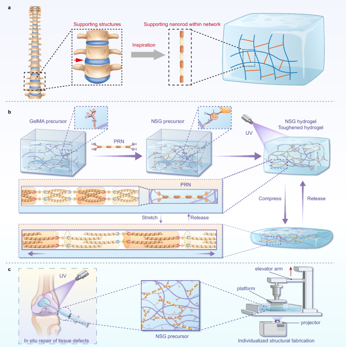
The intervertebral discs in the spine support and stabilize the vertebrae. When subjected to radial forces in the spine, the intervertebral discs deform to provide cushioning and reduce spinal shock. Inspired by the supporting intervertebral discs and their characteristics of deformation cushioning, here we proposed a biocompatible toughening strategy to enhance the compression performance and mechanical stability of GelMA hydrogels by incorporating supporting nanorods (Fig. 1a). The peptide-based rigid nanorods (PRNs) used herein are reported by us previously25, which are physical-covalent hybrid polymers composed of antiparallel homotetrameric peptide bundles and possess extreme rigidity. In this study, we incorporated PRNs into the GelMA hydrogel system. Specifically, PRNs were chemically incorporated into the GelMA precursor solution via thiol groups at both ends of the rods, which were bonded to methacrylate groups on GelMA (Fig. 1b). After gelation, PRNs function as supporting rods within the hydrogel, similar to the disc’s support between vertebrae. When the hydrogel network is subjected to pressure and transmits stress to the PRNs, the peptide bundles within the rods undergo peptide chain extension and reverse movement, resembling the deformation of an intervertebral disc to cushion the stresses (Fig. 1b). This effectively dissipates mechanical stress and inhibits crack propagation, resulting in a 1018% increase in compressive strength and a 508% improvement in toughness of the GelMA hydrogel. The integration of peptide-based rigid nanorods into GelMA chains not only preserves the good biocompatibility of GelMA but also retains the fluidity and photocurable properties. This versatile approach can be applied not only for the in situ curing and repair of load-bearing regions, such as knee osteochondral defects, but also for the additive manufacturing of robust and intricate structures promising for personalized tissue repair applications (Fig. 1c), thereby providing optimized solutions for tissue engineering.

a Resilient disc load-bearing structures inspire the hydrogel toughening strategy in this study. b Schematic representation of the preparation process of NSG hydrogel with PRNs acting as network supports, and the stretching changes that occur in the nanorods when the hydrogel is compressed. c The NSG precursor is highly compatible with the in situ repair of osteochondral defects and additive manufacturing.
Results
Preparation and characterization of PRNs and NSG
PRNs are prepared by two different peptide bundles with unbalanced molar equivalents (10:9) in our previous report, and PRNs with an average length of around 100 nm were synthesized at that time25. Considering that shorter and smaller nanorods are more easily dispersed in solution and may move more violently in solution to react more efficiently with GelMA molecules, we prepared shorter PRNs herein. According to the previous report, two peptide bundles consisting of peptide A (Pa) and peptide B (Pb), which respectively self-assemble into two different peptide bundles, were prepared first (Supplementary Fig. 1)25,26. Pa features a cysteine residue at its N-terminus, while Pb incorporates a maleimide group at its N-terminus (Fig. 2a). Then, the reaction of two peptide bundles with significantly unbalanced equivalents (around 4:3) produced the short PRNs (Supplementary Fig. 2). Atomic force microscopy (AFM) studies revealed that the PRNs possess a diameter of about 2 nm and a length of 31.3 ± 4.7 nm (Fig. 2b). The hydrodynamic radius of the fluid (Rh), as measured by dynamic light scattering (DLS), corresponded to a length of 29.46 ± 5.37 nm for PRNs (Fig. 2c). In addition, small-angle X-ray scattering (SAXS) results indicated that the radius of the PRN is about 1.29 nm and the length is about 32.44 nm (Fig. 2d). Since the length of each peptide bundle is about 4.3 nm (Supplementary Fig. 3), we deduced that each short PRN consists of seven bundles in average and has bundles of Pa at both ends according to the higher feeding ratio of Pa than that of Pb in the synthesis process. Next, we investigated the biocompatibility of PRNs and found that PRNs did not cause changes in cell morphology or a decrease in cell viability (Fig. 2e, f). In fact, they even promoted cell proliferation at a higher concentration (2% PRN) (Fig. 2f).

a Schematic structure of PRN and its constituent peptides. b A representative AFM image and size analysis of PRNs. c Rh of PRN in solutions obtained by DLS. The calculated length of the PRN is ~29.46 ± 5.37 nm. d Scattering from PRN (black squares) and the corresponding rigid cylinder fit (red curve). e Cytoskeletal staining of bone marrow mesenchymal stem cells (BMSCs) incubated with PRNs for 24 h. f Cellular activity of BMSC cultured with PRNs for 1, 3 and 5 days (*p = 0.014, *p = 0.0323, **p = 0.0018). n = 3 biological independent replicates. Data are mean ± SD (One-way analysis of variance (ANOVA), followed by Tukey’s multiple comparison.
Given that the competition between photoinitiated thiol-ene click chemistry and methacrylate radical polymerization during photocrosslinking of the GelMA-PRN mixture, we first prepared the NSG precursor by a one-pot reaction in which the thiol groups at both ends of the PRN underwent a thiol-Michael addition reaction with methacryloyl groups on the GelMA molecules27,28. To verify the successful grafting of PRN to GelMA in the NSG precursor solution, Ellman’s assay was conducted to quantify the free thiol content in NSGx hydrogel precursors29. The results demonstrated that the free thiol content decreased with the increase of reaction time, and when the reaction was carried out for 300 min, the free thiol content of each group decreased to about 5%, indicating that almost all of the PRN was successfully grafted onto the GelMA molecule (Supplementary Fig. 4). Subsequently, the NSG precursor was subjected to photocuring for network formation. As shown by the FT-IR spectroscopy results, the deepened characteristic absorption peak of the C–S bond at 1100 cm−1 indicated that the thiol group was added to the C=C double bond of GelMA30. In addition, the amide bond carried by PRN resulted in enhanced absorption peaks near 1600 cm−1 and 3300 cm−1, proving the successful connection of PRN to GelMA molecules. (Supplementary Fig. 5). Moreover, the viscosity of the NSG precursor was significantly increased compared with that of the simple mixture of GelMA and PRNs (Supplementary Fig. 6), further confirming the successful crosslinking between PRNs and GelMA molecules. Elemental analysis of photo-crosslinked NSG showed both the characteristic sulfur signature of PRNs and their uniform spatial distribution (Supplementary Fig. 7), confirming the successful incorporation and homogeneous dispersion of PRNs in NSG.
Mechanical supporting effects of PRNs and their underlying molecular mechanisms
First, we evaluated the hydrogels obtained by the two preparation methods by compression tests: (1) our developed method involving chemical grafting of PRNs onto GelMA prior to photo-crosslinking (NSG hydrogel), and (2) physical mixing of GelMA and PRNs followed by immediate crosslinking (GelMA-PRN hydrogel). The mechanical testing revealed distinct performance advantages of NSG hydrogels, which exhibited superior deformation tolerance (>80% strain before failure) and achieved significantly higher compressive toughness (508% of pristine GelMA values) (Fig. 3a). In contrast, GelMA-PRN hydrogels showed limited strain tolerance (<70% failure strain) and modest toughness enhancement (up to 2.7-fold increase) under identical testing conditions (Supplementary Fig. 8). Furthermore, morphological characterization demonstrated more uniform pore size distribution in NSG hydrogels, suggesting that the pre-crosslinking conjugation strategy promotes more homogeneous network formation (Supplementary Fig. 9).

a Compressive behavior of NSG hydrogels including stress-strain curves and corresponding toughness. (***p = 0.0006, ****p < 0.0001) n = 3 independent experiments. b Compressive behavior of BSG hydrogels including stress-strain curves and corresponding toughness. (p = 0.38, **p = 0.0012, ****p < 0.0001, *p = 0.0165) n = 3 independent experiments). c The process of BSG and NSG in coarse-grained molecular dynamics simulations. d, e Density variation of bundles of Pa in BSG (d) and PRNs in NSG (e) along the axial stretching direction. The six density peaks correspond to the covalent linkage sites between Pa and Pb bundles (e). f Force-strain curves of BSG and NSG. g Variation of energy dissipation with tensile length in the BSG and NSG systems. h Energetic changes in bond-related interactions and van der Waals attraction in the BSG and NSG systems. i Plots of the components of van der Waals attraction in the BSG and NSG systems. Data are presented as the mean ± SD (One-way ANOVA, followed by Tukey’s multiple comparison).
Next, to compare the supporting effects of PRNs and their constituent unit, the rigid peptide bundle, on GelMA, we also prepared bundle-supported GelMA (BSG) using the same synthesis method. We prepared a series of BSGx and NSGx precursors, where x denotes the mass percentage of bundles or PRNs relative to the GelMA molecules (e.g., 1 represents 1% content). Uniaxial compression tests on the hydrogels revealed that the toughness of both BSG and NSG hydrogels initially increased and then decreased with increasing bundle or PRN content. The NSG hydrogels exhibited significantly superior mechanical properties compared to the BSG hydrogels. The maximum toughness of the BSG hydrogels was 37.8 kJ m−³ (Fig. 3a), while the NSG hydrogels achieved a maximum toughness of 60.7 kJ m−³ (Fig. 3b).
To explore why PRNs offer a greater mechanical enhancement advantage over a single bundle, we conducted coarse-grained molecular dynamics simulations to investigate the behavior changes of PRNs and bundles during the hydrogel compression process. In the simulations, both the PRN and the bundle were anchored at their ends to GelMA molecular, thereby converting the macroscopic compression of the hydrogel into localized network stretching (Supplementary Fig. 10). The results showed that the bundle in BSG and the PRN in NSG undergo elongation under stress (Fig. 3c), accompanied by a decrease in material density (Fig. 3d, e). This may be linked to the unraveling of the tetrameric helices and the reverse movement of the two sets of peptide chains in the homotetrameric bundles. During this process, PRNs can withstand greater forces (Fig. 3f) and absorb more energy (Fig. 3g) compared to the bundles of Pa. Further analysis revealed that the energy from van der Waals interactions significantly outweighed that from bond-related interactions (Fig. 3h). Notably, compared to BSG, the van der Waals energy in the NSG system significantly increased in both peptide-peptide and peptide-water interactions (Fig. 3i). This suggests that the energy absorbed during peptide chain stretching is primarily used to overcome van der Waals interactions between multiple peptide chains and their surrounding environment, which is likely to be stronger than that of a single α-helix peptide chains31 or flexible peptide chains32,33. Collectively, these simulations indicate that the unique molecular configuration of PRNs, along with their higher energy dissipation efficiency, endows NSG with superior energy absorption capacity compared to BSG, resulting in significantly enhanced toughness.
Compressive behavior and mechanisms of toughened NSG hydrogels
We evaluated the compression tests to further elucidate the toughening effect of PRNs on GelMA hydrogels. As shown by the stress-strain curves, GelMA and NSG2 hydrogels have different mechanical properties. The GelMA hydrogel reached its maximum compressive strength at just 66% strain, while the NSG2 hydrogel could withstand over 80% strain, with its stress-strain curve continuing to rise (Fig. 4a, Supplementary Movie 1). Notably, their curves initially exhibited nearly identical slopes, indicating similar elastic moduli (Fig. 4a), but diverged significantly at higher strains. Based on this, we hypothesized that PRNs undergo an activation process during hydrogel compression (Fig. 4b, c). Comparing GelMA and NSG2 hydrogels, GelMA chains remain freely bent before compression and straighten radially around the covalent junctions when compressed. As compression increases, these chains reach critical strains that eventually lead to breakage. In NSG2 hydrogel, force is initially transmitted through GelMA chains, causing PRNs to move as the chains straighten. As the stretching proceeds, the force is transmitted to the PRN, where sacrificial bonds break before the covalent bonds (including overcoming van der Waals interactions), allowing the bundles in the PRN to absorb energy and elongate. This energy dissipation reduces the stress on the GelMA chain and allows it to withstand greater external stresses. Eventually, the NSG2 hydrogel achieved maximum compressive strength and toughness of 712.7 ± 78.6 kPa and 60.8 ± 2.2 kJ m−³, respectively, representing 1018% and 508% increases compared to those of GelMA hydrogels (69.98 ± 7.23 kPa and 11.92 ± 1.12 kJ m−³) (Fig. 4d).

a Compressive stress-strain curves of GelMA and NSG2 hydrogels. Insert reporting the stress-strain curves up to 60% of strain displaying approximately the same modulus of elasticity for GelMA and NSG2. Representative pictures of GelMA (b) and NSG2 (c) hydrogels during compression and schematic representation of the toughening behavior of PRN in hydrogels. d, The toughness and compressive strength of hydrogels (n = 3 independent samples). e Evaluation of the compressive strength and maximum strain of NSG2 and a variety of previously reported nanomaterial-enhanced GelMA hydrogels, including 1–5, carbon and metal-based nanocomposite hydrogels; 6–13, mineral-based nanocomposite hydrogels; and 14-17, polymer-based nanocomposite hydrogels (detailed information of the hydrogels is in Supplementary Table 1). Four consecutive cyclic loading-unloading stress-strain curves of GelMA (f) and NSG2 (g) hydrogels at strains of 65%. h Maximum compressive stress of hydrogels in 4 successive cycles of loading-unloading (****p < 0.0001). Data are presented as the mean ± SD. Statistical analyses were performed with two-tailed unpaired t tests. i 10 consecutive cyclic loading-unloading stress-strain curves of NSG2 hydrogels at strains of 65%. j GelMA hydrogel cannot withstand 70% compressive strain. k 60 consecutive cyclic loading–unloading stress-strain curves of NSG2 hydrogels at strains of 70%. l Stress and stress retention of NSG2 hydrogel during 60 consecutive cycles of loading-unloading.
We compared the compressive performance of the NSG2 hydrogel in this study with other reported nanomaterial-reinforced GelMA systems (Fig. 4e and Supplementary Table 1). These systems were chosen for comparison because they also effectively retain the inherent advantages of GelMA hydrogels. And, this comparison is restricted to single-network GelMA-based hydrogels or systems in which GelMA serves as the primary matrix, incorporating only a minimal amount of additional polymer networks. These traditional nanocomposite hydrogels typically involve adding micro- or nano-fillers (e.g., nanoparticles, nanofibers, or nanosheets) to improve the mechanical properties34,35,36,37,38. However, higher concentrations of fillers tend to disperse unevenly and agglomerate, resulting in the inconsistency of the mechanical prosperities39. Therefore, weaker regions of the hydrogel’s mechanical structure create stress concentration points under pressure, leading to crack formation and accelerating material failure. In comparison, the peptide-based rigid nanorod-reinforced GelMA hydrogel (NSG2) demonstrates significantly superior compressive performance.
We further evaluated the applicability of this nanorod-based energy dissipation mechanism by testing the tensile properties of NSG hydrogels and the compressive properties of other PRN-supported hydrogels. The uniaxial tensile test results showed that the tensile toughness of NSG hydrogel was 3 times higher than that of GelMA hydrogel (Supplementary Fig. 11). And both the more concentrated GelMA hydrogel (20 w/v% GelMA) and another commonly used photocurable biomaterial, methacryloyl hyaluronic acid (HAMA), showed significant enhancement in compressive performance by chemically integrating PRNs (Supplementary Fig. 12 and 13). This suggests that the PRN-based toughening strategy has the potential for universal applicability.
Enhanced structural stability and fatigue resistance in NSG hydrogels
Next, we conducted cyclic compression-unloading tests to evaluate the structural stability and fatigue resistance of NSG2 hydrogels. Given the limited strain tolerance of the GelMA hydrogels, the tests were performed at 65% strain over four consecutive compression-unloading cycles. The results showed differences in structural stability in the hydrogel groups. The GelMA group showed inconsistent hysteresis loops across the four cycles (Fig. 4f), whereas the NSG2 group showed nearly overlapping hysteresis loops (Fig. 4g). Quantitative analysis of stress values showed that the NSG2 group maintained higher stress levels than the GelMA group; however, the stress fluctuations in the NSG2 group were significantly smaller, as evidenced by the more tightly clustered data points in Fig. 4h. The phenomenon can be attributed to the 65% strain exceeding the safe deformation limit of GelMA (~60%), which induces the rupture of local covalent bonds in the hydrogel network. Such irreversible bond breakage then affects the hydrogel’s elastic recovery. In contrast, the NSG2 group benefits from the breakage and rapid reorganization of internal non-covalent bonds within the supporting PRNs, allowing the network to maintain its stability during repeated compression-unloading cycles.
To further evaluate the fatigue resistance of NSG2, we extended the compression-unloading test to 10 cycles. The NSG2 hydrogel maintained good curve reproducibility throughout all cycles (Fig. 4i). Besides, under more rigorous fatigue testing conditions (60 cycles at 70% strain), the GelMA hydrogel fractured before completing the first cycle (Fig. 4j), whereas the NSG2 hydrogel retained over 90% of its initial compressive stress (Fig. 4k, l), underscoring its superior fatigue-resistant properties. In summary, the NSG2 hydrogel demonstrates good mechanical stability and fatigue resistance under cyclic loading conditions. This superior performance stems from the length release mechanism stored within the tetrameric structure of PRNs, which facilitates the transition of fracture towards PRN propagation, providing effective energy dissipation for the hydrogel network.
Biological properties of NSG hydrogels: swelling, degradation, and biocompatibility
A biocompatible toughening strategy should enhance the mechanical properties of the hydrogel while preserving the inherent advantages of GelMA. Accordingly, we investigated their swelling characteristics, degradation behavior, and biocompatibility. The groups with the strongest mechanical properties in BSG and NSG were selected as representatives (i.e., BSG0.75, and NSG2) for comparison with GelMA hydrogel. In swelling tests, all three hydrogel groups reached equilibrium swelling within 24 h, with no statistical difference in the swelling rate (Fig. 5a, b). In vitro and in vivo degradation studies revealed that the BSG and NSG groups exhibited significantly slower degradation rates compared with the GelMA group (Fig. 5c, d, Supplementary Fig. 14), particularly during the initial four weeks after implantation in vivo (Fig. 5e), which may be attributed to their smaller network pore size and denser structure (Supplementary Fig. 15). This characteristic is advantageous for maintaining mechanical support and promoting tissue regeneration in the defect area. In addition, short-term (7-day) in vivo implantation results showed that the hydrogel groups did not induce any significant immune response (Fig. 5f). In addition, compared to the GelMA and BSG groups, the NSG group exhibited decreased CD68+ and iNOS expression, along with an increase in CD206+ expression (Fig. 5g1–g3), which may indicate a faster local tissue repair process40. In addition, long-term (1-month) implantation studies showed no evidence that the hydrogel caused systemic toxicity. (Supplementary Fig. 16).

a Swelling of hydrogels in PBS over 24 h. b Swelling rate of hydrogels at 24 h. c Degradation behavior of hydrogels in vitro. d The in vivo degradation rate of hydrogels. e Hydrogels removed from the dorsal skin of rats after 1, 2, 4, and 8 weeks. f Immunostaining of CD68, iNOS, and CD206 around the hydrogel after implantation of the hydrogel into the dorsal skin for 7 days. g1, g2, g3, Semi-quantification of CD68+, iNOS and CD206+ around hydrogels (normalized to group C).(**p = 0.0086, *p = 0.0262, **p = 0.0064, **p = 0.0051). n = 3 independent samples. h Cellular activity of BMSC seeded on hydrogel surfaces for 1, 4, and 7 day (p = 0.4076, p = 0.2938, p = 0.9267). n = 3 independent samples. i Live/dead staining of BMSC on hydrogel surface after 1, 4, and 7 days of incubation. All of the data are presented as the mean ± SD (n = 3). Statistical analyses were performed with one-way ANOVA, followed by Tukey’s multiple comparison.
Next, we investigated the biocompatibility of the hydrogels for rat BMSCs. Since GelMA is widely used as a scaffold material for tissue defect repair and as a bioink containing cells for additive manufacturing, we assessed cell viability on the hydrogel surface, in hydrogel extracts, and within the hydrogel itself. In 2D culture, all groups of cells were tightly adhered to the hydrogel surface, displaying healthy spindle or fusiform morphologies within 24 h (Supplementary Fig. 17). And after 1, 4, and 7 days of culture, both BSG and NSG groups demonstrated comparable cell viability to the GelMA group, with no statistically significant differences observed among the groups (Fig. 5h, i). Similar results were observed in the extract culture (Supplementary Fig. 18). Furthermore, cells encapsulated within the hydrogels maintained good viability throughout the 7-day culture period (Supplementary Fig. 19a). Although the OD values of the BSG and NSG groups showed a slight decline on day 7, no significant differences in cell viability were observed among the GelMA, BSG, and NSG hydrogels over time (Supplementary Fig. 19b), suggesting that NSG hydrogels are well-suited for use as bioinks. In order to assess the effect of different groups on the migration of BMSCs, we next performed a scratch test. It was found that both BSG and NSG groups tended to promote cell migration compared to the control group, although their effects were not statistically different compared to GelMA (Supplementary Fig. 20). In addition, Alcian blue staining was conducted to evaluate the effect of different hydrogel cultures on chondrogenic differentiation of BMSCs. The results demonstrated enhanced chondrogenic differentiation of BMSCs cultured on BSG and NSG hydrogels, as demonstrated by intense proteoglycan staining (blue) compared to the GelMA control group (Supplementary Fig. 21). These results indicate that the peptide-based nanorod toughening strategy effectively preserves the inherent biocompatibility of GelMA, demonstrating its great potential for applications in tissue repair.
In vivo osteochondral repair efficacy of NSG hydrogels
Based on the enhanced compressive performance and favorable biological properties, we evaluated the potential of NSG hydrogels for repairing load-bearing tissue defects using a rabbit osteochondral defect model41. To investigate the impact of hydrogel strength on repair outcomes, we compared NSG hydrogels with GelMA hydrogels and blank control, conducting repair efficacy analyses at 6 and 12 weeks post-surgery (Fig. 6a). At 6 weeks post-surgery, the defect in both the blank and GelMA groups remained clearly visible, while in the NSG group, a significant amount of newly formed tissue had already developed around the defect region (Fig. 6b). At 12 weeks, the surgical area in the NSG group was fully covered by smooth, regenerated tissue resembling native cartilage, with no visible defects. In contrast, the other two groups, although showing some reduction in the defect area, displayed no significant cartilage formation (Fig. 6b). The International Cartilage Repair Society (ICRS) scoring of NSG was higher than that of the blank and GelMA groups (Fig. 6c). In addition, micro-CT scanning was employed to assess subchondral bone regeneration. While noticeable voids persisted in the defect areas of both the blank and GelMA groups, the NSG group exhibited smooth, well-formed bone tissue in the defect region (Fig. 6d). Quantitative analysis showed that the bone volume/total volume (BV/TV) and trabecular thickness (Tb.Th) in the NSG group were significantly higher than those in the blank group (Fig. 6e), indicating substantial osteogenic capacity. In contrast, the blank and GelMA groups displayed higher trabecular separation (Tb.Sp), suggesting more pronounced bone resorption. Next, we performed histological and immunohistochemical staining to study the effects of the three treatments on tissue regeneration in cartilage. Hematoxylin and eosin (H&E) staining revealed the formation of fibrous-like tissue in the control group, with discontinuous structures and evident porous regions, indicating limited cartilage repair capability. In contrast, both the GelMA and NSG groups generated cartilage-like tissue (Fig. 6f1). Notably, the NSG group exhibited the formation of a continuous, smooth, and thick cartilage layer, where proteoglycans in the extracellular matrix were stained blue by Toluidine blue, covering a significantly larger area compared to the GelMA group (Fig. 6f2). Safranin O-fast green (SO-FG) staining further confirmed that the NSG group displayed the largest area of red-stained basophilic cartilage tissue among all groups (Fig. 6f3). As a critical chondrocyte maturation marker, collagen type II (Col II) immunohistochemistry showed more intense positive staining at the cartilage surface of the NSG group than in the blank and GelMA groups (Fig. 6f4, g).

a Surgical procedure and evaluation timeline for rabbit osteochondral repair. Created by Fig.draw (www.figdraw.com). b Macroscopic appearance of rabbit osteochondral defects at 6 and 12 weeks postoperation. c ICRS scores of cartilage repair at 6 and 12 weeks postoperative (*p = 0.0122, *p = 0.0242, *p = 0.0472, ***p = 0.0008). n = 3 or 4 independent samples. d Micro-CT images of rabbit osteochondral defects at 12 weeks postoperative. e, Bone-related assessment metrics include BV/TV, Tb.Th, Tb.N and Tb.Sp. (for BV/TV: **p = 0.0026, **p = 0.0033, ****p < 0.0001; for Tb.Th: *p = 0.0345, *p = 0.0284, ***p = 0.0003; for Tb.N: p = 0.5553; for Tb.Sp: **p = 0.0046, **p = 0.0033, ****p < 0.0001). n = 3 or 4 independent samples. All of the data are presented as the mean ± SD. Statistical analyses were performed with one-way ANOVA, followed by Tukey’s multiple comparison. f1-f4, Histologic staining included H&E staining (f1), toluidine blue staining (f2), SO-FG staining (f3), and immunohistochemical staining for Col II (f4). g Semi-quantification for Col II (p = 0.3724, **p = 0.0077, *p = 0.0392). n = 3 independent samples. All of the data are presented as the mean ± SD. Statistical analyses were performed with one-way ANOVA, followed by Tukey’s multiple comparison. h PCA plot illustrating distinct clustering of GelMA and NSG groups. i, Volcano plot of differentially expressed genes between GelMA and NSG groups. j Upregulated terms in enriched GO analysis of NSG vs. GelMA. k, Upregulated terms in enriched KEGG analysis of NSG vs. GelMA. l1-l4, Relative gene expression levels for Il6 (l1), Tnf-α (l2), Il10 (l3), and Col II (l4) by RT-qPCR (*p = 0.0278, **p = 0.0020, **p = 0.0031, *p = 0.0316). n = 3 independent samples. All of the data are presented as the mean ± SD. Statistical analyses were performed with two-tailed unpaired t tests.
Transcriptomic analysis of early repair mechanisms modulated by NSG hydrogels
Given that the NSG hydrogel exhibited a faster osteochondral defect repair effect at an early stage (Fig. 6b, c), we further explored the potential repair mechanisms. Based on the advantage of genetic analysis in rats, we established a rat model of knee osteochondral defects and treated the defects with GelMA and NSG hydrogels, respectively. At 4 weeks postoperatively, tissue samples from the defect site were collected for transcriptome analysis. Principal component analysis (PCA) revealed distinct clustering of the GelMA and NSG group samples, forming two clearly separate clusters (Fig. 6h), indicating that the two hydrogel treatments resulted in significantly different tissue transcriptomic profiles. Volcano plot analysis further revealed 425 differentially expressed genes (DEGs) between the NSG and GelMA groups, with 253 genes significantly upregulated and 172 genes significantly downregulated (Fig. 6i). Gene Ontology (GO) enrichment analysis indicated that the upregulated genes in the NSG group were primarily associated with inflammatory response and immune response (Fig. 6j), highlighting the significant role of NSG in modulating early-stage inflammation and immune reactions, which is consistent with the immunofluorescence results of immune markers presented earlier (Fig. 5g1–g3). Kyoto Encyclopedia of Genes and Genomes (KEGG) pathway analysis further revealed that the differentially regulated genes by NSG were significantly enriched in the chemokine signaling pathway and cytokine and growth factors pathways (Fig. 6k), indicating that NSG promoted tissue repair by facilitating immune cell migration and intercellular signaling. To validate the role of NSG in immune regulation and tissue repair, we conducted quantitative reverse transcription polymerase chain reaction (RT-qPCR) analysis. The results revealed a significant reduction in the expression of pro-inflammatory factors Il-6 and Tnf-α in the NSG group (Fig. 6l1, 2), accompanied by a marked upregulation of the anti-inflammatory factor Il-10 (Fig. 6l3). Furthermore, the expression of Col II was significantly increased in the NSG group (Fig. 6l4). These findings suggested that NSG hydrogels significantly modulated the inflammation and immune responses, and promoted collagen synthesis, thereby accelerating bone-cartilage tissue regeneration.
Applicability of the PRN-based toughening strategy in additive manufacturing
The good performance of NSG hydrogel in compressive mechanical properties, biocompatibility, and in situ tissue repair highlights its great potential for biomedical applications. Given that GelMA is one of the most commonly used hydrogels for additive manufacturing, the NSG hydrogel developed in this study should retain this advantage while further enhancing its potential for personalized tissue repair. Specifically, it aims to maintain good biocompatibility and photo-responsiveness while improving the structural stability and mechanical strength of intricate designs. We have demonstrated that NSG exhibited good biocompatibility for 3D cell culture (Supplementary Fig. 19). Here, we first compared the rheological properties of NSG and GelMA. It was shown that NSGx and GelMA precursors quickly converted into hydrogels upon UV irradiation, as indicated by the transient increase in their corresponding modulus, suggesting that the PRN-methacryloyl interactions did not significantly impede the radical polymerization process. (Fig. 7a). The storage modulus of the hydrogels initially increased and then decreased as the PRN content increased, with the highest storage modulus observed for NSG2 (Fig. 7b, c), which was ~7 times that of GelMA, suggesting that the PRN-based toughening strategy not only retains the fast photo-responsiveness of GelMA but also improves the elasticity of hydrogels.

a Time sweeping of various sets of hydrogel precursors, accompanied by UV irradiation halfway through. b Storage modulus of NSGx and GelMA hydrogels after UV radiation. c Storage modulus of NSGx and GelMA hydrogels (****p < 0.0001, ****p < 0.0001, ****p < 0.0001, n = 3 independent biological replicates). The average storage modulus for each group is displayed on the bar. Data are presented as the mean ± SD. Statistical analyses were performed with one-way ANOVA, followed by Tukey’s multiple comparison. d Schematic diagram of DLP. e Multi-channel cylinder of NSG2 and GelMA prepared using DLP technology. f, g Representative photographs and stress-strain curves of multi-channel cylindrical NSG2 and GelMA hydrogels during compression. h, i 10 consecutive cyclic load-unload curves of NSG2 cylindrical hydrogels at 55% strain. j Round and square porous structures of NSG2 hydrogel fabricated by DLP can withstand deformation over 50% strain.
Since the inherent structural fragility of GelMA hydrogels, the structure fabricated by additive manufacturing is also susceptible to rupture under stress. Here, we employed digital light processing (DLP) to fabricate complex structures composed of NSG2 and GelMA hydrogels, respectively (Fig. 7d), and compared their mechanical properties to validate the applicability of the PRN-based toughening strategy for additive manufacturing. We fabricated cylindrical structures, each containing four channels (Fig. 7e). In the compression-unloading tests, the GelMA hydrogel exhibited visible cracks at 50% strain, which rapidly propagated into the rupture, leading to structural failure (Fig. 7f, g and Supplementary Movie 2). In contrast, the NSG2 hydrogel was able to withstand a higher strain (~58%) (Supplementary Movie 3), and maintained structural integrity through 10 consecutive compression-unloading cycles (Fig. 7h, i), highlighting its good fatigue resistance. This significantly expands the potential applications of hydrogels in additive manufacturing42. Additionally, we printed more complex architectures, such as flattened hollow balls and lattice rectangles, which also withstood over 50% strain and fully recovered their original morphology (Fig. 7j). These results demonstrate the applicability of the PRN-based toughening strategy in additive manufacturing, highlighting the potential of NSG hydrogel in enhancing personalized structures and its promise for clinical translation.
Discussion
The development of biomaterials with enhanced mechanical properties is critical for advancing applications in tissue engineering and additive manufacturing. Hydrogels, particularly GelMA hydrogel, are widely employed due to their biocompatibility, photocrosslinking capability, and adaptability to additive manufacturing technologies. However, their inherent mechanical fragility limits their functionality in load-bearing applications. Here, we introduced a biocompatible nanorod reinforcement strategy by covalently incorporating a small amount of PRNs (2 w/v%) into the GelMA network, creating NSG hydrogels with substantially improved mechanical performance and biological functionality, while maintaining the intrinsic crosslinking efficiency of the GelMA matrix.
Our mechanical testing demonstrated that optimal NSG hydrogel achieved over 1000% improvement in compressive strength and 500% enhancement in compressive toughness compared to pure GelMA hydrogel. Furthermore, the NSG hydrogel can endure repeated compressive cycles without significant breakage, which highlights its good fatigue resistance. The superior mechanical performance of NSG hydrogels can be attributed to the role of PRN in connecting the hydrogel chains, and its ability to radial elongation under stress. This is analogous to intervertebral discs in the spine, which provide structural support while deforming to maintain overall integrity.
This toughness enhancement strategy, based on chemically bonded rigid peptide nanorods, offers several advantages. First, this toughening strategy is biocompatible, as the fabrication of NSG hydrogels requires only GelMA, PRNs, and a minimal amount of photoinitiator—all of which have been verified for biocompatibility. Hydrogel crosslinking is initiated simply by UV light exposure, ensuring its suitability for biomedical applications. Additionally, our strategy keeps the hydrogel porosity within a range suitable for cell viability43, making NSG hydrogels ideal candidates for bioinks44. This is superior to strategies that increase network density at the cost of significantly reducing pore size45,46. Second, the PRN-based toughening strategy is implemented during the precursor stage, allowing the structure to achieve mechanical reinforcement immediately after photocuring or additive manufacturing. This reduces or eliminates the need for post-processing step7, which may risk damaging the structure during transfer or handling. Third, the PRNs are soluble and covalently bonded to the matrix material, such as GelMA, ensuring uniform distribution within the solution. Unlike physically mixed fillers, which may precipitate during preparation and lead to uneven mechanical properties47,48, NSG hydrogels maintain consistent material strength. In addition, NSG hydrogel can be used to promote osteochondral defect repair. Our data demonstrate that NSG hydrogels’ superior osteochondral repair performance, which may be attributed to their exceptional mechanical properties, as enhanced mechanical signaling can promote M2 macrophage polarization, enhance secretion of IL-10 and other repair cytokines, and promote tissue repair response49,50. These advantages enable this strategy to seamlessly integrate with both in situ repair of load-bearing tissues and additive manufacturing, offering exceptional compatibility and efficiency. Moreover, our strategy is adaptable to other polymer-based hydrogel systems. PRNs can be integrated into hydrogels through methacrylate-thiol Michael addition, extending the applicability of this approach beyond GelMA-based systems. This offers possibilities for biomaterials tailored to specific biomedical applications, such as drug delivery, wound healing, and organoid development. Despite these advancements, certain limitations warrant further investigation. The long-term stability and biodegradation rates of NSG hydrogels in different physiological environments need to be systematically studied to ensure their suitability for chronic applications. Additionally, while our study focused on cartilage and bone tissue repair, the potential of NSG hydrogels in other soft and hard tissues remains to be explored.
In conclusion, our work presented a biocompatible PRN-based toughening strategy inspired by the supporting intervertebral discs in the spine. This strategy not only addresses a critical limitation of GelMA hydrogels but also provides a versatile framework for the development of next-generation biomaterials. By bridging the gap between material science and biomedical engineering, NSG hydrogels offer transformative potential for tissue engineering and advancing additive manufacturing, paving the way for more effective and durable therapeutic solutions.
Methods
Ethics statement
All animal experiments in this study were performed in accordance with the ordinance of the Ethical Committee of Sichuan University. All animal experimental protocols were approved by Sichuan University’s Institutional Animal Care and Use Committee in Chengdu, China (Approval No. WCHSIRB-D-2024-701).
Preparation of GelMA
We synthesized GelMA according to the following method, specifically: a 0.25 mol L−1 carbonate buffer (CB) was prepared and the pH was adjusted to 9.0. Next, 4 g of gelatin was dissolved in 30 mL of CB and heated to 60 °C until fully dissolved, resulting in a homogeneous gelatin solution. Next, 400 µL of methacrylic anhydride (MA) was added dropwise to the gelatin solution at 50 °C. After stirring for 2 h, the reaction was terminated by adding 5 times the volume of deionized water. The mixture was then dialyzed in deionized water using a 12–14 kDa dialysis bag for 3 days. Finally, the product was freeze-dried for 48 h to obtain a white, porous GelMA foam. The MA substitution degree (DS) of GelMA was determined by 1H NMR and analyzed by MestReNova 12.0. The results showed that the DS of MA of the prepared GelMA was ~90% (Supplementary Fig. 22).
Preparation and purification of peptides
The sequences of Pa and Pb have previously been denoted peptide 225 and peptide 525, respectively. As reported in our previous research25, we first synthesized the components of PRNs, Pa, and Pb, using the solid-phase peptide synthesis (SPPS) technique. The detailed procedure is as follows:
We synthesized the peptides with specific amino acid sequences using the Liberty Blue microwave peptide synthesizer (CEM Corporation). The synthesis was carried out in N,N’-dimethylformamide (DMF) as the solvent, with ethyl cyanoglyoxylate-2-oxime and N,N’-diisopropylcarbodiimide (DIC) as coupling reagents. A 20% piperidine solution in DMF was used for deprotection.
After peptide synthesis, the peptide-resin conjugate was cleaved for 3 h using a cleavage solution composed of 92.5% TFA, 2.5% TIPS, 2.5% 1,2-ethanedithiol (EDT), and 2.5% deionized water (by volume), to separate the peptide from the resin. The product was dried under a nitrogen atmosphere to prevent oxidation, and then washed by centrifugation with diethyl ether, repeating the process 3 times. Pa was then dissolved in a mixture of deionized water and acetonitrile, and purified by reverse-phase HPLC using a C18 column (Shimadzu, LC-16, Japan). The mobile phases for the reverse-phase HPLC consisted of solvent A (0.1% TFA in water) and solvent B (0.1% TFA in acetonitrile). Pa was purified with a linear gradient of solvent B, from 20 v/v% to 80 v/v% over 24 min. Purified Pa is lyophilized at -80°C and stored at -20°C away from light.
The preparation of Pb involves additional steps. After synthesizing the peptide using solid-phase peptide synthesis, 4-maleimide butyric acid (0.3 mmol), benzotriazol-N,N,N’,N’-tetramethylurea hexafluorophosphate (HBTU, 1.2 mmol), and N,N-diisopropylethylamine (DIPEA, 522 μL) were dissolved in DMF (9.6 mL) to prepare the reaction solution. This solution was then added to the resin after solid-phase peptide synthesis and incubated with agitation for 1 h. The same solution was added again to achieve double coupling, ensuring the maleimide group was successfully attached to the N-terminus. Afterward, the product was cleaved by adding a cleavage solution composed of 95 v/v% TFA, 2.5 v/v% TIPS, and 2.5 v/v% deionized water and reacting for 3 h to cleave the peptide from the resin. The resulting peptide was washed by centrifugation with diethyl ether and finally purified by reverse-phase HPLC. Pb was purified with a linear gradient of solvent B, from 10 v/v% to 90 v/v% over 30 min. Mass spectrometry data for both peptides were acquired on an ISQ-EM mass spectrometer (Thermo Scientific).
Preparation of PRNs
We weighed pure Pa and Pb (molar ratio 4:3), dissolved them in ultrapure water to prepare a 10 g L−1 peptide solution, and added 0.2 equivalents of 50 mM tris(2-carboxyethyl) phosphine (TCEP). The mixture was then placed on a shaker and incubated at room temperature for 24 h to form rod-like structures. Then the reaction mixture was dialyzed for 24 h using a 500 Da dialysis bag and freeze-dried at -80 °C for 72 h to obtain the pure powder. The resulting powder was stored at -20 °C in light-protected conditions.
Theoretical length measurement of peptides
The amino acid sequences of Pa and Pb were entered into the Alphafold website (https://alphafoldserver.com/), where the maleimide group of Pb was omitted from the omission because it is not an amino acid component51. Then, the corresponding peptide structures were generated. We imported the generated structures into Mol* Viewer (https://molstar.org/viewer/) and measured the theoretical peptide lengths52.
1H NMR
Pa and Pb were individually dissolved in D₂O, and then both peptides were mixed and dissolved in D₂O containing 50 mM TCEP for reaction. The three sample groups were then detected using ¹H NMR and analyzed with MestReNova 12.0.
Atomic force microscope (AFM)
The morphology of the peptide bundles was detected using a Bruker AFM (Bruker Dimension Icon). In brief, 5 µL of a 0.01 mM peptide solution was deposited onto a clean mica substrate. The sample was then gently rinsed with deionized (DI) water to remove any remaining salts. After standing for at least 24 h, the sample was imaged using the AFM. The PRN in the AFM images was observed with NanoScope Analysis 3.00 (Bruker) and analyzed using ImageJ (Version 1.46r).
DLS
A 4 mL sample of 1 g L−1 PRN solution was collected, and the size of the PRN was analyzed using a Malvern Zetasizer (Nano ZS, Malvern, UK). Dynamic measurements yield a size called the hydrodynamic radius, Rh. The length (L) of a rod particle was calculated via the equation:
$${{rm{L}}}=R{{rm{h}}}times 5.11$$
(1)
SAXS analysis
A 10 mg sample of PRN was dissolved in 3 mL of Tris-HCl buffer (pH=8) and analyzed using SAXS. The measurements were performed over a 2θ range of 0.1 to 3.2 degrees using a Xenocs 3.0 C system with a GeniX 3D Cu microfocus source. The results were analyzed using SasView 5.0.6 (Supplementary Table 2).
Hydrogel preparation
We prepared the NSG and BSG precursor solutions according to the reported literature27,53. Specifically, a Tris-HCl buffer solution (pH 8) containing 0.5 w/v% LAP was prepared for dissolving Pa and PRN. Additionally, a GelMA solution was prepared using the same buffer. The Pa and PRN solutions were then separately mixed with the GelMA solution at varying ratios, and the pH was adjusted to 8. The mixture was then sonicated at 40 °C for 5 h. The final products were BSG solutions with 0.25%, 0.5%, 0.75%, and 1% Pa (named BSG0.25, BSG0.5, BSG0.75, and BSG1), as well as NSG solutions with 1%, 2%, and 4% PRN (named NSG1, NSG2, and NSG4). For GelMA-PRN hydrogel fabrication, GelMA and PRN solutions containing 0.5 w/v% LAP were first prepared. Then, 20 w/v% of GelMA solution was uniformly mixed with 2 w/v%, 4 w/v% and 8 w/v% of PRN solution at a volume ratio of 1:1, and then immediately subjected to photo-crosslinking to form GelMA-PRN1, GelMA-PRN2 and GelMA-PRN4 hydrogels, respectively.
Ellman’s assay
To quantify the free thiol content in NSGx hydrogel precursors (NSG1, NSG2, NSG4), we performed Ellman’s assay using corresponding PRN solutions as controls. Samples were incubated in sodium phosphate buffer (0.1 M, pH 7.4, containing 1 mM EDTA) with 0.2 mM Ellman’s reagent for 6 h in the dark. After incubation, 200 μL of supernatant from each sample was transferred to a 96-well plate, followed by absorption measurement at 412 nm. The percentage of free thiol groups was calculated using the following equation:
$${{rm{Free}}}; {{rm{thiol}}}; {{rm{groups}}},[%]=frac{{{rm{A}}}({{rm{NSGx}}}) – {{rm{A}}}({{rm{GelMA}}})}{{{rm{A}}}({{rm{PRN}}}; {{rm{control}}})-{{rm{A}}}({{rm{buffer}}})}*100%$$
(2)
Viscosity measurement of NSG
The viscosity of NSG and G-PRN was measured using an MCR302 rheometer (Anton Paar) equipped with a PPTD200 measuring cell and CP252 spindle (plate diameter 25 mm). A 300 μL aliquot of hydrogel precursor was placed on the rheometer platform, and a 25 mm stainless steel parallel plate was used, with the gap height set to 500 μm during measurement. At 25 °C, the shear viscosity (Pa·s) was monitored over a shear rate range of 0.01 to 100 s⁻¹.
Hydrogel characterization
The freeze-dried hydrogel samples were analyzed using FT-IR spectroscopy (Nicolet 6700). Additionally, the cross-sections of the hydrogels were observed with a scanning electron microscope (SEM, HITACHI S800, Japan), and the pore sizes in the images were analyzed using ImageJ. Elemental mapping and compositional analysis were also performed.
Rheological measurements
Rheological measurements were performed using an MCR302 rheometer (Anton Paar). First, 300 μL of hydrogel precursor solution was placed on the rheometer platform, and a 25 mm stainless steel parallel plate was lowered to achieve a gap height of 500 μm. Time sweeps were performed at a frequency of 1 Hz and a strain of 1%, with UV exposure applied halfway through the measurement to monitor changes in the storage modulus (G′) and loss modulus (G′′) of the sample. The GelMA group was the control.
Mechanical properties of hydrogels
Mechanical testing was performed using a dynamic mechanical analyzer (DMA, TA-Q800, USA) and recorded via Origin 2021 (version 9.80.200). The untreated hydrogel was set as a control group, e.g., GelMA, HAMA.
Compression sample preparation
The hydrogel precursor was introduced into an acrylic mold and exposed to UV LED light (365 nm, 10 W cm−2) for 30 s to solidify the hydrogel. Cylindrical hydrogels with a height of 4 mm and a diameter of 4 mm were prepared, and multi-channel hollow cylindrical hydrogels were fabricated using DLP printing. The samples can either be tested for compression immediately after preparation or stored in sealed EP tubes for short-term preservation before subsequent compression testing.
Compression testing method
For uniaxial compression tests, samples were compressed to 15 N starting at 3 N min−1 until they reached the desired compression limit.
For the 4 consecutive cyclic compression-unloading tests, the sample was compressed to a predetermined strain at a rate of 50% min−1.
For 10 and 60 consecutive loading-unloading cycles, the sample was compressed to a predetermined strain at a rate of 100% min−1.
The stress-strain curves obtained from compression testing were used to calculate the compressive toughness (kJ m−³) of the hydrogels from the area under the curve.
Sample preparation for uniaxial tensile tests
Dumbbell-shaped tensile samples were fabricated using molds. The ends of the samples were securely adhered to a polyacrylic plate, which was then fixed in the DMA clamps for tensile tests. The samples were stretched at a rate of 1 mm min−1. The stress-strain curves were used to calculate the tensile toughness (kJ m−3) of the hydrogels from the area under the curve.
Molecular dynamics simulations
Molecular dynamics simulations were performed by Phadcalc (www.phadcalc.com) using the GROMACS software package (Version 5.1.1). Modeling was performed using AlphaFold 2, Martinize, Packmol, and VMD, with Python employed for data analysis. Visualization and plotting were done using Python and VMD. The system architecture consisted of two groups: one formed by GelMA molecular chains and Pa bundles, and the other formed by GelMA molecular chains and PRNs. The GelMA chain was composed of 20–30 collagen basic units, with each unit containing 83 amino acid residues. These basic units were connected and polymerized via maleic acid reactive sites. GelMA, Pa bundles, and PRNs were first modeled using an all-atom model, followed by energy minimization and equilibration. Then, they were converted to the Martini coarse-grained model using the Martinize coarse-graining scheme. The two system configurations were placed in orthogonal cubic water boxes (20 nm × 20 nm × 50 nm), with water modeled using the Martini BP4 model. At the top and bottom of the Z-axis, there is one GelMA chain, with Pa bundles and PRNs connected to the GelMA chains at the respective ends of the system.
The simulation was performed using umbrella sampling-based enhanced sampling methods to thoroughly explore the phase space during the peptide chain elongation process. During the production simulation, a soft constraint based on umbrella sampling was applied to the lower GelMA chain, maintaining the center of mass approximately fixed while allowing the chain to stretch and bend flexibly. A soft driving force, also based on umbrella sampling, was applied to the upper GelMA chain, allowing it to stretch and bend flexibly while gradually and uniformly moving its center of mass away from the lower GelMA chain. During this process, the peptide chain was extended under force, generating stress.
Swelling test
Hydrogel samples with a diameter of 4 mm and height of 5 mm were weighed (W₀) and immersed in 10 mL of PBS at room temperature. At specified time points, the samples were taken out, excess liquid was blotted with filter paper and reweighed (Wt). The GelMA group was set as a control. The swelling ratio was calculated using the following formula:
$${mathrm{Swelling; Ratio}},[%]=frac{W{{rm{t}}}}{W{{rm{o}}}}*100%$$
(3)
In vitro degradation
Hydrogel samples of the same volume from each group were freeze-dried and weighed. The GelMA group was set as a control. The samples were then immersed in PBS and collected at specified time points. After freeze-drying, the samples were reweighed. The remaining weight percentage was calculated using the following formula:
$${mathrm{Remaining; Weight}},[%]=frac{W({mathrm{before}})}{W({mathrm{after}})}*100%$$
(4)
DLP fabrication
Both the GelMA and NSG2 precursor solutions contained 0.5% LAP (EFL-Tech Co., Ltd) as the photoinitiator and 0.5% tartrazine (Macklin) as the UV-blocking agent. The DLP fabrication process was performed using a commercial digital light projection printer (EFL-BP8601 Pro, China). The light intensity of the printer was set to 13 mW cm–² with an exposure time of 8 s. After the solution was poured into the resin tank of the DLP, the model was printed according to the pre-designed structure. Once printing was complete, the model was washed in warm water (50–60 °C) to remove any excess uncured liquid. The GelMA group was the control.
In vivo hydrogel degradation and biocompatibility assessment
Fifteen 8-week-old male Sprague-Dawley (SD) rats were anesthetized by intraperitoneal injection of 3% sodium pentobarbital. After shaving the dorsal skin and disinfecting with povidone-iodine, a 1 cm incision was made to separate the skin from the fascia. Sterilized hydrogel samples (GelMA, BSG, and NSG) with a diameter of 10 mm and a thickness of 2 mm were implanted subcutaneously in the rats. The incision was sutured and disinfected again. The blank group underwent surgery without hydrogel implantation.
At 1, 2, 4, and 8 weeks post-surgery, hydrogel samples were retrieved from the dorsal tissue, photographed, freeze-dried, weighed, and assessed for degradation in vivo. Additionally, at 1 week and 1 month, tissue samples from the surgical site and internal organs were collected, fixed in 4% paraformaldehyde, and processed for both frozen and paraffin sections. Frozen sections were stained for CD68+, iNOS, and CD206+ to assess the local immune response and inflammation, which were observed by using a spinning disk confocal super resolution microscope (Olympus SpinSR10) and analyzed with imageJ. The antibodies used are as follows: anti-iNOS, Rabbit, HUABIO, ER1706-89, 1:200 (IF); anti-CD206, Rabbit, HUABIO, HA722892, 1:500 (IF); anti-CD68, Rabbit, HUABIO, HA722285, 1:200 (IF); Alexa Fluor 647 anti-rabbit (Invitrogen, A-31573) (dilution 1:300). Paraffin sections were stained with H&E to evaluate the in vivo toxicity of the hydrogels, and observed using a pathology slice scanner (NanoZoomer S360).
Isolation and culture of BMSCs
BMSCs were isolated from 10-day-old SD rat pups and cultured in α-MEM medium supplemented with 10% fetal bovine serum and 1% penicillin-streptomycin. The cells were cultured at 37 °C in a 5% CO2 incubator. Cells at passage 2 (P2) were cultured to 80-90% confluence, and then collected for subsequent experiments.
Biocompatibility assessment
To evaluate the biocompatibility of PRNs, BMSCs were seeded at a density of 8000 cells per well in a 96-well plate. After cell adhesion, the complete medium containing different concentrations of PRN (0.5%, 1%, and 2%) was introduced, and then the cell viability was assessed using the cell counting kit-8 (CCK-8) assay kit on days 1, 3, and 5. Additionally, 20,000 cells were seeded in confocal dishes and cultured for 24 h, followed by fixation with 4% paraformaldehyde and fluorescence staining with FITC-labeled cytochalasin D (CA1620, Solarbio). Cell morphology was then observed under a confocal microscope (Zeiss, LSM880, Germany) and analyzed with ZEN software (Carl Zeiss Microscopy GmbH, Version 2.3.69.1000).
To comprehensively assess the impact of the hydrogel on cell viability, we employed three different BMSC culture methods: co-culturing with hydrogel extract (extract culture), seeding cells on the hydrogel surface (2D culture), and encapsulating cells within the hydrogel (3D culture).
Extract culture
The groups were set as follows: control, GelMA, BSG, and NSG. The sterilized lyophilized hydrogel was soaked in a complete culture medium (50 g L−1) and incubated at 37 °C for 24 h to prepare the extract, which was used for subsequent experiments. P2 cells were trypsinized, resuspended in a complete medium, and adjusted to a concentration of 8000 cells per well for culture. The control group was cultured in a complete medium, while the other groups were cultured in their respective hydrogel extracts.
2D culture
The experimental groups were designated as GelMA, BSG, and NSG. The hydrogel precursor solutions were sterilized by filtration, and 50 μL was added into each well of a 96-well plate, followed by UV exposure for 10 s. Subsequently, 200 μL of the cell suspension was added to each well.
3D culture
P2 cells were centrifuged and resuspended in hydrogel precursor solution, with a cell density of 10⁶ cells mL−1. 100 μL of this suspension was added to each well, followed by UV light curing for 10 s. A complete medium was then added for culture. The groups were the same as in the 2D culture model.
For all three culture models, at least three replicates per group were set up. The cells were cultured overnight at 37 °C in a 5% CO2 incubator. After 1, 4, and 7 days of culture, cell viability was evaluated using the CCK-8 assay kit and live/dead staining.
CCK-8 assay
At the designated time points, prepare a 10% CCK-8 working solution. After removing the original culture medium, add 100 μL of CCK-8 reagent to each well and gently shake to ensure even distribution. Incubate for 1–2 h, or until a noticeable color change occurs. Measure the absorbance at 450 nm using a microplate reader.
Live/dead cell staining
Cell grouping and culture times were the same as for the CCK-8 assay. At the designated time points, live/dead cell staining was performed using the Calcein/PI cell viability and cytotoxicity assay kit (C2015S, Beyotime). Before staining, wash the cells with sterile PBS (pH = 7.4) to remove the culture medium. Prepare the staining solution according to the manufacturer’s instructions and add it to the wells. Incubate at 37 °C for 30 min. After incubation, observe the cells under a fluorescence microscope (Leica DMi8 M). Live cells emit green fluorescence, while dead cells emit red fluorescence.
Scratch assay in vitro
To evaluate the effect of different hydrogel leachates on BMSC migratory capacity, a scratch assay was performed as follows: BMSCs were cultured in 6-well plates in a complete growth medium. When BMCSs reached 90% confluency, uniform linear scratches were made across the cell monolayer using a sterile 200 μL pipette tip. After detached cells were removed by gentle PBS washing, cells were treated with hydrogel leachate prepared in serum-free medium (serum-free medium as control). Scratch areas were imaged at 0 h and 24 h, and migration was quantified via ImageJ.
Alcian blue staining
For chondrogenic differentiation analysis, 100 μL of sterile hydrogel precursor solution was added to 24-well plates and photo-crosslinked. Then, BMSCs were seeded at a density of 1 ×1 05 cells per well onto the hydrogel surfaces and cultured in rat BMSC chondrogenic induction medium (OriCell®, RAXMX-90041) with medium changes every 2 days. After 14 days of induction, chondrogenic differentiation was assessed by Alcian blue staining and subsequent microscopic examination (Leica DMi8 M).
Cell adhesion on hydrogels
Two hundred microliters of hydrogel precursor solution was added to a confocal dish and solidified under light exposure. A BMSC cell suspension (100,000 cells mL−1) was then added to the hydrogel surface, and the dish was incubated at 37 °C with 5% CO₂.
After 24 h of culture, remove the original culture medium and fix the cells with 4% paraformaldehyde. The cells were then permeabilized with 0.5% Triton X-100 for 5 min, followed by PBS washing. FITC-labeled cytochalasin D was diluted in PBS to a 0.25 v/v% staining solution and applied to the cells in the dark for 30 min. After staining, the cells were washed with PBS for 10 min. Finally, a thin layer of anti-fade mounting medium containing DAPI (S2110) was applied to cover the surface. Fluorescence images were captured using a confocal microscope (Zeiss, LSM880, Germany).
In vivo evaluation of osteochondral regeneration in rabbits
Eighteen male New Zealand white rabbits (2.5–3.0 kg, about 3 months old) were randomly divided into three groups (blank group, GelMA group, and NSG group) for in vivo osteochondral repair evaluation. After intravenous injection of 3% sodium pentobarbital (30 mg kg−1) for anesthesia, the rabbits were fixed in a supine position on the surgical table. The fur around the knee joint was shaved, and the area was disinfected and covered with a sterile drape. A 1.5 cm long incision was made on the medial side of the knee joint, and the patella was dislocated to expose the trochlear groove. Using a motorized bone drill, a 3.5 mm diameter, 4 mm deep osteochondral defect was created at the center of the trochlear groove in both knees. Then, the extravasated blood was carefully absorbed using sterile cotton swabs, followed by rapid injection of the hydrogel precursor solution into the cleared defect and immediate photo-crosslinking (Supplementary Movie 4). In the blank group, the wound was sutured without further intervention, while the other groups received hydrogel injection followed by light curing, after which the incisions were sutured. All rabbits were administered intramuscular penicillin postoperatively. At 6 and 12 weeks post-surgery, the rabbits were euthanized, and the knee joint regions were photographed. Knee joint heads were harvested for fixation. Cartilage repair was assessed through gross behavior observation, micro-CT analysis, and histological analysis to evaluate the efficacy of different hydrogels in promoting osteochondral defect healing.
Micro-CT analysis
After the rabbits were sacrificed, the musculature around the knee was isolated. The joint tissue was fixed in 4% paraformaldehyde for 2 days. For micro-CT scanning (μCT50, Scanco Medical AG, Switzerland), the knee side was immobilized in foam and placed in the sample tube. The samples were scanned using 70 kVp, 114 μA, X-ray energy, and 20 µm resolution (voxel size). Then, the scanned subchondral bone was reconstructed and analyzed.
Histological evaluation
The harvested samples were decalcified in 10% EDTA for 2 months, embedded in paraffin, and cut into 5-μm-thick sections. These sections were then stained with H&E, toluidine blue, SO-FG staining and Col II, which were scanned using a pathology slide scanner (NanoZoomer S360), observed using NDP.view 2 (Version 2.9.29, Hamamatsu), and analyzed using imageJ (Version 1.46r).
In vivo evaluation of knee joint repair in rats
Thirty 8-week-old male SD rats were used to establish osteochondral defect models in this study. The guidelines were strictly adhered to at every stage of the experiment. Anesthesia was administered using 3% sodium pentobarbital via intraperitoneal injection. After the anesthesia took effect, rats were fixed in the supine position on the operating table, and a longitudinal incision was made on the medial side of the knee joint, pivoting the knee capsule from the medial side to the lateral side of the knee joint and completely exposing the femoral hoof groove.
A cylindrical osteochondral defect (2 mm in diameter and 2 mm in depth) was drilled into the femoral trochanter with a steel drill. Hydrogel precursor solution was injected and UV light illumination was applied for 10 s for adequate curing in the GelMA and NSG groups, respectively. Finally, the wound is sutured, and postoperative antibiotics are used to prevent infection. The samples were harvested at 4 weeks.
RNA-sequencing
Tissue samples (2 mm in diameter and 2 mm in height) were collected from the surgical sites of rats and preserved in RNALater™ animal tissue RNA stabilization solution. The samples were sent to Hangzhou Kaitai Biotechnology Co., Ltd. (Hangzhou, China) for transcriptomic sequencing. Libraries were prepared using the U-mRNAseq Library Prep Kit (AT4221, KAITAI-BIO) and the Ribo-off rRNA Depletion Kit (Bacteria) (N407, Vazyme), followed by sequencing on the Illumina NovaSeq platform. The mapped reads for each sample were assembled in a reference-guided manner using StringTie (v1.3.6). Expression levels were normalized using FPKM. Genes with FDR < 0.05 and |log2 fold change | > 1 were identified as differentially expressed genes. Finally, functional enrichment analyses, including GO and KEGG pathway analyses, were performed to identify key biological attributes.
RT-qPCR
After 4 weeks, tissues were collected from the surgical area of the rat knee joint and preserved in RNALater™ animal tissue RNA stabilization solution (Beyotime, R0118). For tissue RNA extraction, we utilized a magnetic bead-based total RNA extraction kit (Cat No. 18605ES60; Yeasen, Shanghai, China) following the manufacturer’s protocol. After RNA extraction, the concentration and quality of the RNA were assessed using a NanoDrop spectrophotometer. Reverse transcription to cDNA was performed using the PrimeScript™ FAST RT Reagent Kit with gDNA Eraser (Takara, No. RR092S). For qPCR, TB Green® Premix Ex Taq™ II FAST qPCR (Takara, No. CN830A) was used to perform the RT-qPCR according to Supplementary Table 3.
Statistical analysis
Data represent the mean ± SD of at least three replicates. Statistical analysis was conducted by using GraphPad Prism 9 statistical software. Statistical significance was determined using Student’s t test or one-way ANOVA, followed by Tukey’s multiple comparison test. *p < 0.05, **p < 0.01, ***p < 0.001, and ****p < 0.0001 were regarded as statistically significant. In addition, “ns” denoted no significant difference.
Reporting summary
Further information on research design is available in the Nature Portfolio Reporting Summary linked to this article.
Data availability
All data supporting the findings of this study are available within the article and its supplementary files. Raw sequencing data generated in this study have been deposited in the NCBI SRA database under accession number “PRJNA1242972” (https://www.ncbi.nlm.nih.gov/sra/PRJNA1242972). Source data are provided with this paper.
References
-
Li, X. & Gong, J. P. Design principles for strong and tough hydrogels. Nat. Rev. Mater. 9, 380–398 (2024).
-
Kuang, X., Arican, M. O., Zhou, T., Zhao, X. & Zhang, Y. S. Functional tough hydrogels: design, processing, and biomedical applications. Acc. Mater. Res. 4, 101–114 (2022).
-
Zhang, H. J. et al. Tough physical double-network hydrogels based on amphiphilic triblock copolymers. Adv. Mater. 28, 4884–4890 (2016).
-
Takeshita, S. & Ono, T. Biopolymer-polysiloxane double network aerogels. Angew. Chem. Int. Ed. 62, e202306518 (2023).
-
Allen, M. J. et al. Multimorphic materials: spatially tailoring mechanical properties via selective initiation of interpenetrating polymer networks. Adv. Mater. 35, e2210208 (2023).
-
Rajawasam, C. W. H. et al. Carbodiimide-driven toughening of interpenetrated polymer networks. Angew. Chem. Int. Ed. 63, e202400843 (2024).
-
Fang, Z. et al. 3D printable elastomers with exceptional strength and toughness. Nature 631, 783–788 (2024).
-
Sun, X., Mao, Y., Yu, Z., Yang, P. & Jiang, F. A Biomimetic “salting out-alignment-locking” tactic to design strong and tough hydrogel. Adv. Mater. 36, 2400084 (2024).
-
Choi, S. et al. Bone-adhesive anisotropic tough hydrogel mimicking tendon enthesis. Adv. Mater. 35, e2206207 (2023).
-
Kim, J., Zhang, G., Shi, M. & Suo, Z. Fracture, fatigue, and friction of polymers in which entanglements greatly outnumber cross-links. Science 374, 212–216 (2021).
-
Zhu, R., Zhu, D., Zheng, Z. & Wang, X. Tough double network hydrogels with rapid self-reinforcement and low hysteresis based on highly entangled networks. Nat. Commun. 15, 1344 (2024).
-
Ma, J. et al. Designing ultratough single-network hydrogels with centimeter-scale fractocohesive lengths via inelastic crack blunting. Adv. Mater. 36, e2311795 (2024).
-
Wang, Z. et al. Toughening hydrogels through force-triggered chemical reactions that lengthen polymer strands. Science 374, 193–196 (2021).
-
Xue, B. et al. Strong, tough, rapid-recovery, and fatigue-resistant hydrogels made of picot peptide fibres. Nat. Commun. 14, 2583 (2023).
-
Li, X. et al. High-Strength and nonfouling zwitterionic triple-network hydrogel in saline environments. Adv. Mater. 33, e2102479 (2021).
-
Yue, K. et al. Synthesis, properties, and biomedical applications of gelatin methacryloyl (GelMA) hydrogels. Biomaterials 73, 254–271 (2015).
-
Zhang, Y. et al. Gelatin-based injectable hydrogels loaded with copper ion cross-linked tannic acid nanoparticles for irregular wound closure repair. Acs Appl. Nano Mater. 6, 21775–21787 (2023).
-
Guzzi, E. A. et al. Hierarchical biomaterials via photopatterning-enhanced direct ink writing. Biofabrication 13, 044105 (2021).
-
Das, S., Jegadeesan, J. T. & Basu, B. Gelatin methacryloyl (GelMA)-based biomaterial inks: process science for 3D/4D printing and current status. Biomacromolecules 25, 2156–2221 (2024).
-
Jiang, T. et al. Untangling the response of bone tumor cells and bone forming cells to matrix stiffness and adhesion ligand density by means of hydrogels. Biomaterials 188, 130–143 (2019).
-
Lin, S. et al. Anti-fatigue-fracture hydrogels. Sci. Adv. 5, eaau8528 (2019).
-
Subbiah, R. et al. Nanoscale mineralization of cell-laden methacrylated gelatin hydrogels using calcium carbonate-calcium citrate core-shell microparticles. J. Mater. Chem. B 9, 9583–9593 (2021).
-
Wu, S. et al. Poly(vinyl alcohol) hydrogels with broad-range tunable mechanical properties via the hofmeister effect. Adv. Mater. 33, e2007829 (2021).
-
Yuan, X. et al. Tough gelatin hydrogel for tissue engineering. Adv. Sci. 10, e2301665 (2023).
-
Wu, D. et al. Polymers with controlled assembly and rigidity made with click-functional peptide bundles. Nature 574, 658–662 (2019).
-
Zhang, H. V. et al. Computationally designed peptides for self-assembly of nanostructured lattices. Sci. Adv. 2, e1600307 (2016).
-
Fan, P.-R., Zhao, X., Wei, Z.-H., Huang, Y.-P. & Liu, Z.-S. Robust immobilized enzyme reactor based on trimethylolpropane trimethacrylate organic monolithic matrix through “thiol-ene” click reaction. Eur. Polym. J. 124, 109456 (2020).
-
Deng, Y., Shavandi, A., Okoro, O. V. & Nie, L. Alginate modification via click chemistry for biomedical applications. Carbohydr. Polym. 270, 118360 (2021).
-
Göckler, T. et al. Tuning superfast curing thiol-norbornene-functionalized gelatin hydrogels for 3d bioprinting. Adv. Healthc. Mater. 10, e2100206 (2021).
-
Zheng, Y. et al. Neuro-regenerative imidazole-functionalized GelMA hydrogel loaded with hAMSC and SDF-1α promote stem cell differentiation and repair focal brain injury. Bioact. Mater. 6, 627–637 (2021).
-
Liu, P., Zhang, Y., Guan, Y. & Zhang, Y. Peptide-crosslinked, highly entangled hydrogels with excellent mechanical properties but ultra-low solid content. Adv. Mater. 35, e2210021 (2023).
-
Cai, H. et al. Injectable interface-bonded fiber-reinforced thiolated chitosan hydrogels for enhanced cellular activities and cartilage regeneration. Carbohydr. Polym. 347, 122643 (2025).
-
Vinikoor, T. et al. Injectable and biodegradable piezoelectric hydrogel for osteoarthritis treatment. Nat. Commun. 14, 6257 (2023).
-
Xin, T. et al. Inorganic strengthened hydrogel membrane as regenerative periosteum. ACS Appl. Mater. Interfaces 9, 41168–41180 (2017).
-
Shin, S. R. et al. Carbon nanotube reinforced hybrid microgels as scaffold materials for cell encapsulation. ACS Nano 6, 362–372 (2012).
-
Paul, A. et al. Nanoengineered biomimetic hydrogels for guiding human stem cell osteogenesis in three dimensional microenvironments. J. Mater. Chem. B 4, 3544–3554 (2016).
-
Zhou, Y. et al. Unusual multiscale mechanics of biomimetic nanoparticle hydrogels. Nat. Commun. 9, 181 (2018).
-
Kurian, A. G., Singh, R. K., Patel, K. D., Lee, J.-H. & Kim, H.-W. Multifunctional GelMA platforms with nanomaterials for advanced tissue therapeutics. Bioact. Mater. 8, 267–295 (2022).
-
Sakr, M. A., Siddiqua, S., Shin, S. R. & Kim, K. Synthesis and characterization of a nanoclay reinforced gelatin-based hybrid hydrogel. J. Biomed. Mater. Res. A 113, e37870 (2025).
-
Pajarinen, J. et al. Mesenchymal stem cell-macrophage crosstalk and bone healing. Biomaterials 196, 80–89 (2019).
-
Hayashi, K. et al. Wood-derived hydrogels for osteochondral defect repair. ACS Nano 19, 520–534 (2025).
-
Ren, C. et al. Reinforcing gelatin hydrogels via in situ phase separation and enhanced interphase bonding for advanced 3D fabrication. Adv. Mater. 37, e2416432 (2025).
-
Zauchner, D. et al. Synthetic biodegradable microporous hydrogels for in vitro 3D culture of functional human bone cell networks. Nat. Commun. 15, 5027 (2024).
-
Ying, G., Jiang, N., Yu, C. & Zhang, Y. S. Three-dimensional bioprinting of gelatin methacryloyl (GelMA). Bio-Des. Manuf. 1, 215–224 (2018).
-
Fu, L. et al. Cartilage-like protein hydrogels engineered via entanglement. Nature 618, 740–747 (2023).
-
Yang, Y. et al. Ultra-durable cell-free bioactive hydrogel with fast shape memory and on-demand drug release for cartilage regeneration. Nat. Commun. 14, 7771 (2023).
-
Urruela-Barrios, R., Ramírez-Cedillo, E., Díaz de León, A., Alvarez, A. J. & Ortega-Lara, W. Alginate/gelatin hydrogels reinforced with TiO₂ and β-TCP fabricated by microextrusion-based printing for tissue regeneration. Polymers 11, 457 (2019).
-
Chimene, D., Kaunas, R. & Gaharwar, A. K. Hydrogel bioink reinforcement for additive manufacturing: a focused review of emerging strategies. Adv. Mater. 32, e1902026 (2020).
-
Yu, T. et al. Mechanically robust hydrogels facilitating bone regeneration through epigenetic modulation. Adv. Sci. 9, e2203734 (2022).
-
Yang, W. et al. Mechanical stimulation of anti-inflammatory and antioxidant hydrogels for rapid re-epithelialization. Adv. Mater. 36, e2312740 (2024).
-
Abramson, J. et al. Accurate structure prediction of biomolecular interactions with AlphaFold 3. Nature 630, 493–500 (2024).
-
Sehnal, D. et al. Mol* Viewer: modern web app for 3D visualization and analysis of large biomolecular structures. Nucleic Acids Res. 49, W431–W437 (2021).
-
Karuppasamy, K. et al. A rapid one-pot synthesis of novel high-purity methacrylic phosphonic acid (PA)-based polyhedral oligomeric silsesquioxane (POSS) frameworks via thiol-ene click reaction. Polymers 9, 192 (2017).
Acknowledgements
This study was supported by National Nature Science Foundation of China (82271034) (J.W.), Natural Science Foundation of Sichuan Province (2023NSFSC2000) (J.W.), Research and Development Program (West China Hospital of Stomatology, Sichuan University) (RD-03-202107) (J.W.), National Nature Science Foundation of China (22275128) (D.D.W.) and Research Funding from West China School/Hospital of Stomatology Sichuan University (QDJF2021-2) (D.D.W.). We acknowledge FigDraw (www.figdraw.com) for illustration support.
Ethics declarations
Competing interests
The authors declare no competing interests.
Peer review
Peer review information
Nature Communications thanks Yong He, Yi Cao, and Bin Xue for their contribution to the peer review of this work. A peer review file is available.
Additional information
Publisher’s note Springer Nature remains neutral with regard to jurisdictional claims in published maps and institutional affiliations.
Supplementary information
Source data
Rights and permissions
Open Access This article is licensed under a Creative Commons Attribution-NonCommercial-NoDerivatives 4.0 International License, which permits any non-commercial use, sharing, distribution and reproduction in any medium or format, as long as you give appropriate credit to the original author(s) and the source, provide a link to the Creative Commons licence, and indicate if you modified the licensed material. You do not have permission under this licence to share adapted material derived from this article or parts of it. The images or other third party material in this article are included in the article’s Creative Commons licence, unless indicated otherwise in a credit line to the material. If material is not included in the article’s Creative Commons licence and your intended use is not permitted by statutory regulation or exceeds the permitted use, you will need to obtain permission directly from the copyright holder. To view a copy of this licence, visit http://creativecommons.org/licenses/by-nc-nd/4.0/.
About this article

Cite this article
Zhu, J., Wei, Y., Yang, G. et al. Peptide-based rigid nanorod-reinforced gelatin methacryloyl hydrogels for osteochondral regeneration and additive manufacturing. Nat Commun 16, 7090 (2025). https://doi.org/10.1038/s41467-025-62540-0
-
Received:
-
Accepted:
-
Published:
-
DOI: https://doi.org/10.1038/s41467-025-62540-0