
- Perspective
- Open access
- Published:
Subjects
Abstract
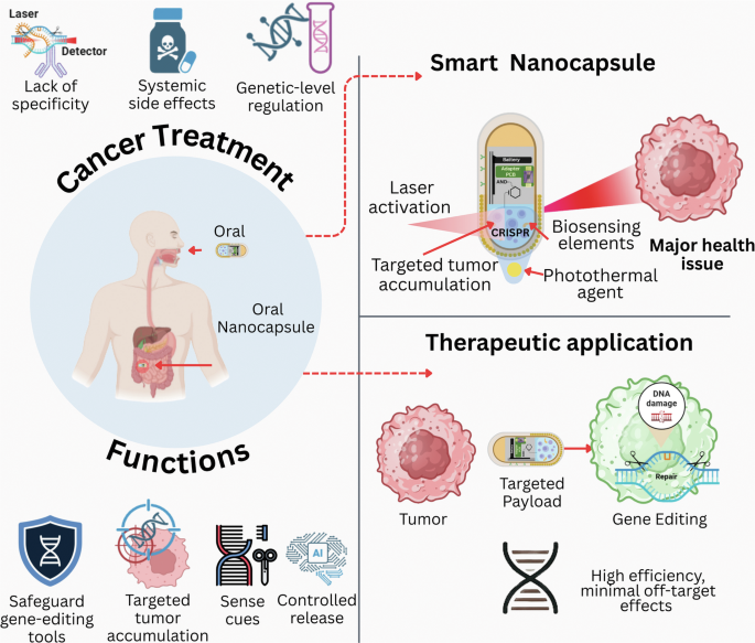
Current therapeutic techniques for cancer often lack specificity. They also cause systemic toxicity and lack genetic control. Thus, cancer ranks among the most complex and crucial global health issues. The novel concept of smart nanocapsules is discussed in this Perspective. These oral medications modify genes using CRISPR technology and integrate biosensing and laser-guided activation to enable more personalized cancer therapies. The creation of these versatile nanocapsules is driven by three objectives. First, they aim to enable controlled gene editing in the gastrointestinal tract. Second, they deliver treatments to specific target areas. Third, they detect tumors in real time. Nanocapsules equipped with biosensing components provide microenvironmental input. An external laser can trigger the release of light-absorbing agents. Moreover, these features reduce off-target effects and allow spatiotemporal precision, thhe enteric-coated architecture ensures oral stability. Surface functionalization enhances selective tumor accumulation. AI-guided control algorithms can manage diagnostic interpretation and activation. The CRISPR-based cancer medicines offer the potential for improved safety, specificity, and translational use in the future. Combining advanced nanotechnology, gene editing, and AI-guided control could create innovative solutions.
Introduction
Cancer gene therapy represents a major shift in oncology, aiming not merely to destroy malignant cells but to correct the genetic errors that drive tumor growth. These treatments directly target the molecular components responsible for oncogenesis, offering the potential for durable and precise intervention. However, achieving effective in vivo gene editing remains difficult due to low editing efficiency, limited delivery to tumor sites, and pronounced genetic heterogeneity. Tumors differ not only between patients but also within the same lesion, where distinct subpopulations of cancer cells often carry divergent mutations that complicate uniform therapeutic response [1]. This complexity highlights the importance of developing tailored, mutation-specific therapeutic approaches, as it makes the development of standardized treatment procedures challenging [2]. Due to the huge number of concurrent genetic mutations in advanced solid tumors, effective intervention frequently requires multi-targeted techniques. Advanced solid tumors, in particular, provide extra difficulties in achieving this goal [3]. Nanotechnology and molecular engineering have recently made great strides, allowing for the creation of smart nanocarriers, tiny, engineered structures designed to transport drugs that can deliver therapeutic chemicals to specific areas, detect biological responses, and release their payload under regulated conditions. One potential method for targeted, non-invasive cancer treatment is the use of swallowable, laser-guided nanocapsules that combine biosensing capabilities with CRISPR-based gene editing (a technique for precise alteration of DNA). Degradation during gastrointestinal transit, unintended effects on non-target cells (off-target effects), and inadequate control over where and when gene-editing activity occurs (spatiotemporal control) are some of the important shortcomings that these multifunctional platforms try to improve [4, 5]. This Perspective focuses on smart, swallowable nanocapsules for CRISPR-mediated cancer gene therapy. It aims to assess and summarize recent developments in this area. The main attention areas are: (I) methods for stable oral distribution; (II) biosensing element integration for real-time tumor monitoring; (III) laser-guided activation for precise spatial and temporal control; and (IV) AI-guided optimization of treatment efficacy. It covered specific aspects of nanocarrier design and CRISPR delivery, swallowable systems with integrated sensing, gene editing, and controlled activation.
Traditional methods can limit therapeutic success since tumors are different from one another, and most cancers require genetic medications. Furthermore, in the design of methods for moving genes, including the use of viral vectors, safety should always come first [6]. Viruses can also infect hosts by inserting themselves into the host’s DNA before the immune system starts to fight them off. Polymeric nanoparticles, which are biocompatible and non-viral carriers, can help with these problems. It is possible to lower the immunogenicity of viruses while still allowing for regulated release [7]. Despite its many advantages, the limited dispersion effectiveness makes consistency of therapeutic results difficult. Delivery mechanisms that are incapable of distinguishing between normal and malignant cells may make therapeutic medications more hazardous or less effective [8]. Smart materials that respond to the acidic pH or high enzyme levels in the tumor microenvironment might be the solution to this issue. In effect, through changing the release in response to these signals, it is feasible to improve targeted accuracy and treatment safety. To avoid off-target consequences, it is very important to precisely control when and where therapeutic genes are turned on. Possible ways to do this are to control how therapeutic medicines are released based on how enzymes respond or pH. Thus, gene therapy may now focus on certain locations thanks to targeted delivery. Researchers are trying to establish a balance between control, efficiency, and specificity in gene therapy [9, 10].
Theranostics, a platform that combines diagnostics and therapeutics, is an attractive option for improving the efficacy of cancer treatments. Patients are more likely to take their gene treatments orally rather than through injections or infusions; therefore, a biocompatible delivery system that can carry these medications might increase compliance. A promising innovation in this field is the use of biodegradable nanocarriers, which can encapsulate nucleic acids and ensure their stability, enabling the oral delivery of gene treatments [11,12,13,14]. Hence, the development of innovative delivery systems capable of transporting CRISPR components with high specificity and control is urgently required, given these constraints. Another reported discussion, CRISPR/Cas9, enables precise genome alterations to eliminate or correct cancer-causing mutations, thereby restoring cells to a normal state [15]. However, delivery challenges make this technique challenging to implement, especially when attempting to access the nucleus of tumor cells in complex and dense microenvironments. As a result, the treatment efficacy will be diminished, and uptake will be decreased [16]. A critical safety problem is the risk of off-target modifications, which can cause negative consequences due to accidental changes [6]. One potential answer to these difficulties is systems based on smart nanocapsules. The acid-responsive releasing capabilities of the linear-dendritic polymer nanocapsules developed for combination therapy in clinical trials [17]. In combination with oral administration, precision targeting, and CRISPR-based gene editing, this smart nanocapsule represents a paradigm shift in cancer treatment. Using this technology, cancer therapies become highly efficient, personalized, and minimally invasive, as instruments are protected, tumor microenvironments are monitored, and laser-controlled release can be carried out, as shown in Fig. 1.

Figure parts generated with BioRender.com.
To selectively release CRISPR components, such as Cas9 and guide RNA, into the acidic tumor microenvironment, these nanocapsules can encapsulate them. A single system that combines imaging and therapy, allowing both treatment and real-time monitoring, may be created by modifying these carriers to include theranostic properties. Biodegradable nanocapsules loaded with CRISPR payloads have the potential to enhance the identification and infiltration of immune cells, thereby improving their therapeutic efficacy [18]. Furthermore, laser activation enhances accuracy by utilizing additional triggers to improve safety and minimize systemic exposure. Near-infrared irradiation can be used to selectively regulate the release of CRISPR components at tumor locations. This Perspective argues that smart, swallowable nanocapsules integrating CRISPR-based gene editing, biosensing, and laser-guided activation represent the most promising direction for advancing precision cancer therapy. Contrary to other evaluations, we deliver an in-depth analysis of recent breakthroughs, identify key technology and translation gaps, and establish a theoretical structure by dividing the field into actionable components. To guide the next stage of precision cancer gene therapy, it stresses the need for translational strategies, improved biocompatibility, and AI-driven optimization. By merging critical assessment with future-focused insights, this work aims to accelerate the clinical development of CRISPR-enabled nanotherapeutics.
Engineering and functional design of smart nanocapsule
Oral nanocapsule formulations have been developed as an alternative to injections and infusions, aiming to provide more patient-friendly therapies. This shift is especially significant in cancer care due to the beneficial impacts on compliance and patient comfort resulting from fewer and less painful hospitalizations (see Fig. 2). Moreover, this is an area where biodegradable nanocapsules have demonstrated great promise. A feature that reflects the tumor microenvironment is that poly(lactic-co-glycolic acid) capsules containing doxorubicin are stable at physiological pH but release their contents in acidic conditions [19]. Hence, these results provide more evidence that oral nanocapsules have the potential to be modified for targeted medication delivery to sick areas. Researchers are now focusing on nanocapsules that respond to environmental cues, in addition to pH-responsive devices. By demonstrating that thermo-responsive carriers can be activated by near-infrared light, Xie et al. showed the ability to regulate drug release while simultaneously enhancing imaging capabilities [20].

a Structural design integrating an enteric coating, biodegradable polymer shell, targeting ligands, and internal biosensing module; b CRISPR-mediated cancer gene editing therapy enabled by integrated sensing and laser guidance; (c) incorporation of AI-driven monitoring and feedback to enhance precision and personalization of treatment. Figure parts generated with BioRender.com.
CRISPR gene editing has added a unique aspect to this area that enables accurate and direct alteration of disease-causing genetic circuits using biodegradable nanocapsules. Research has shown that CRISPR parts can be used along with small-molecule drugs or RNA molecules. The design of the nanocapsules’ surface is also highly essential. Nanocapsules that respond to environmental signals may take in more and be more accurate. Combining these designs with CRISPR payloads allows genes to modify at the cellular level with a high level of precision. Moreover, the result could lead to a reduction in side effects and an increase in therapeutic advantages. The advances show that biodegradable nanocapsules, CRISPR technology, and oral formulations can be combined to provide a single platform for therapeutic effects. Thus, the patient-centered strategy combines targeted delivery with controlled release and theranostic potential for the future of personalized medicine [21].
Enteric coating for oral stability
In cancer gene therapy, safety considerations should be included in the design and construction of oral drug delivery systems. In addition, these procedures should make sure that the GI tract stays stable and functions correctly. Therefore, the design needs enteric coatings to keep the drugs safe from the acidic environment of the stomach. Enteric coatings are particularly significant because they keep medicinal compounds safe from the harmful effects of stomach acid and digestive enzymes. It makes sure that drugs go to the right place in the stomach. This protection is very crucial in gene therapy because genetic material is very fragile and can break down in the harsh environment of the upper gastrointestinal system [22]. Hence, this is why there has been a lot of study on pH-responsive polymers like Eudragit, which can stay stable in acidic environments and dissolve in the small intestine’s neutral pH, making it a great drug release inhibitor. The acidic secretions in the stomach maintain the digestive enzyme Eudragit FS intact, while the less acidic environment of the intestines breaks it down. Tabare et al. found that the therapeutic effectiveness of bacteriophages in medication formulations was greatly diminished in the stomach when this defense was lacking [23]. Thus, Enteric coatings like Eudragit make therapy far more successful by making drugs more stable and allowing them to be delivered more directly to the site of action.
Researchers are looking into other ways to get even more exact distribution than what is possible with standard systems. Zhang et al. used a hybrid approach that mixes polymethacrylic acid with other biocompatible polymers to make oral delivery systems that are specific to the colon. Their research intended to enhance the targeted release of therapeutic chemicals in the colon while still within the gastrointestinal tract by integrating micro- and nanoencapsulation with enteric coatings. Local delivery is particularly important for treating ulcerative colitis and other related diseases [24].
Enteric coatings that contain several polymers work better to control release and offer protection. It is possible to create interpolymer complexes by combining polysaccharides with Eudragit polymers. These combinations allow for the stabilization of the therapeutic payload as well as the adjustment of release timing and location [25]. Thus, oral medicine delivery devices for gene therapy require enteric coatings. In such a setting, they are more effective, reliable, and accurate since they prevent the stomach from breaking down drugs and release them only at specific sites. Gene therapy formulations are steadily improving in terms of efficacy and reliability. One important aspect of this evolution is the creation of new materials, such as Eudragit and creative polymer combinations.
Biodegradable polymer shell (PLGA/PEG layer)
Biodegradable polymer shells, such as poly(lactic-co-glycolic acid) (PLGA) and polyethylene glycol (PEG), are essential to modern drug delivery systems, especially in targeted gene therapy. In addition, these materials are biocompatible, have a long circulation time, and release therapeutic compounds in a regulated manner through calibrated biodegradation profiles. The PEG outer layer of PEG helps increase the time in circulation of PLGA-based delivery systems, which enables proteins to be absorbed slowly and be detected by macrophages. Therefore, these are the primary measures to take to get out of circulation as soon as possible. Thus, when the pharmaceutical component reaches its target site, it is more bioavailable. Since Kalele et al. focused on exosomes in oral cancer rather than polymer-based nanoparticles, it is inappropriate. In addition, there is a lack of tangible data to support these assertions regarding stealth in the current literature on PEG-PLGA systems [26]. To address therapeutic demands, researchers can modify the biodegradation behavior of PLGA and PEG to create customised systems. The rate of PLGA breakdown can be controlled by adjusting the polymer’s molecular weight, and the lactic-to-glycolic acid ratio will influence the release kinetics of the encapsulated medicine [27]. As pH and enzyme activity, among other environmental factors, further alter these release patterns, chances for individualized delivery methods that coordinate medication release with physiological settings emerge [28]. Upgrading pH-sensitive devices could enhance their precision in targeting specific sections of the GI tract for drug delivery. Formulations based on Eudragit demonstrate site-specific release in response to pH changes, according to Yu et al. [29]. Systems involving PLGA and PEG can benefit from this. Colon cancer treatment improves with pH-sensitive coatings and PLGA. According to Heikal et al., the coatings facilitate targeted medication release, enhancing delivery to the colon while minimizing systemic exposure. Biodegradable polymer systems using PLGA and PEG help distribute drugs efficiently through controlled breakdown and quick release [30]. Adjusting responsive polymers and PLGA can lead to more adaptable drug delivery systems. However, further research is needed to confirm the effectiveness of these devices as targeted therapies, particularly their capacity to blend in, degrade properly, and stay undetected.
Targeting ligands on the surface
A crucial advancement in developing more precise and effective cancer treatments is adding targeting ligands to the surfaces of drug delivery systems. These systems can identify tumor biomarkers and modify their surfaces with biological molecules such as peptides, aptamers, and antibodies to specifically interact with cancer cells. This approach allows targeted drug delivery directly to tumors, reducing damage to healthy tissues and enhancing treatment effectiveness away from the target. Monoclonal antibodies play a central role in targeted therapy because of their high affinity for antigens on cancer cell surfaces. In addition, they allow for precise targeting of cancer cells while avoiding healthy tissue. Antibody-drug conjugates function by merging antibodies’ capacity to identify cancer cells with the delivery of strong therapeutic agents directly to tumors. Research by Tajau et al. [31] demonstrated that antibody-conjugated nanoparticles effectively targeted breast cancer cells, leading to better treatment results with lower systemic toxicity [31].
In addition, aptamers offer extremely selective biomarkers to tumor biomarkers; these oligonucleotides are short and may fold into unique three-dimensional structures. The benefits of low immunogenicity and good stability are coupled with their ease of synthesis, which is comparable to that of antibodies. Research has shown that nanoparticles functionalized with aptamers can increase the transport of therapeutic drugs to tumor cells, leading to more effective therapy with fewer side effects associated with conventional treatments [32]. Peptides, a kind of ligand that shows promise, are engineered to attach to certain receptors that are overexpressed in tumor cells, including integrin αvβ3 or HER2/neu. Their integration into delivery systems enhances the internalization of therapeutic drugs by cancer cells through receptor-mediated uptake. Parra-Nieto et al. and Ghosh et al. found that peptide-modified nanoparticles can increase the concentration of doxorubicin in hypoxic tumor areas, leading to more targeted administration of the medicine and better overall therapy results [33].
Proteins, glycoproteins, or RNA sequences that are overexpressed in cancer cells are examples of tumor-specific biomarkers that must be identified for these targeted approaches to be effective. Moreover, this is demonstrated by the fact that biomarkers like carcinoembryonic antigen (CEA) and prostate-specific antigen (PSA) have been used in targeted therapy for different types of carcinomas and prostate cancer, respectively, by Dattilo et al. [34]. In addition, these biomarkers are critical for developing area-specific targeting systems [34]. Using nanotechnology as a unifying mechanism, theranostics integrates diagnostic and therapeutic applications. Imaging and drug-delivering nanoparticles allow for continuous tracking of therapeutic efficacy [35]. Cancer treatments are made far more efficient and successful by attaching ligands like peptides, aptamers, and antibodies to drug delivery devices. Molecular signatures optimize treatment results while reducing systemic adverse effects by using tumor biomarker molecular signatures. As a result, cancer therapies may be tailored to suit the unique biological traits of each patient.
Internal biosensing module
Improving treatment results in cancer gene therapy by continuously monitoring the tumor microenvironment is made possible by an implanted biosensing module. This system may self-diagnose and react to tumor growth by utilizing aptamer-based platforms and molecular signaling technologies to detect biochemical changes in real time and adjust its activity accordingly. Aptamers are perfect biosensing components because they are short oligonucleotides that bind selectively to molecular targets like metabolites and proteins. As of today, they are powerful diagnostic tools thanks to their ability to identify tumor biomarkers. Gong et al. [36] emphasized their possible use in cancer diagnostics, whereas Qian et al. [37] revealed the construction of DNAzyme molecular machines that can govern cell behavior in situ and monitor tumor conditions [36, 37]. These results demonstrate the potential of aptamer-based biosensors for early tumor identification and guidance of therapy response. Molecular beacons enhance biosensing through their stem-loop structure, which emits fluorescence when fused with a specific nucleic acid. As a direct measure of cellular activity, this process enables the identification of RNA or DNA sequences in the tumor microenvironment. Such designs can monitor disease development and therapy efficacy in real-time, as demonstrated by Dey et al. [38] in relation to cancer-associated genomic expression. Biological processes, including apoptosis, proliferation, and metastasis, may be captured by real-time tumor monitoring [38]. It is possible to tailor treatment approaches to the changing biology of the tumor by monitoring biomarkers such as microRNAs or protein levels [39, 40]. Wu et al. [41] drew attention to miRNA-21 as a clinically relevant biomarker for tumor development. Moreover, it showed how biosensing systems can utilize miRNA-21 detection to provide rapid diagnostic feedback [41]. Cancer gene therapy gains a double-edged sword when it incorporates an integrated biosensing module that combines aptamers with molecular beacons. Advancements in the field of personalized, adaptive, and more effective cancer therapies are being made possible by such systems, which enable continuous monitoring of the tumor microenvironment and adjust therapeutic responses [42].
Therapeutic payload (CRISPR/Cas9)
Oncogenes are precisely targeted by the Cas9 ribonucleoprotein (RNP) complex in conjunction with the highly specialized guide RNA. Cas9 endonuclease and guide RNA are components of the Cas9 RNP, which directs the enzyme to the target genomic area for editing. This technology offers a simple and efficient way to change genes by inserting double-strand breaks at accurate spots [43, 44]. One major advantage of temporarily delivering Cas9 RNPs for therapeutic purposes versus plasmid-or viral-based administration is the decreased likelihood of off-target activation. Han et al. [45] provided more evidence of the therapeutic potential of Cas9 RNPs by showing that these particles exhibit high editing efficiency in mesenchymal stem cells [45].
Significant headway has also been made in developing delivery systems that enhance the therapeutic efficacy of Cas9 RNPs. Wei et al. [46] showed that the Cas9 RNP can be systemically delivered using nanoparticles, enabling successful genome editing across a variety of tissues, including skeletal muscle and brain. The versatility of this approach demonstrates the potential for precision medicine to cure illnesses or tissues by altering particular regions of the genome [46]. If the guide RNA is of poor quality, the CRISPR/Cas9 platform will not be effective in treating cancer. Oncogenes include DNA sequences that are complementary and direct the synthesis of RNAs that may be identified. According to Chen et al. [47], the protospacer adjacent motif (PAM) sequence can be used to identify genomic loci that Cas9 can bind and cleave. Among the several things made feasible by sequence specificity are the reactivation of mutant oncogenes and the restoration of tumor suppressor functions. To ensure guide RNA is highly precise and prevents unintended modifications, its design must be improved [47].
Using well-planned guide RNAs makes gene editing in cancer-related genes far more successful and specific. It includes survivin in 4T1 tumor cells. It is very important to make guide sequences correctly so that they can strike the right balance between being effective as a treatment and causing fewer side effects. Using the Cas9 RNP complex and properly crafted guide RNAs, it is feasible to make advanced cancer therapies that target specific genes [48]. The technology for modifying and delivering medications is getting better all the time. Consequently, this implies that medicines can be made safer and more effective. Cas9 RNPs might be a novel way to treat cancer because they work well on many forms of cancer, can be changed to fit different situations, and don’t have many side effects [48].
Laser-responsive nanomaterials
Nanomaterials that respond to light and heat, such as carbon dots and gold nanoparticles, effectively treat cancer. Lasers absorb light at specific wavelengths and convert it into heat in targeted areas, providing precise control over treatment. Gold nanoparticles are enhanced by their surface plasmon resonance, which makes them more efficient at absorbing light and generating heat. Thus, it allows photothermal treatment agents to selectively target tumor cells, destroying them while minimizing damage to surrounding healthy tissues [49]. Zhu et al. [50] demonstrated that by customizing nanoparticles to specific optical absorption spectra, it is possible to maximize efficiency while minimizing thermal injury to nearby tissues [51].
For laser-responsive nanomaterials, the ability to precisely target specific areas is the key benefit. Nanoparticles in the tumor microenvironment can be targeted with laser beams and delivered energy. Systemic adverse effects are reduced, and treatment effectiveness is increased using this strategy. As size-dependent plasmon resonance considerably enhances thermal conversion efficiency compared to spherical particles, Zhou et al. [52] demonstrated that nanoparticle shape is critical for photothermal performance, particularly in gold nanorods [52]. Therefore, one of the most important ways to maximize therapeutic effects is to optimize the size and form of nanoparticles. However, when it comes to precision cancer therapy using laser-responsive nanomaterials, two of the most promising classes are carbon dots and gold nanoparticles. As a result of their photothermal conversion capabilities, they can precisely ablate tumors with minimal collateral damage.
Smart nanocarrier methods to improve CRISPR/Cas9 cancer treatment delivery are listed in the (Table 1). Lipid nanoparticles and cationic liposomes are popular for encapsulating genetic elements and responding to temperature and pH. Further, this responsiveness improves endosomal escape and targeted delivery, which are essential for gene editing efficacy [53, 54]. Biocompatibility and toxicity are issues for gold nanoparticles, despite their high loading capacity and light manipulation [55]. Polymeric nanocarriers like PLGA can be surface modified for improved targeting, but their complex production can limit clinical scalability [56].
DNA-based nanocarriers can be customized to convey specific cargo, but their stability in physiological contexts is an issue [57]. Although exosome-like nanoparticles are natural and harmless, their uncontrolled cargo release is a drawback [58]. In addition, micelles, amphiphilic block copolymers, temperature, pH, and enhanced solubility in hydrophobic drugs [59]. As a result, nanocarrier types are improving, but stability, biocompatibility, and physiological delivery mechanisms remain issues. To optimize CRISPR/Cas9 cancer medicines for clinical effectiveness, continuous research is needed.
Tissue penetrability (how well light passes through tissue), thermal safety (the potential for overheating and damaging tissue), and optical absorption (how materials absorb light) are three areas where laser-responsive nanomaterials are currently lacking in clinical development, despite their promising potential for targeted cancer treatment. Many studies found that while near-infrared (NIR, light with wavelengths between 650-950 nanometers) penetrates tissues more effectively than visible light, it is still not enough for deep-seated malignancies [60, 61].
Functionalized with LK peptides, bumpy Au triangular nanoprisms (BATrisms) showed higher cellular uptake, increased photothermal conversion to around 86%, and enhanced near-infrared absorption. With minimum tissue damage, triple-negative breast tumors were effectively destroyed in vivo using a modest dosage of 2.5 μg Au under an 808 nm, 0.25 W/cm² laser [62]. The NA1020-NO@PLX nanocomposite was able to efficiently convert light into heat and penetrate deep into tissues due to its NIR-II absorption (Λmax = 1020 nm). This technique showed promise for targeted therapeutic applications by enabling low-temperature, traumatic PTT with improved tumor apoptosis in deep-tissue osteosarcoma [63]. However, traditional PDT and PTT methods also face limitations, including shallow light penetration (<10 mm) and increased thermal damage risk, which restrict their effectiveness for deep-seated tumors [64]. Additionally, light transmission is limited by biological factors like hemoglobin and water absorption, and many conventional photothermal agents have poor near-infrared (NIR) absorption [65].
Adjuncts and imaging components
Integrating imaging components into cancer treatment has made it much more precise and effective. It employs both chemotherapeutic drugs and immune modulators. Cancer treatments could benefit from a combination of the two methods, and patients might get better care with real-time monitoring and better imaging technologies [66]. To improve the efficacy of therapies, including regular chemotherapy, immune modulators are essential. Combining immune-based treatments with chemotherapy can assist in overcoming resistance mechanisms that metastatic tumors commonly acquire. Evidence suggests that immunological modulation and cytotoxic drugs can work together. In hormone-positive breast cancer, immune checkpoint inhibitors combined with traditional chemotherapy can slow tumor growth and train the immune system to identify and destroy cancer cells [67]. In recent years, Xia et al. [68] found that these fluorophores’ strong tissue penetration and negligible background interference make them ideal for tumor visualization. Consequently, this enables precise monitoring of treatment distribution and efficacy [68].
Multifunctional magnetic nano agents, as demonstrated by Ding et al. [69], have a wide range of potential therapeutic applications, including bioimaging, drug administration, and localized photothermal treatment simultaneously [69]. Therefore, an adaptable framework for cancer treatment is provided by integrating immune modulators, chemotherapeutic agents, and cutting-edge imaging technologies. Better patient outcomes result from the introduction of this integrated strategy’s real-time monitoring and personalized treatment regimens, which in turn enhance the efficacy of both conventional and immune-based medicines.
Mechanistic workflow of the smart nanocapsule for targeted cancer therapy
Several sequential steps comprise the mechanistic process of developing nanocapsules for targeted cancer treatment using CRISPR technology, as shown in Fig. 3. These steps guarantee the efficient and regulated delivery of genetic editing tools to tumor locations. Upon oral ingestion, the nanocapsules undergo transit through the gastrointestinal tract. Gastric acid and digestive enzymes cannot break them down route due to an enteric coating. The coating is dissolved upon reaching the neutral pH of the small intestine, allowing for systemic absorption [70]. Carriers such as PLGA and PEG increase the probability of accumulation at tumor sites by extending circulation times and enhancing bioavailability after nanocapsules are absorbed through the intestinal epithelium [71]. The Enhanced Permeability and Retention (EPR) effect enables the targeted delivery of functionalized ligands, such as antibodies or peptides, to the surface of the capsule. Subsequently, this improves accumulation by enabling selective localization inside tumors via the detection of biomarkers unique to tumors [72]. The tumor microenvironment may be monitored for changes in pH levels or biomarker expression using nanocapsules equipped with biosensing devices like aptamers and molecular beacons. Moreover, this enables the nanocapsules to deliver self-diagnostic feedback, which may then be employed to dynamically modify therapy approaches [73].

a Representation of oral delivery, enabling systemic distribution and tumor accumulation. b Biosensing of the microenvironment with self-diagnosis. c The laser-triggered CRISPR release, followed by safe biodegradation. Figure parts generated with BioRender.com.
Li et al. [74] report that localized laser irradiation triggers the release of the payload. As photothermal agents, gold nanoparticles convert absorbed light into heat, enabling the controlled release of CRISPR/Cas9 components at tumor sites with little systemic exposure [74]. After being released, the CRISPR/Cas9 system may alter genes by removing tumor suppressor genes or oncogenes, which can restrict tumor growth, cause cell death, and even cause tumors to shrink [75]. The nanocapsule, composed of biodegradable polymers such as PLGA, breaks down hydrolytically into harmless byproducts that are naturally flushed out by the body according to the engineered degradation rate [76]. This integrated process outlines a complex strategy for enhancing precision cancer treatment with CRISPR-based genomic interventions, encompassing the oral delivery of nanocapsules, their distribution throughout the body, targeted tumor specificity, biosensing, controlled activation, gene editing, and clearance of the nanocapsules [77].
Artificial intelligence–enabled nanocapsule for theranostic applications
Nanomedicine with AI has changed the game for theranostic systems, improving accuracy, imaging in real-time, and tailored therapy results [50, 78, 79]. Building on these advancements, researchers found that AI-driven platforms, such as photo-triggered, MRI-traceable, and nanoarchitectonic-based nanoparticles, enhance therapeutic control, adaptive response, and targeting accuracy. Collectively, these developments demonstrate how AI connects on the lab bench to real-world applications in cancer and cardiovascular care, molding the future of precision nanomedicine [80, 81].
As biological signals dictate when and where to administer drugs, nanocarriers’ sensing capabilities allow for on-demand delivery of therapeutic chemicals based on these environmental cues [82]. Imaging methods like near-infrared fluorophores offer real-time therapy tracking. As the nanocarrier interacts with the tumor environment, it senses triggers medication release and provides instantaneous cancer response data. Thus, it permits fast treatment strategy revisions if needed [83, 84]. In addition to response mechanisms, imaging components like near-infrared fluorophores offer real-time therapy monitoring. This capability allows clinicians to monitor medicine distribution and efficacy in real time, helping them customize treatment programs, according to Liv et al. (2023). Having diagnostic and therapeutic capabilities on one nanoplatform improves treatment accuracy and flexibility [85]. By dynamically adapting treatment plans to the specific molecular features of each patient’s cancer, this theranostic method heralds a new era in personalized medicine, with the promise of improved patient outcomes. An AI-guided nanocapsule for the detection and editing of cancer cells is illustrated in Fig. 4a.

a illustration of AI-enabled theranostic nanocapsules integrating diagnostic sensing and targeted gene-editing functions. b Overview of applications, including cancer detection and genome editing, as well as broader therapeutic uses in neurology, infectious diseases, autoimmune disorders, tissue regeneration, and vaccine development. Figure parts were generated with BioRender.com.
AI-guided nanocapsules provided medical promise due to their adaptable delivery, personalized design, and accurate sensing capabilities [86]. It can enable the delivery of RNA, neuroprotective drugs, and blood-brain barrier-crossing CRISPR tools to treat neurodegenerative illnesses such as Alzheimer’s, Parkinson’s, and Huntington’s. Additionally, early detection of neural damage becomes possible with biosensing, which works in tandem with targeted medicine delivery. AI-optimized gene editing nanocapsules further expand treatment options, target latent HIV reservoirs or antibiotic-resistant bacteria to cure infections. Similarly, by sensing inflammatory signals and releasing immunomodulatory medicines locally, these nanocapsules help minimize conditions like multiple sclerosis and rheumatoid arthritis. As a result of oxidative stress or lipid accumulation, nanocapsules designed through artificial intelligence may also benefit cardiovascular medicine. Sensing features in regenerative medicine are used to release factors that promote tissue repair in organs such as the liver, kidney, or skin. As vaccine platforms, nanocapsules could revolutionize cancer and infectious disease immunotherapy by using AI algorithms to select optimal antigen combinations and delivery strategies based on individual immune profiles. For metabolic disorders such as diabetes and obesity, they could function as artificial pancreas-like devices that monitor glucose or insulin levels and release therapeutic agents, accordingly, representing an important step toward dynamic, personalized treatments (see Fig. 4b).
Challenges and considerations
The clinical translation of swallowable, laser-guided CRISPR nanocapsules requires coordinated solutions across biostability, delivery efficiency, optical access, immunological safety, and regulation. Reviews and preclinical analyses document low systemic nucleic-acid bioavailability after oral/GI passage, typically reported around 15–30%, and attribute losses to gastric acid and enzymatic degradation [30, 87]. Enteric polymers, such as Eudragit S100/FS, have been shown to enhance payload protection compared to uncoated forms and exhibit pH-dependent dissolution that must be optimized to intestinal transit, with dissolution windows typically near pH 6.8-7.2, coinciding with absorption windows of approximately 3 hours [23]. Hybrid micro/nano-encapsulation strategies have been reported to reduce premature leakage, though they introduce batch-critical variables that can influence Cas9/RNP activity retention [70, 88]. Therefore, integrated formulation workflows that incorporate quantitative release-rate mapping and accelerated stability testing are essential for preserving editing activity and enabling regulatory-grade translation [70, 88].
Stability begins before dosing and ends only after controlled release at the disease site. Cas9 RNPs and guide RNAs are sensitive to acid, nucleases, shear, temperature, and interfacial stress during fabrication and storage. Enteric architectures such as Eudragit S100/FS and hybrid micro-/nanoencapsulation shield the payload through the stomach and proximal intestine, but they shift dissolution profiles and add batch-critical variables that must be locked by process controls; release kinetics should be tuned to local pH and enzyme activity to avoid premature leak or incomplete unloading [22,23,24]. PLGA-based and other pH-responsive systems support site-selective opening, yet polymer ratio, molecular weight, residual solvent, and lyophilization buffers must be optimized so that Cas9 retains activity after processing and reconstitution; otherwise, apparent “stability” in transit can still yield subtherapeutic editing at the target [30, 70].
Delivery precision dictates both efficacy and risk: viral vectors achieve high in vitro transduction but carry well-documented immunogenic and insertional risks [87, 89]; in contrast, non-viral polymeric carriers (PLGA, LNPs) generally report lower editing yields (typical ranges ∼20–40%) while offering improved safety and manufacturability [70, 90]. Transient Cas9 ribonucleoprotein (RNP) dosing shortens nuclease exposure from days to hours, substantially lowering off-target cutting relative to plasmid delivery [88, 91]. Optical gating with near-infrared (NIR) light confines editing to millimeter scales but remains depth-limited by tissue attenuation and scattering, constraining in vivo penetration [92, 93]. Photothermal actuators amplify optical control; gold nanorods exhibit markedly higher photothermal conversion than spherical nanoparticles, improving heating efficiency, while hybrid Au–carbon-dot or multilayered architectures enable uniform heating (±≈1 °C) at clinically relevant fluences (<0.8 W·cm−2), thereby reducing collateral injury [94,95,96].
Editing precision governs clinical risk. Transient RNP delivery shortens nuclease exposure compared with plasmid or viral vectors, reducing the window for unintended cuts, while optical gating restricts activity in space and time. These advantages only materialize with a disciplined guide design that enforces PAM requirements, models mismatch tolerance, and uses orthogonal off-target assays across relevant cells or organoids; tumor heterogeneity and context-dependent DNA repair still favor rare large deletions, rearrangements, or selection of edited clones, which necessitates longitudinal genomic surveillance and predefined stopping rules in early trials [43,44,45, 97].
Optical actuation adds a precise, external control handle but is bounded by tissue optics. Near-infrared and NIR-II windows increase penetration and enable image guidance, yet attenuation, scattering, vascular heat sinks, and fibrotic stroma constrain achievable fluence in deep lesions. Photothermal transducers must therefore be tuned in size, aspect ratio, and resonance to maximize conversion efficiency at safe irradiance, while real-time fluorophores support closed-loop dosimetry to minimize off-target heating. Practical deployment often pairs non-invasive beams for superficial disease with interstitial or endoscopic fibers for deep sites, each requiring validated dose models in heterogeneous tissue [52, 98, 99].
PEGylation (e.g., PEG–PLGA) can extend nanoparticle circulation from minutes to hours, but repeated dosing can provoke anti-PEG-mediated accelerated blood clearance in animal models, necessitating immune monitoring during chronic regimens [100]. Therefore, successful translation into clinical applications requires quantitative organ-level retention mapping, analysis of degradation products, and adaptive immune surveillance across repeated cycles to detect bioaccumulation or immunogenic metabolites [101]. Imaging-capable biodegradable tracers, such as polymer-functionalized upconversion nanoparticles and NIR-II biodegradable nanoprobes, should be utilized for whole-body mass balance and clearance kinetics studies to determine whether polymer shells and metallic cores are eliminated within clinically acceptable time frames and to quantify the risks associated with reticuloendothelial system (RES) retention [71, 93]. Integrating these quantitative imaging endpoints into preclinical safety protocols will inform acceptable exposure windows and regulatory safety criteria [101]. Biocompatibility and immune response reflect the full construct, not just the polymer. PEG/PLGA shells can prolong circulation and improve tumor exposure, but they also reshape the protein corona, may elicit anti-PEG responses with repeat dosing, and can delay renal–hepatobiliary elimination.
Designs should quantify organ retention and degradation products, characterize ligand-driven tropism beyond EPR assumptions, and measure innate and adaptive responses to Cas9, nucleic acids, photothermal agents, and surface ligands under clinically relevant schedules. Imaging-capable, biodegradable platforms enable mass-balance studies and inform iteration toward full biodegradation or renal-tunable clearance that avoids long-term persistence of inorganic cores [75, 76, 102].
A three‑stage regulatory roadmap, preclinical optimization, animal validation, and regulatory integration, must bind quantitative release, manufacturing, immunotoxicity, and optical‑dosimetry endpoints into standardized validation workflows [7, 90]. Preclinical goals should enforce tight process control (e.g., ≤5% relative standard deviation in capsule size and ≥90% payload encapsulation) and orthogonal specificity assays (deep sequencing for off-target indels ≤0.1% plus gastrointestinal-simulating in vitro/ex vivo digestion) to preserve Cas9 ribonucleoprotein activity during enteric processing [45, 88]. Animal validation must combine whole-body biodistribution and immunogenicity mapping with image-guided laser dosimetry to benchmark therapeutic index versus systemic CRISPR (aiming for ≥3× on-target: off-target gains) using near-infrared photonic control platforms and plasmonic nanohybrids [92, 93]. Finally, Chemistry, Manufacturing, and Controls (CMC) alignment with gene-therapy precedents and longitudinal genomic surveillance are essential for regulatory acceptance and post-market safety monitoring [90, 103].
Regulatory and ethical requirements complete the envelope. As a combination product, the capsule must present GMP-ready chemistry-manufacturing-controls for the carrier, RNP, targeting ligands, and optical interface; stability-indicating assays for payload integrity, release, and photothermal response; and verified device–drug–biologic interactions. Clinical programs should remain in somatic contexts with robust consent, genomic surveillance, and data governance, align with gene-therapy precedents for quality and safety documentation, and stage first-in-human studies around image-based pharmacology and predefined safety gates [7, 77, 97]. Together, these considerations define the feasible design space and the evidentiary standard for a swallowable, laser-guided CRISPR theranostic intended for clinical use.
Table 2 summarizes quantitative benchmarks across current CRISPR delivery platforms and emerging smart nanocapsule systems. The data reveal that polymer-based and pH-responsive capsules offer up to a five-fold improvement in oral stability over lipid nanoparticles, while laser-guided architectures promise sub-percent off-target activity with comparable cost per milligram to advanced polymer carriers. These comparative metrics provide an objective basis for evaluating translational potential and highlight where further optimization, particularly in photothermal tuning and reproducible ligand density, is needed.
Future perspectives
The integration of nanotechnology and gene-editing tools such as CRISPR/Cas9 continues to expand the possibilities of precision oncology, but progress remains incremental rather than revolutionary. Recent work combining biodegradable nanocarriers, AI-assisted formulation design, and non-viral genome-editing systems indicates measurable improvements in targeting accuracy and pharmacokinetic control, though many reported efficiencies remain below those needed for clinical efficacy (typically < 50% editing in solid-tumor models [43, 45, 46]).
Artificial-intelligence frameworks provide opportunities to optimize capsule geometry, surface chemistry, and release kinetics by analyzing experimental datasets that link polymer ratios, particle size, and surface potential to in-vivo biodistribution. However, these models are still limited by the scarcity of standardized datasets and by an incomplete understanding of how microenvironmental parameters—pH, enzyme activity, stromal density—affect oral nanocarrier performance. Future progress will depend on generating quantitative design–response maps and validating them experimentally across multiple cancer types. Combination strategies that couple CRISPR nanocapsules with immunotherapy or checkpoint inhibitors are promising for overcoming tumor immune evasion, yet they also raise safety and regulatory complexities. Synergistic effects between immune activation and genome editing have been demonstrated in vitro and in small-animal studies [104, 105], but dose balancing, cytokine release control, and long-term immune memory require systematic evaluation before human translation.
Emerging non-invasive activation modalities—including magneto-thermal and ultrasound-responsive systems—offer complementary spatial control without optical attenuation limits. Their energy-to-heat conversion efficiencies are modest (5–15%) compared with near-infrared photothermal systems [51, 52, 98, 102], and reproducible calibration under heterogeneous tissue conditions remains an unresolved technical constraint. Further investigation into dual-mode activation (optical + acoustic) could enhance safety margins while maintaining local precision.
A credible translational roadmap will require (i) standardized large-scale manufacturing with batch variation <5%; (ii) good-manufacturing-practice (GMP)–grade polymer and guide-RNA synthesis; (iii) comprehensive biodistribution and immunogenicity studies in large-animal models; and (iv) alignment with regulatory pathways established for combination gene-therapy products. Multi-institutional consortia integrating materials scientists, clinicians, and data-science specialists will be essential to meet these criteria. The swallowable, laser- or stimulus-responsive CRISPR nanocapsule should be regarded as a promising but still experimental platform within the broader continuum of precision-delivery technologies. Its success will depend on converting conceptual integration into quantitatively validated performance and regulatory reproducibility rather than on speculative expectations of clinical transformation.
According to Li et al. [106], neoadjuvant therapy can improve treatment outcomes in locally advanced gastric cancer by changing tumor clonal evolution. Additionally, combined therapeutic strategies can target resistant tumor clones, which can help optimize treatment [106]. Sun [107] reviewed the progress in gene therapy for hematological malignancies and emphasized how CRISPR and gene editing might transform blood cancer therapies [107]. The significance of non-viral vectors in circumventing conventional delivery obstacles was highlighted by Luiz et al. [108], who explored the development of liposomal systems for the safe and effective delivery of genes in cancer treatment [108]. Personalized cancer treatments that target genetic and immune-related tumor complexity have been developed, and Youssef et al. (2025) examined gene therapy techniques that combine with immune regulation for comprehensive cancer treatments [109]. Xu et al. [110] investigated the potential of targeted siRNA delivery using carbon-based nanomaterials, which could significantly enhance the efficacy of gene therapy [110].
Cesur-Ergün and Dora [111] summarized gene therapy methodologies and their authorized clinical uses in cancer treatment, providing clarity on the development of these medicines and directing future studies [111]. Hasbullah and Musa [112] evaluated potential gene therapies for colorectal cancer that target p53 and KRAS mutations, highlighting the importance of precision medicine in developing such interventions [112]. Through their discussion of cytochrome P450 enzyme design, Carrera-Pacheco et al. [2] shed light on safer therapeutic techniques that can enhance the specificity and efficacy of cancer gene therapies [2]. After reviewing the current state of cancer gene therapy, Katare et al. (2023) identified key areas that require improvement in both safety and effectiveness [113]. In his discussion of gene therapy’s role in lung cancer treatment, Liu [114] reaffirms the promise of this approach in addressing the unique challenges associated with this illness [114].
As a novel approach to overcoming resistance, Kordkheyli et al. (2021) have explored the application of gene editing, utilizing CRISPR technology, for controlling drug-resistant lung cancer [115]. To improve individualized treatment and direct patient care, Shoji et al. [116] assessed therapeutic approaches for ovarian cancer patients with a BRCA mutation [116]. To facilitate the development of targeted therapies, Fois et al. [117] investigated the frequency of actionable mutations in non-small cell lung cancer [117]. To inform precision medicine approaches, Passarelli et al. [118] reviewed recent developments in our understanding of molecular characteristics and targets for non-muscle-invasive bladder cancer [118]. In their work on the state of the art and potential future research areas, Aggarwal and Mittal [119] outlined the successes and failures of cancer gene therapy methods [119]. Finally, Yamano et al. [120] explored the potential uses of gene therapy in various oral disorders, expanding the treatment horizon and confirming the therapy’s importance in multiple clinical settings [120].
The evolution of cancer gene therapy and the relationship between different treatment approaches can be better grasped with the help of this table. Personalized medicine, immunotherapy combinations, and targeted therapy are the foci of the highlighted sources, which examine a variety of gene therapy applications (see Table 3). The therapeutic landscape is enhanced by the expertise of each contributor, which will inform future oncology clinical practices and enable the development of novel cancer therapies. The significance of continuing research into cancer biology and the development of targeted, effective treatment options for patients.
Conclusion
This Perspective has outlined the conceptual and experimental basis for oral, laser-activated nanocapsules that combine CRISPR-based genome editing with biosensing and controlled release within a single delivery architecture. The capsule described here should be regarded not as a finished therapeutic product but as an evolving framework that connects already established elements such as biodegradable polymers, photothermal agents, transient ribonucleoprotein delivery, enteric protection, and tumor-specific surface ligands into one coordinated system. Current reports show progress in payload stability, targeting precision and remote activation, but important gaps remain, including preservation of nuclease activity through the gastrointestinal tract, reproducible and complete release at the target site, quantitative mapping of off target edits in heterogeneous tumors, and validation of light penetration and thermal safety in vivo. In parallel, requirements for large-scale manufacturing, control of innate and adaptive immune responses, and regulatory assessment as a combination product will have to be met before clinical use can be justified. The potential to pair such capsules with artificial intelligence-guided design, with immunotherapy, or with alternative noninvasive triggers such as magnetothermal or ultrasound systems is clear, but each of these options will need standardized testing, scalable production routes, and harmonized safety analytics. The real value of a swallowable, sensing, and editing capsule will therefore depend on quantitative head-to-head comparisons with existing CRISPR delivery platforms and on coordinated work across polymer chemistry, optical engineering, and genomic toxicology to generate evidence that is consistent and transferable. If these conditions are satisfied, the concept can mature from a promising integration of known technologies into a practical route toward more precise, minimally invasive, and patient-adapted cancer treatment.
References
-
Ning L, Xi J, Zi Y, Chen M, Zou Q, Zhou X, et al. Prospects and challenges of CRISPR/Cas9 gene-editing technology in cancer research. Clin Genet. 2023;104:613–24.
-
Carrera-Pacheco SE, Mueller A, Puente-Pineda JA, Zúñiga-Miranda J, Guamán LP. Designing cytochrome P450 enzymes for use in cancer gene therapy. Front Bioeng Biotechnol. 2024;12:1405466.
-
Belete TM. The current status of gene therapy for the treatment of cancer. Biol Targets Ther. 2021;15:67–77.
-
Rauf MA, Rao A, Sivasoorian SS, Iyer AK. Nanotechnology-based delivery of CRISPR/Cas9 for cancer treatment: a comprehensive review. Cells. 2025;14:1–22.
-
Zhou S, Li Y, Wu Q, Gong C. Nanotechnology-based CRISPR/Cas9 delivery system for genome editing in cancer treatment. MedComm – Biomater Appl, 2024;3: e70.
-
Dai X, Blancafort P, Wang P, Sgro A, Thompson EW, Ostrikov KK. Innovative precision gene-editing tools in personalized cancer medicine. Adv Sci. 2020;7:1902552.
-
Paunovska K, Loughrey D, Dahlman JE. Drug delivery systems for RNA therapeutics. Nat Rev Genet. 2022;23:265–80.
-
Madry H, Venkatesan JK, Carballo-Pedrares N, Rey-Rico A, Cucchiarini M. Scaffold-Mediated Gene Delivery for Osteochondral Repair. Pharmaceutics. 2020;12:930.
-
Tasset A, Bellamkonda A, Wang W, Pyatnitskiy I, Ward D, Peppas N, et al. Overcoming barriers in non-viral gene delivery for neurological applications. Nanoscale. 2022;14:3698–719.
-
Shende P, Trivedi R. 3D printed bioconstructs: regenerative modulation for genetic expression. Stem Cell Rev Rep. 2021;17:1239–50.
-
Jeleriu RM, Hajaj RK, Trăilă IA, Zaharie M, Puiu M. Theoretical models and simulations of gene delivery with polyurethane: the importance of polyurethane as a vector in personalized therapy. Biomed. 2025;13:692.
-
Taha BA, Addie AJ, Saeed AQ, Haider AJ, Chaudhary V, Arsad N. Nanostructured photonics Probes: A transformative approach in neurotherapeutics and brain circuitry. Neuroscience https://doi.org/10.1016/j.neuroscience.2024.10.046 (2024).
-
Taha BA, Addie AJ, Chahal S, Haider AJ, Rustagi S, Arsad N, et al. Unlocking new frontiers in healthcare: The impact of nano-optical biosensors on personalized medical diagnostics. J Biotechnol. 2025;400:29–47.
-
Taha, BA, Kadhim, AC, Addie, AJ, Al-Jubouri, Q, Azzahrani, AS, Haider, AJ, et al. Optical spectroscopy of cerebral blood flow for tissue interrogation in ischemic stroke diagnosis. ACS Chem Neurosci https://doi.org/10.1021/acschemneuro.4c00809 (2025).
-
Huang CH, Lee KC, Doudna JA. Applications of CRISPR-Cas enzymes in cancer therapeutics and detection. Trends Cancer. 2018;4:499–512.
-
Zhang D, Wang G, Yu X, Wei T, Farbiak L, Johnson LT, et al. Enhancing CRISPR/Cas gene editing through modulating cellular mechanical properties for cancer therapy. Nat Nanotechnol. 2022;17:777–87.
-
Liu K, Xiang J, Wang G, Xu H, Piao Y, Liu X, et al. Linear-dendritic polymer-platinum complexes forming well-defined nanocapsules for acid-responsive drug delivery. ACS Appl Mater Interfaces. 2021;13:44028–40.
-
Liu K, Cui JJ, Zhan Y, Ouyang QY, Lu QS, Yang DH, et al. Reprogramming the tumor microenvironment by genome editing for precision cancer therapy. Mol Cancer. 2022;21:1–23.
-
Zhao J, Yang H, Li J, Wang Y, Wang X. Fabrication of pH-responsive PLGA(UCNPs/DOX) nanocapsules with upconversion luminescence for drug delivery. Sci Rep. 2017;7:1–11.
-
Xie Y, Yang Z, Shen H, Chen J, Weitz DA, Chen D, et al. Interfacial engineering of biocompatible nanocapsules for near-infrared-triggered drug release and photothermal therapy. Adv Sci 2025;12:2410844.
-
Naveed M, Tabassum N, Aziz T, Shabbir MA, Alkhateeb MA, Alghamdi S, et al. CRISPR-Cas9 guided RNA-based model for the silencing of spinal bulbar muscular atrophy: A functional genetic disorder. Cell Mol Biol. 2024;70:74–80.
-
Liu K, Chen YY, Li XY, Li QM, Pan LH, Luo JP, et al. Hydrolytic Quinoa protein and cationic lotus root starch-based micelles for co-delivery of Quercetin and Epigallo-catechin 3-Gallate in Ulcerative Colitis Treatment. J Agric Food Chem. 2022;70:15189–201.
-
Tabare E, Dauchot T, Cochez C, Glonti T, Antoine C, Laforêt F, et al. Eudragit® FS microparticles containing bacteriophages, prepared by spray-drying for oral administration. Pharmaceutics. 2023;15:1602.
-
Zhang L, Li M, Zhang G, Gao C, Wang S, Zhang T, et al. Micro- and Nanoencapsulated Hybrid Delivery System (MNEHDS): a novel approach for colon-targeted oral delivery of Berberine. Mol Pharm. 2021;18:1573–81.
-
Bukhovets AV, Fotaki N, Khutoryanskiy VV, Moustafine RI. Interpolymer complexes of eudragit® copolymers as novel carriers for colon-specific drug delivery. Polymer. 2020;12:1–16.
-
Kalele K, Nyahatkar S, Mirgh D, Muthuswamy R, Adhikari MD, Anand K. Exosomes: a cutting-edge theranostics tool for oral cancer. ACS Appl Bio Mater. 2024;7:1400–15.
-
Soares LA, Crema E. Study of a delayed-release system for hard and soft capsules coated with eudragit® s100 acrylic polymers. Acta Sci – Heal Sci. 2020;42:1–10.
-
Souto EB, da Ana R, Souto SB, Zielińska A, Marques C, Andrade LN, et al. In vitro characterization, modelling, and antioxidant properties of polyphenon-60 from green tea in Eudragit S100-2 Chitosan Microspheres. Nutrients. 2020;12:967.
-
Yu S, Piao H, Rejinold NS, Lee H, Choi G, Choy JH. pH-responsive inorganic/organic nanohybrids system for controlled nicotinic acid drug release. Molecul. 2022;27:6439.
-
Heikal EJ, Kaoud RM, Gad S, Mokhtar HI, Alattar A, Alshaman R, et al. Development of novel pH-sensitive eudragit coated beads containing curcumin-mesalamine combination for colon-specific drug delivery. Gels 2023;9:264.
-
Tajau R, Rohani R, Abdul Hamid SS, Adam Z, Mohd Janib SN, Salleh MZ. Surface functionalisation of poly-APO-b-polyol ester cross-linked copolymers as core–shell nanoparticles for targeted breast cancer therapy. Sci Rep. 2020;10:1–17.
-
Hapuarachchige S, Artemov D. Theranostic pretargeting drug delivery and imaging platforms in cancer precision medicine. Front Oncol. 2020;10:1–8.
-
Parra-Nieto J, del Cid MAG, de Cárcer IA, Baeza A. Inorganic porous nanoparticles for drug delivery in antitumoral therapy. Biotechnol J. 2021;16:2000150.
-
Dattilo M, Patitucci F, Prete S, Parisi OI, Puoci F. Polysaccharide-based hydrogels and their application as drug delivery systems in cancer treatment: a review. J Funct Biomater. 2023;14:55.
-
Ciolacu DE, Nicu R, Ciolacu F. Cellulose-based hydrogels as sustained drug-delivery systems. Materials. 2020;13:5270.
-
Gong H, Dai Q, Peng P. Cell-membrane-anchored DNA logic-gated nanoassemblies for in situ extracellular bioimaging. ACS Appl Mater Interfaces. 2022;14:43026–34.
-
Qian RC, Zhou ZR, Wu Y, Yang Z, Guo W, Li DW, et al. Combination cancer treatment: using engineered DNAzyme molecular machines for dynamic inter- and intracellular regulation. Angew Chem – Int Ed. 2022;61:e202210935.
-
Dey S, Ahmed E, Somvanshi PS, Sina AA, Wuethrich A, Trau M. An electrochemical and raman scattering dual detection biosensor for rapid screening and biomolecular profiling of cancer biomarkers. Chemosensors 2022;10:93.
-
Su J, Liu W, Chen S, Deng W, Dou Y, Zhao Z, et al. A carbon-based DNA framework nano–bio interface for biosensing with high sensitivity and a high signal-to-noise ratio. ACS Sens. 2020;5:3979–87.
-
Chen YL, Sun X, He JW, Xin MK, Liu D, Li CY. Light-driven and metal–organic framework synergetic loaded DNA tetrahedral amplifier for Exonuclease III-powered all-in-one biosensing and chemotherapy in live biosystems. ACS Appl Mater Interfaces. 2023;15:34311–20.
-
Wu Q, Wei X, Chen F, Huang M, Zhang S, Zhu L, et al. Aptamer-assisted blockade of the immune suppressor sialic acid-binding immunoglobulin-Like Lectin-15 for cancer immunotherapy. Angew Chem Int Ed. 2023;62:e202312609.
-
Zeng Z, Qi J, Wan Q, Zu Y. Aptamers with self-loading drug payload and ph-controlled drug release for targeted chemotherapy. Pharm. 2021;13:1221.
-
Liang Y, Han W, Xu C, Wang J, Zhang J, An J, et al. Cascade-Responsive “Oxidative Stress Amplifiers” Simultaneously Destroy Lysosomes and Co-Deliver CRISPR/Cas9 to Enhance Oxidative Damage in Tumor. Adv Funct Mater. 2023;33:2301256.
-
Qi Y, Liu Y, Yu B, Hu Y, Zhang N, Zheng Y, et al. A Lactose-Derived CRISPR/Cas9 delivery system for efficient genome editing in vivo to treat orthotopic hepatocellular carcinoma. Adv Sci. 2020;7:2001424.
-
Han AR, Shin HR, Kweon J, Lee SB, Lee SE, Kim EY, et al. Highly efficient genome editing via CRISPR-Cas9 ribonucleoprotein (RNP) delivery in mesenchymal stem cells. BMB Rep. 2024;57:60–5.
-
Wei T, Cheng Q, Min YL, Olson EN, Siegwart DJ. Systemic nanoparticle delivery of CRISPR-Cas9 ribonucleoproteins for effective tissue specific genome editing. Nat Commun. 2020;11:1–12.
-
Chen X, Chen Y, Xin H, Wan T, Ping Y. Near-infrared optogenetic engineering of photothermal nanoCRISPR for programmable genome editing. Proc Natl Acad Sci USA. 2020;117:2395–405.
-
Liu H, Mu M, Hou Y, Gong Y, Wang C, Ma G, et al. A Novel CRISPR/Cas9-encapsulated biomimetic manganese sulfide nanourchins for targeted magnetic resonance contrast enhancement and self-enhanced chemodynamics-gene-immune synergistic tumor therapy. Adv Funct Mater. 2024;34:2401370.
-
Alrahili M, Peroor R, Savchuk V, McNear K, Pinchuk A. Morphology dependence in photothermal heating of gold nanomaterials with near-infrared laser. J Phys Chem C. 2020;124:4755–63.
-
Zhu L, Mao H, Yang L. Advanced iron oxide nanotheranostics for multimodal and precision treatment of pancreatic ductal adenocarcinoma. Wiley Interdiscip Rev Nanomed Nanobiotechnol. 2022;14:1–39.
-
Zhu K, Qian S, Guo H, Wang Q, Chu X, Wang X, et al. pH-activatable organic nanoparticles for efficient low-temperature photothermal therapy of ocular bacterial infection. ACS Nano. 2022;16:11136–51.
-
Zhou W, Yao Y, Qin H, Xing X, Li Z, Ouyang M, et al. Size dependence of gold nanorods for efficient and rapid photothermal therapy. Int J Mol Sci 2024;25:2018.
-
Song N, Chu Y, Tang J, Yang D. Lipid-, Inorganic-, Polymer-, and DNA-based nanocarriers for delivery of the CRISPR/Cas9 system. ChemBioChem. 2023;24:e202300180.
-
Hasanzadeh A, Noori H, Jahandideh A, Haeri Moghaddam N, Kamrani Mousavi SM, Nourizadeh H, et al. Smart Strategies for Precise Delivery of CRISPR/Cas9 in Genome Editing. ACS Appl Bio Mater. 2022;5:413–37.
-
Lyu Y, Yang C, Lyu X, Pu K. Active delivery of CRISPR system using targetable or controllable nanocarriers. Small. 2021;17:2005222.
-
Liu Q, Yang J, Xing Y, Zhao Y, Liu Y. Development of delivery strategies for CRISPR-Cas9 genome editing. BMEMat 2023;1:e12025.
-
Kim T, Nam K, Kim YM, Yang K, Roh YH. DNA-assisted smart nanocarriers: progress, challenges, and opportunities. ACS Nano. 2021;15:1942–51.
-
Hillman T. The use of plant-derived exosome-like nanoparticles as a delivery system of CRISPR/Cas9-based therapeutics for editing long non-coding RNAs in cancer colon cells. Front Oncol. 2023;13:1–17.
-
Sabir F, Zeeshan M, Laraib U, Barani M, Rahdar A, Cucchiarini M, et al. DNA-based and stimuli-responsive smart nanocarrier for diagnosis and treatment of cancer: applications and challenges. Cancers. 2021;13:3396.
-
Aldalawi AA, Suardi N, Ahmed NM, Al-Farawn MA, Dheyab MA, Jebur WI, et al. Comparison of wavelength-dependent penetration depth of 532 nm and 660 nm lasers in different tissue types. J Lasers Med Sci. 2023;14:e28.
-
Ismail M, Michel T, Heysselaer D, Houeis S, Peremans A, Vanheusden A, et al. Absorption, Thermal relaxation time, and beam penetration depth of laser wavelengths in ex vivo porcine gingival tissues. Dent J. 2025;13:1–17.
-
Ha M, Nam SH, Sim K, Chong SE, Kim J, Kim Y, et al. Highly efficient photothermal therapy with cell-penetrating peptide-modified bumpy au triangular nanoprisms using low laser power and low probe dose. Nano Lett. 2021;21:731–9.
-
Fang Z, Zhang J, Shi Z, Wang L, Liu Y, Wang J, et al. A Gas/phototheranostic Nanocomposite Integrates NIR-II-Peak Absorbing Aza-BODIPY with thermal-sensitive nitric oxide donor for atraumatic Osteosarcoma Therapy. Adv Mater. 2023;35:2301901.
-
Viswanath D, Won Y-Y. Combining Radiotherapy (RT) and Photodynamic Therapy (PDT): Clinical Studies on Conventional RT-PDT Approaches and Novel Nanoparticle-Based RT-PDT approaches under preclinical evaluation. ACS Biomater Sci Eng. 2022;8:3644–58.
-
Zhang C, Chen X. Photothermal-therapy-based targeting thrombolytic therapy. ACS Appl Bio Mater. 2025;8:1820–34.
-
Hussain N, Naeem M, Pinato DJ. Concomitant medications and immune checkpoint inhibitor therapy for cancer: causation or association? Hum Vaccines Immunother. 2021;17:55–61.
-
O’Farrell J, Lapp C, Kuznia H, Afzal MZ. The role of immunotherapy and immune modulators in hormone-positive breast cancer: implications for localized and metastatic disease. J Clin Med. 2025;14:1–12.
-
Xia Y, Duan S, Han C, Jing C, Xiao Z, Li C. Hypoxia-responsive nanomaterials for tumor imaging and therapy. Front Oncol. 2022;12:1–15.
-
Ding Y, Zeng L, Xiao X, Chen T, Pan Y. Multifunctional magnetic nanoagents for bioimaging and therapy. ACS Appl Bio Mater. 2021;4:1066–76.
-
Fang T, Cao X, Ibnat M, Chen G. Stimuli-responsive nanoformulations for CRISPR-Cas9 genome editing. J Nanobiotechnol. 2022;20:1–28.
-
Yang RQ, Wang PY, Lou KL, Dang YY, Tian HN, Li Y, et al. Biodegradable Nanoprobe for NIR-II fluorescence image-guided surgery and enhanced breast cancer radiotherapy efficacy. Adv Sci. 2022;9:1–18.
-
Lyles ZK, Tarannum M, Mena C, Inada NM, Bagnato VS, Vivero-Escoto JL. Biodegradable silica-based nanoparticles with improved and safe delivery of Protoporphyrin IX for the in vivo photodynamic therapy of breast cancer. Adv Ther. 2020;3:1–16.
-
Zhuo C, Kong H, Yi K, Chi CW, Zhang J, Chen R, et al. Magnetic-activated nanosystem with liver-specific CRISPR nonviral vector to achieve spatiotemporal liver genome editing as Hepatitis B Therapeutics. Adv Funct Mater. 2023;33:2210860.
-
Li J, Yuan J, Li H, Guo J, Li M, Zhang T, et al. Advances in Nanocarriers for Delivering Therapeutic Agents Against Hepatitis B Virus. Adv NanoBiomed Res. 2024;4:2300132.
-
Chen C, Huang B, Zhang R, Sun C, Chen L, Ge J, et al. Surface ligand-regulated renal clearance of MRI/SPECT dual-modality nanoprobes for tumor imaging. J Nanobiotechnol. 2024;22:1–15.
-
Jenifer J, Upputuri RTP. In vitro release mechanism and cytotoxic behavior of curcumin loaded casein nanoparticles. Braz J Pharm Sci. 2022;58:e19801.
-
Huang J, Zhou Y, Li J, Lu A, Liang C. CRISPR/Cas systems: Delivery and application in gene therapy. Front Bioeng Biotechnol. 2022;10:1–21.
-
Christodoulou RC, Papageorgiou PS, Pitsillos R, Woodward A, Papageorgiou SG, Solomou EE, et al. A Narrative Review of Theranostics in Neuro-Oncology: Advancing Brain Tumor Diagnosis and Treatment Through Nuclear Medicine and Artificial Intelligence. Int J Mol Sci. 2025;26:1–19.
-
Taha BA, Addie AJ, Haider AJ, Kadhim AC, Azzahrani AS, Arsad N Needle-free targeted injections using bubble laser technology in therapeutics. Langmuir https://doi.org/10.1021/acs.langmuir.4c03513 (2024).
-
Đorđević S, Gonzalez MM, Conejos-Sánchez I, Carreira B, Pozzi S, Acúrcio RC, et al. Current hurdles to the translation of nanomedicines from bench to the clinic. Drug Deliv Transl Res. 2022;12:500–25.
-
Soltani M, Moradi Kashkooli F, Souri M, Zare Harofte S, Harati T, Khadem A, et al. Enhancing clinical translation of cancer using nanoinformatics. CancerS. 2021;13:2481.
-
Abdellatif AA, Mohammed AM, Saleem I, Alsharidah M, Al Rugaie O, Ahmed F, et al. Smart injectable chitosan hydrogels loaded with 5-fluorouracil for the treatment of breast cancer. Pharmaceutics. 2022;14:661.
-
Ma LL, Li CY, Pan JT, Ji YE, Jiang C, Zheng R, et al. Self-assembled liquid crystal architectures for soft matter photonics. Light Sci Appl. 2022;11:270.
-
Taha BA, Abbas EM, Kadhim AC, Azzahrani AS, Haider AJ, Chaudhary V, et al. Needle scattered light guided chiplets-interfaced with AI for advanced biomedical application. Microelectron Eng. 2024;292:112228.
-
Lv Q, Zhang Y, Yang R, Dai Y, Lin Y, Sun K, et al. Photoacoustic imaging endometriosis lesions with nanoparticulate polydopamine as a contrast agent. Adv Healthc Mater. 2024;13:2302175.
-
Bae H, Ji H, Konstantinov K, Sluyter R, Ariga K, Kim YH, et al. Artificial intelligence-driven nanoarchitectonics for smart targeted drug delivery. Adv Mater, 2025;37:e102.
-
Zu H, Gao D. Non-viral vectors in gene therapy: recent development, challenges, and prospects. AAPS J. 2021;23:78.
-
Zhang S, Shen J, Li D, Cheng Y. Strategies in the delivery of Cas9 ribonucleoprotein for CRISPR/Cas9 genome editing. Theranostics. 2020;11:614–48.
-
Bulcha JT, Wang Y, Ma H, Tai PWL, Gao G. Viral vector platforms within the gene therapy landscape. Signal Transduct Target Ther. 2021;6:53.
-
Li T, Yang Y, Qi H, Cui W, Zhang L, Fu X, et al. CRISPR/Cas9 therapeutics: progress and prospects. Signal Transduct Target Ther. 2023;8:36.
-
Yang Y, Xu J, Ge S, Lai L. CRISPR/Cas: advances, limitations, and applications for precision cancer research. Front Med 2021;8:649896.
-
Zeng J, Huang X, Yang Y, Wang J, Shi Y, Li H, et al. Near-infrared optogenetic nanosystem for spatiotemporal control of CRISPR-Cas9 gene editing and synergistic photodynamic therapy. ACS Appl Mater Interfaces. 2024;17:701–10.
-
Zhang L, Jin D, Stenzel MH. Polymer-functionalized upconversion nanoparticles for light/imaging-guided drug delivery. Biomacromolecules. 2021;22:3168–201.
-
Yin B, Ho WK, Xia X, Chan CK, Zhang Q, Ng YM, et al. A multilayered mesoporous gold nanoarchitecture for ultraeffective near-infrared light-controlled chemo/photothermal therapy for cancer guided by SERS Imaging. Small. 2023;19:2206762.
-
Odion RA, Liu Y, Vo-Dinh T. Nanoplasmonics enabling cancer diagnostics and therapy. Cancers. 2022;14:5737.
-
Zheng Z, et al. Light-induced dynamic assembly of gold nanoparticles in a lab-on-fiber platform for surface-enhanced raman scattering detection. ACS Appl Nano Mater. 2022;5:8005–11.
-
Wang SW, Gao C, Zheng YM, Yi L, Lu JC, Huang XY, et al. Current applications and future perspective of CRISPR/Cas9 gene editing in cancer. Mol Cancer. 2022;21:1–27.
-
Candreva A, De Rose R, Perrotta ID, Guglielmelli A, La Deda M. Light-induced clusterization of gold nanoparticles: a new photo-triggered antibacterial against E. coli proliferation. Nanomaterials. 2023;13:1–12.
-
Zhao H, Chen K, Liu M, Wang Z, Li L, Li M, et al. A mitochondria-targeted NIR-II molecule fluorophore for precise cancer phototheranostics. J Med Chem. 2024;67:467–78.
-
Dong JH, Ma Y, Li R, Zhang WT, Zhang MQ, Meng FN, et al. Smart MSN-drug-delivery system for tumor cell targeting and tumor microenvironment release. ACS Appl Mater Interfaces. 2021;13:42522–32.
-
Cao D, Chen L, Zhang Z, Luo Y, Zhao L, Yuan C, et al. Biodegradable nanomaterials for diagnosis and therapy of tumors. J Mater Chem B. 2023;11:1829–48.
-
Liu S, Liao Y, Zhang Y, Shu R, Zhang M, Luo X, et al. Engineered Collaborative Size Regulation and Shape Engineering of Tremella-Like Au-MnOx for Highly Sensitive Bimodal-Type Lateral Flow Immunoassays. Small. 2023;19:2301598.
-
Cetin B, Erendor F, Eksi YE, Sanlioglu AD, Sanlioglu S. Advancing CRISPR genome editing into gene therapy clinical trials: progress and future prospects. Expert Rev Mol Med. 2025;27:1-96.
-
Zhou Y, Guo Y, Chen L, Zhang X, Wu W, Yang Z, et al. Co-delivery of phagocytosis checkpoint and STING agonist by a Trojan horse nanocapsule for orthotopic glioma immunotherapy. Theranostics. 2022;12:5488–503.
-
Chen X, Xu Z, Li T, Thakur A, Wen Y, Zhang K, et al. Nanomaterial-encapsulated STING agonists for immune modulation in cancer therapy. Biomark Res. 2024;12:1–22.
-
Li S, Yu W, Xie F, Luo H, Liu Z, Lv W, et al. Neoadjuvant therapy with immune checkpoint blockade, antiangiogenesis, and chemotherapy for locally advanced gastric cancer. Nat Commun. 2023;14:8.
-
Sun Y. Current status and perspectives of gene therapy in the treatment of haematological cancers. Theor Nat Sci. 2023;23:243–7.
-
Luiz MT, Dutra JA, Tofani LB, de Araújo JT, Di Filippo LD, Marchetti JM, et al. Targeted liposomes: a nonviral gene delivery system for cancer therapy. Pharmaceutics 2022;14:821.
-
Youssef E, Fletcher B, Palmer D. Enhancing precision in cancer treatment: the role of gene therapy and immune modulation in oncology. Front Med. 2024;11:1–24.
-
Xu H, Chang J, Wu H, Wang H, Xie W, Li Y, et al. Carbon dots with guanidinium and amino acid functional groups for targeted small interfering RNA delivery toward tumor gene therapy. Small. 2023;19:2207204.
-
Cesur-Ergün, B & Demir-Dora, D Gene therapy in cancer. J Gene Med. 2023;25:e3550.
-
Hasbullah HH, Musa M. Gene therapy targeting p53 and kras for colorectal cancer treatment: A myth or the way forward? Int J Mol Sci. 2021;22:11941.
-
Niraj TK, Ajay GM, Vaibhav P, Uplanchiwar KSB, Vaishnavi BP. Gene therapy for the cancer treatment. Int J Pharm Sci Drug Anal. 2023;3:109–18.
-
Liu H. Research on the gene therapy and prevention of lung cancer. Theor Nat Sci. 2023;23:103–7.
-
Akbari Kordkheyli V, Rashidi M, Shokri Y, Fallahpour S, Variji A, Nabipour Ghara E, et al. CRISPER/CAS system, a novel tool of targeted therapy of drug- resistant lung cancer. Adv Pharm Bull. 2022;12:262–73.
-
Shoji T, Kikuchi K, Kogita H, Jonai N, Tomabechi H, Kudoh A, et al. New treatment strategy for ovarian cancer with a BRCA gene mutation. Eur J Gynaecol Oncol. 2021;42:1–9.
-
Fois SS, Paliogiannis P, Zinellu A, Fois AG, Cossu A, Palmieri G. Molecular epidemiology of the main druggable genetic alterations in non-small cell lung cancer. Int J Mol Sci. 2021;22:1–19.
-
Passarelli R, Pfail J, Golombos D, Jang TL, Packiam VT, Ghodoussipour S. Targeted Therapy in Non-Muscle Invasive Bladder Cancer-Clinical Updates. Arch Pharmacol Ther. 2024;6:43–9.
-
Aggarwal A, Mittal R. Gene therapy: recent advancement and challenges. Asian J Biochem Genet Mol Biol. 2022;10:1–7.
-
Yamano S, Inoue K, Taguchi Y. Application of gene therapy to oral diseases. Pharmaceutics. 2025;17:1–28.
-
Sun Y, Chatterjee S, Lian X, Traylor Z, Sattiraju SR, Xiao Y, et al. In vivo editing of lung stem cells for durable gene correction in mice. Science. 2024;384:1196–202.
-
Jadlowsky JK, Chang JF, Spencer DH, Warrington JM, Levine BL, June CH, et al. Regulatory considerations for genome-edited T-cell therapies. Cancer Immunol Res. 2024;12:1132–5.
-
Nguyen NNY, Choi TG, Kim J, Jung MH, Ko SH, Shin Y, et al. A 70-gene signature for predicting treatment outcome in advanced-stage cervical cancer. Mol Ther – Oncol. 2020;19:47–56.
-
Vasquez JL, Lai Y, Annamalai T, Jiang Z, Zhang M, Lei R, et al. Inhibition of base excision repair by natamycin suppresses prostate cancer cell proliferation. Biochimie. 2020;168:241–50.
-
Yadav, AC & Gopisankar, MG Gene therapy. Introduction to Basics Pharmacology Toxicology. Vol. 1 Gen. Mol. Pharmacol. Princ. Drug Action 319–28 https://doi.org/10.1007/978-981-32-9779-1_23 (2019).
Acknowledgements
The authors acknowledge thank Universiti Kebangsaan Malaysia, Al-Imam University College in Balad, Iraq, and the University of Technology in Baghdad, Iraq, for their valuable support and contributions to this work.
Ethics declarations
Competing interests
The authors declare no competing interests.
Additional information
Publisher’s note Springer Nature remains neutral with regard to jurisdictional claims in published maps and institutional affiliations.
Rights and permissions
Open Access This article is licensed under a Creative Commons Attribution-NonCommercial-NoDerivatives 4.0 International License, which permits any non-commercial use, sharing, distribution and reproduction in any medium or format, as long as you give appropriate credit to the original author(s) and the source, provide a link to the Creative Commons licence, and indicate if you modified the licensed material. You do not have permission under this licence to share adapted material derived from this article or parts of it. The images or other third party material in this article are included in the article’s Creative Commons licence, unless indicated otherwise in a credit line to the material. If material is not included in the article’s Creative Commons licence and your intended use is not permitted by statutory regulation or exceeds the permitted use, you will need to obtain permission directly from the copyright holder. To view a copy of this licence, visit http://creativecommons.org/licenses/by-nc-nd/4.0/.
About this article

Cite this article
Taha, B.A., Addie, A.J., Haider, A.J. et al. Perspective of smart nanocapsule swallowable laser-guided for integrated sensing and crispr-mediated cancer gene editing. Cancer Gene Ther (2026). https://doi.org/10.1038/s41417-026-01004-z
-
Received:
-
Revised:
-
Accepted:
-
Published:
-
Version of record:
-
DOI: https://doi.org/10.1038/s41417-026-01004-z