Introduction
Eucalyptus camaldulensis Dehnh. (Myrtaceae), commonly known as river red gum, is one of the most widely planted Eucalyptus species in arid and semi-arid regions, spanning over 90 countries, due to its exceptional drought tolerance and fast growth1,2,3,4. Only 5% of the tree is used for pool construction, and the majority of its industrial use (70–80%) is in the paper industry5, followed by carbon production (10–15%). This kind of industrial exploitation produces waste in the form of leaves and branches, which could potentially be a good source of bioactive compounds4. Beyond its agroforestry value, this species has long been utilized in traditional medicine—especially in Aboriginal Australian, North African, and South Asian practices—for treating wounds, gastrointestinal disorders, and microbial infections6,7,8. This tree is prized for its ecological relevance and therapeutic characteristics in addition to its timber and aesthetic appeal9,10. The food, pharmaceutical, and cosmetic industries have been particularly concerned with the chemical composition and biological properties of Eucalyptus extracts and essential oils11,12.
Two anatomically and biochemically distinct phytochemical reservoirs stand out: the green-branch bark, a lignified protective tissue, and the dark brown viscous exudates known as “kinos”, a defense secretion produced in response to injury or pathogen attack4. Despite ethnopharmacological prominence, comparative phytochemical and functional analyses of these matrices using modern hyphenated techniques remain underexplored, particularly concerning antifungal potential in the context of rising resistance to conventional agents13.
Numerous studies have examined the biological activities of bioactive chemicals isolated from the bark of E. camaldulensis against various pathogens, including bacteria, fungi, and viruses3,7,14,15. This activity is facilitated by the presence of phenolic compounds and essential oils, which make Eucalyptus bark a promising material for the production of natural antimicrobial agents. Several investigations were conducted on the utilization of E. camaldulensis extracts as natural antioxidants and anticancer agents1,16,17. The bark and leaves of E. camaldulensis have garnered attention in recent years due to their rich chemical composition, which includes a variety of bioactive compounds18,19,20,21.
Phenolic exudates or kinos often are mixed with terpenoid materials (the building block of exudates known as resins) and carbohydrates (the building block of exudates known as gums)22. Kinos from Eucalyptus trees are a trunk exudate that contains high levels of potentially useful polyphenols23,24.
Flavonoids have also been identified in kinos and the honey of Eucalyptus species25,26. They have played an important role in the traditional medicines of Australian Aboriginal people and were also a valued source of antibacterial and astringent agents for early European settlers22,24. All kino extracts from Corymbia terminalis in various solvents demonstrated bactericidal activity against some wound-associated bacterial strains, with the methanolic and crude aqueous extracts producing the largest inhibition zones27.
Phenolic and flavonoid compounds are the most bioactive chemicals in the extracts from Eucalyptus. A wide variety of polyphenolic compounds, such as hydrolyzable tannins, proanthocyanidins, flavanone glycosides, and formylated phloroglucinol compounds, were found in the extracts from the wound-associated wood that formed 17 months after artificial xylem injury in Eucalyptus globulus and Eucalyptus nitens28. There are significant quantities of flavonoids, with quercetin and catechin being the specific flavonoids identified in the extracts1. Gallic acid, p-hydroxybenzoic acid, syringic acid, and vanillic acid are among the phenolic acids that have been found, especially in polar extracts like the methanol extract1. Additional phenolic chemicals, such as kinotannic acid, catechol, and pyrocatechol, were revealed to be present in the kino exudates4. Saponins, flavonoids, tannins, and volatile oils were found in the crude methanol extract of E. camaldulensis stem bark from Bangladesh, but anthraquinones, hydrolyzable tannins, alkaloids, and glycosides were absent29. Candy that was made using a natural brown dye derived from eucalyptus bark was measured for its durability by assessing the amount of caffeic acid in the candies30.
E. camaldulensis is mostly used in the manufacturing of wood, paper, and charcoal; its leaves and branches are regarded as byproducts of these processes. Nonetheless, bioactive chemicals from these plant byproducts could be utilized to create new and enhanced consumer products31. Because of their substantial content of tannins, flavonoids, and phenolic compounds, which have strong antimicrobial, antioxidant, and anti-inflammatory properties, the green-branch bark and kinos (red gum exudate) from E. camaldulensis have a great deal of potential for a variety of applications. Therefore, to add potential significance for the uses of green-branch bark and kinos from E. camaldulensis, they were used as a wood-biofungicide to protect Pinus halepensis wood from the growth of some molds isolated from the same tree.
Pines are considered one of the most important components of natural forests, and the most commercially significant trees in the world, particularly in the Mediterranean region. Due to its adaptability, ease of cultivation, and lack of environmental requirements. Pine trees are susceptible to many diseases because their thin external tissues make them vulnerable to the entry of bacteria, viruses, nematodes, viroids, and fungi32. The pinewood fungus Fusarium circinatum and Pythium tardicrescens, which cause pitch canker and damping-off, respectively, are the most significant fungal infections33,34.
In light of this, the present study undertakes an integrated approach: detailed phytochemical profiling of E. camaldulensis green-branch bark and kino via orthogonal HPLC-DAD and GC–MS, with quantification of key markers, along with an evaluation of their antifungal activity when applied to wood samples.
Materials and methods
Preparation of extracts
This study has complied with relevant institutional, national, and international guidelines and legislation. This study does not contain any studies with human participants or animals performed by any of the authors. The green-branch bark and the brown gum exudates “kinos” from Eucalyptus camaldulensis (Fig. S1) were collected during 2025 from the tree growing at the nursery of Floriculture, Ornamental Horticulture and Garden Design, Faculty of Agriculture (El-Shatby), Alexandria University, Alexandria, Egypt. The plant was identified under the voucher number ME891 by Dr. Mervat EL-Hefny (Department of Floriculture, Ornamental Horticulture and Garden Design, Faculty of Agriculture (El-Shatby), Alexandria University, Alexandria, Egypt. The plant was further identified and deposited at the Herbarium of the Plant Production Department, Faculty of Agriculture (Saba Basha), Alexandria University, Alexandria, Egypt.
The gathered green-branch bark (GB) samples were air-dried at room temperature35. The dried bark was ground into a coarse particle size (20 mesh) using a small laboratory mill. Approximately 50 g of coarse particles of E. camaldulensis bark GB powder was macerated with 200 mL of methanol in a 2-L conical flask and shaken for 2 h. Then, it was left under laboratory conditions for three days to complete the extraction process. The mixture was then filtered using Whatman filter paper No. 1 to obtain the E. camaldulensis green-branch bark extract (GBE). The brown exudates (10 g) were dissolved in methanol (50 mL) and filtered through Whatman filter paper No. 1. The resultant kino extract was then dried by evaporating the methanol using a rotary evaporator at 60 °C. To complete the drying, the GBE was poured into Petri dishes under laboratory conditions.
HPLC conditions for phytochemical analysis
The HPLC-DAD analysis of extracts from E. camaldulensis GB and kinos was carried out using an Agilent 1260 series device. The separation was performed using a Zorbax Eclipse Plus C8 column (4.6 mm × 250 mm, id, 5 μm film thickness). The mobile phase consisted of water (A) and 0.05% trifluoroacetic acid in acetonitrile (B) at a flow rate of 0.9 mL/min. A mobile phase linear gradient program was implemented with a step size of 1 min and durations of 5, 8, 12, 15, 16, and 20 min, using (A) at the concentrations of 82, 80, 60, 60, 82, 82, and 82%, respectively. The multi-wavelength detector was monitored at 280 nm. The injection volume was 5 µL for each sample solution (redissolved in acetone)36. The column temperature was maintained at 40 °C. Standard HPLC-grade phenolic and flavonoid compounds were used, including gallic acid, chlorogenic acid, catechin, methyl gallate, caffeic acid, syringic acid, pyrocatechol, rutin, ellagic acid, p-coumaric acid, vanillin, ferulic acid, naringenin, rosmarinic acid, daidzein, quercetin, cinnamic acid, kaempferol, and hesperetin. The identification of compounds was confirmed by comparing their retention time with the standard. All chemical standards (high-performance liquid chromatography (HPLC grade) were from Sigma‒Aldrich (St. Louis, MO, USA)37.
Analysis of extracts by GC–MS
The possible chemical compounds that could be found in the GBE and kinos from E. camaldulensis were identified using Gas chromatography-mass spectrometry (GC–MS). A Trace GC-TSQ mass spectrometer (Thermo Scientific, Austin, TX, USA) with a direct capillary column TG–5MS (30 m × 0.25 mm × 0.25 μm film thickness) was used38,39. The column oven’s temperature was first kept at 50 °C, then increased by 5 °C/min to 250 °C for 2 min, and then increased by 30 °C/min to 300 °C for 2 min. The injector and MS transfer line were kept at 260 and 270 °C, respectively. Helium was used as a carrier gas at a constant flow rate of 1 mL/min. After a 2 min solvent delay, 1 µL diluted samples in n-hexane were automatically injected using the Autosampler AS1300 paired with GC in split mode. EI mass spectra were acquired at 70 eV ionization voltages in the m/z 50–650 range using full scan mode. The ion source’s temperature was set at 200 °C. The components were identified by matching their mass spectra to those of the NIST 14 and WILEY 09 mass spectral databases40. The Xcalibur 3.0 data system and threshold settings of the GC–MS were used to confirm that all of the mass spectra for the identified compounds were matched to the library. Additionally, the measurement match factor (MF) with values > 650 was used to confirm the identified chemicals41.
Antifungal bioassay
The molecularly identified fungal isolates from the diseased root samples from Pinus halepensis (Mill.) (Fig. S2): Fusarium circinatum and Pythium tardicrescens (accession numbers PV636492 and PV636491), respectively33, were used for the antifungal activity. The prepared sapwood samples from P. halepensis with a dimension of 2 × 2 cm, with or without the concentrated extracts (125, 250, 500, and 1000 µg/mL) from the GBE, and kinos of E. camaldulensis were subjected to antifungal evaluation against the growth of two molds: F. circinatum, and P. tardicrescens. Each wood sample received 100 µL of the prepared concentrations of the extracts.
A week-old PDA medium was created for each type of fungus. One hundred microliters of the generated concentrations were given to each wood sample. In a Petri dish containing 15 mL of PDA medium, each fungal disc (5 mm in diameter) was introduced to the treated samples and controls. After that, the samples were grown at 28 °C for 7 days. Three duplicates of each fungus were utilized42,43. The inhibition percentage of fungal growth (IPFG) = [(Control growth – Growth in treatment)/Growth in control] × 100 was obtained and recorded for both treated and untreated woods against each fungus, using recommendations from previously published works37,44. The minimum inhibitory concentrations (MICs) of the GBE and kinos that were prepared at concentrations of 15.6 to 250 µg/mL were assessed using the broth dilution method according to CLSI45. Using the poisoned food approach, the positive control, Cure-M 72% WP (Mancozeb 64% + Metalaxyl 8%), was tested for antifungal activity at the suggested dosage (2 g/L) 46.
Statistical analysis
The fungal inhibition percentages measured from the treated wood with the extracts were statistically analyzed. The statistical method was performed using two-way ANOVA (analysis of variance) in SAS software (SAS Institute, Release 8.02, Cary, North Carolina, USA), and the means were compared to the control treatment. The means of the treatments were compared using Duncan’s Multiple Range Test at the level of probability p < 0.05.
Results
Chemical compounds in the green-branch bark and kino extracts from E. camaldulensis
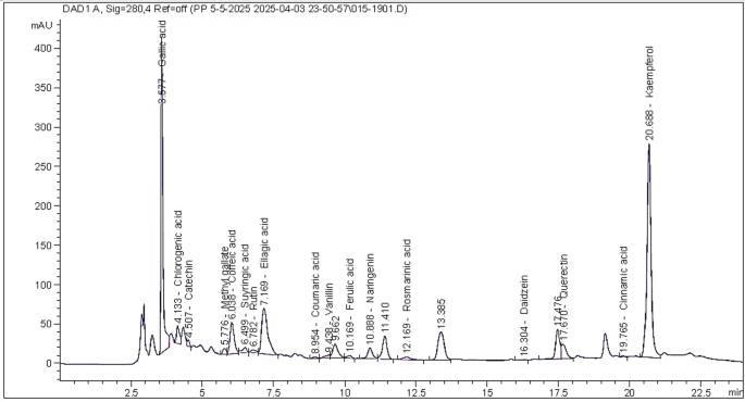
The green-branch bark extract (GBE) was analyzed by HPLC analysis (Table 1; Fig. 1), where the main compounds were kaempferol (14043.15 µg/g extract), gallic acid (7021.37 µg/g extract), ellagic acid (4983.92 µg/g extract), quercetin (1447.17 µg/g extract), caffeic acid (1211.46 µg/g extract), chlorogenic acid (1143.84 µg/g extract), naringenin (799.73 µg/g extract), and catechin (546.32 µg/g extract).
The kinos material previously dissolved in the methanol solvent were analyzed by the HPLC analysis (Table 1; Fig. 2). The main compounds belong to phenolic and flavonoid types were chlorogenic acid (12511.35 µg/g extract), gallic acid (12443.92 µg/g extract), ellagic acid (8147.54 µg/g extract), rutin (2025.87 µg/g extract), rosmarinic acid (925.31 µg/g extract), ferulic acid (722.79 µg/g extract), and hesperetin (519.01 µg/g extract).

The HPLC chromatogram peaks of the identified compounds in the methanol extract from Eucalyptus camaldulensis green-branch bark extract.

The HPLC chromatogram peaks of the identified compounds in Eucalyptus camaldulensis kinos.
The main chemical compounds found in the GBE (Table 2; Fig. 3) were p-cymene (31.91%), spathulenol (26.56%), crypton (11.60%), terpinen-4-ol (5.80%), cuminaldehyde (3.34%), eucalyptol (3.02%), D-limonene (2.31%), phellandral (2.26%), α-pinene (1.35%), trans-4-(isopropyl)-1-methylcyclohex-2-en-1-ol (1.06%), isospathulenol (1.05%), and p-(dimethoxymethyl)-isopropylbenzene (1.03%).

The GC–MS chromatographic peaks of the detected chemical compounds in the Eucalyptus camaldulensis green-branch bark extract.
The main chemical compounds in the kinos from Eucalyptus camaldulensis by the GC–MS analysis (Table 3; Fig. 4) were spathulenol (19.61%), isoaromadendrene epoxide (9.13%), α-acorenol (4.71%), patchoulane (4.68%), methyl 5,7-hexadecadiynoate (3.71%), 3-ethyl-3-hydroxy-(5à)-androstan-17-one (3.56%), 6-methyl-cyclodec-5-enol (3.56%), doconexent (3.10%), 4-(2-methyl-3-oxocyclohexyl)butanal (2.81%), 7-hydroxyfarnesen (2.42%), epiglobulol (2.28%), aromadendrene oxide-(2) (2.06%), ledene oxide-(II) (1.95%), Z-(13,14-epoxy)tetradec-11-en-1-ol acetate (1.94%), 7-oxo-2-oxa-7-thiatricyclo[4.4.0.0(3,8)]decan-4-ol (1.82%), estra-1,3,5(10)-trien-17á-ol (1.81%), 6,9,12-octadecatrienoic acid methyl ester (1.55%), retinal (1.44%), docosa-6,9,12,15-tetraenoate (1.36%), 9,10-secochola-5,7,10(19)-trien-24-al, 3-hydroxy-, (3β,5Z,7E)- (1.29%), 2-[4-methyl-6-(2,6,6-trimethylcyclohex-1-enyl)hexa-1,3,5-trienyl]cyclohex-1-en-1-carboxaldehyde (1.25%), 2-(7-heptadecynyloxy)tetrahydro-2 H-pyran (1.23%), 2,2,4-trimethyl-4-(2-methyl-2-propenyl)hexahydrocyclopropa[cd]pentalene-1,3-dione (1.14%), undec-10-ynoic acid octadecyl ester (1.13%), 1-(cyclopropyl-nitro-methyl)-cyclopentanol (1.07%), tetrahydroactinidiolide (1.06%), and ascaridole epoxide (1.03%).

The GC–MS chromatographic peaks of the detected chemical compounds in the Eucalyptus camaldulensis kinos.
Antifungal activity
Figures 5 and 6 show the visual observations of the antifungal activity of the GBE and kinos, respectively, when applied to P. halepensis wood against the growth of F. circinatum, and P. tardicrescens at the concentrations of 125, 250, 500, and 1000 µg/mL. These were compared to the negative control (10% DMSO) and the positive control, Cure-M 72% WP (Mancozeb 64%+Metalaxyl 8%), at the recommended dosage (2 g/L).
In Table 4, at a concentration of 1000 µg/mL, GBE and kinos demonstrated the highest activity against the growth of F. circinatum, with fungal inhibition percentage (FIP) values of 71.85% and 71.11%, respectively. These were followed by the kinos extract and GBE at 500 µg/mL, with FIP values of 66.66 and 66.29%, respectively. Additionally, GBE and kinos exhibited an FIP value of 61.11% at 250 µg/mL, which was higher than the value (FIP 58.88%) observed from the positive control (Cure-M 72% WP) at 2 g/L.
The GBE at 1000, 500, and 250 µg/mL showed the highest antifungal activity against the growth of P. tardicrescens with FIP values of 39.62, 35.55, and 32.96%, respectively. But these values are lower than those of the positive control (46.29%).
The minimum inhibitory concentrations (MICs) calculated for the treatments of E. camaldulensis GBE and kinos ranged between 15.6 and 62.5 µg/mL for both fungal isolates. The MIC calculated for the treatments of E. camaldulensis GBE was 15.6, and kinos was 31.3 µg/mL for F. circinatum isolate. The same result was observed with E. camaldulensis GBE and kinos. It was 62.5 µg/mL for P. tardicrescens isolate. The results showed that E. camaldulensis GBE had a better effect on F. circinatum than on P. tardicrescens compared to kinos.

Visual observation of the antifungal activity of the green-branch bark extract when applied to Pinus halepensis wood against the growth of (A) Fusarium circinatum, and (B) Pythium tardicrescens at the concentrations of (B1) 1000 µg/mL, (B2) 500 µg/mL, (B3) 250 µg/mL, and (B4) 125 µg/mL.

Visual observation of the antifungal activity of the kino extract when applied to Pinus halepensis wood against the growth of (A) Fusarium circinatum, and (B) Pythium tardicrescens at the concentrations of (K1) 1000 µg/mL, (K2) 500 µg/mL, (K3) 250 µg/mL, and (K4) 125 µg/mL.
Discussion
The chemical composition of E. camaldulensis bark is complex; however, its extracts are particularly rich in phenolic compounds, flavonoids, tannins, essential oils, and other secondary metabolites, which contribute to its antioxidant properties. The specific composition varies by factors such as clone, but major constituents include condensed tannins, phenolic acids (like gallic and syringic acids), and flavonoids (such as catechin). Polar extracts are also rich in polysaccharides, including glucans and xylans, while non-polar extracts can contain fatty acids like palmitic acid47. These compounds contribute to the tree’s defense mechanisms against pathogens and herbivores, as well as its adaptability to environmental stressors48,49.
In the present work, several phenolic and flavonoid compounds like gallic acid, chlorogenic acid, catechin, methyl gallate, caffeic acid, syringic acid, rutin, ellagic acid, coumaric acid, vanillin, ferulic acid, naringenin, rosmarinic acid, daidzein, quercetin, cinnamic acid, and kaempferol were detected in the green-branch bark (GBE) and kino extracts.
In E. camaldulensis, various phenolic acids, such as gallic acid, caffeic acid, and ferulic acid, have been identified50,51. These compounds display a variety of biological activities, including antimicrobial, anti-inflammatory, and anticancer effects. The level of phenolic compounds can change depending on environmental factors such as soil type, climate, and the age of the tree. The ethyl acetate fraction from leaf extracts led to the identification of six compounds: gallic acid, taxifolin, methyl gallate, quercetin, luteolin, and hesperidin51. Common flavonoids found in Eucalyptus bark include quercetin, kaempferol, and myricetin. The color of the bark and leaves, pollinator attraction, and UV protection are all influenced by flavonoids48. Numerous pharmacological effects, such as anti-inflammatory and anticancer properties, have been connected to their existence in Eucalyptus bark52,53.
Individual nonvolatile compounds have been isolated from various Eucalyptus species using GC and HPLC techniques, either alone or in combination with an auxiliary spectroscopic technique such as MS or NMR54. The phenolic compounds, such as gallic acid, protocatechuic acid, and ellagic acid55,56, have been identified in Eucalyptus extracts57,58.
The exudates extract analyzed by the HPLC showed the main compounds chlorogenic acid, gallic acid, ellagic acid, rutin, rosmarinic acid, ferulic acid, and hesperetin. Monomeric flavonoids and other phenolic chemicals have been found in kinos, with the bulk of them being intermediates that polymerize into tannins59. However, it is believed that these unique monomeric components are what give kinos their diverse physicochemical characteristics and, thus, their distinctive classification into the three “Maiden-groups” that were previously addressed.
Kinos from E. largiflorens showed the presence of methylated derivatives of gallic acid with other aromatic compounds, including 1,3,5-trimethoxybenzene and 1,3,5-trimethoxy-2-methyl-benzene60. Hydrolyzable tannins undergo methylation and hydrolysis to produce methylated derivatives of gallic acid and deoxy-sugar acids. The existence of varying methylation 3,6-deoxy-hexonic acid methyl esters in the pyrograms61, where gallic acid is connected to sugars via ester linkages, indicates the presence of sugars in kinos. Kaempferol was isolated from the kino of Eucalyptus citriodora25. Additionally, the narrow-leaved ironbark (E. crebra) honey identified tricetin, quercetin, luteolin, and kaempferol26. The wound-associated tissue of E. nitens, E. globulus, and E. obliqua contained a complex array of secondary metabolites, including hydrolyzable tannins, proanthocyanidins, flavonone glycosides, stilbene glycosides, formylated phloroglucinol compounds, volatile terpenes, and phenols62.
By the GC–MS analysis, several compounds, including p-cymene, spathulenol, crypton, terpinen-4-ol, cuminaldehyde, eucalyptol, D-limonene, phellandral, and α-pinene were found in the bark extract. Furthermore, the kino compounds analyzed by the GC–MS showed some bioactive compounds including spathulenol, isoaromadendrene epoxide, α-acorenol, patchoulane, methyl 5,7-hexadecadiynoate, 3-ethyl-3-hydroxy-(5à)-androstan-17-one, 6-methyl-cyclodec-5-enol, doconexent, 4-(2-methyl-3-oxocyclohexyl)butanal, 7-hydroxyfarnesen, epiglobulol, and aromadendrene oxide-(2).
GC–MS is essential for characterizing volatile and semi-volatile compounds, especially monoterpenes and sesquiterpenes linked to membrane disruption and efflux pump inhibition, in addition to polar metabolite identification63,64,65. Eucalyptus species are mainly known for their essential oils (EOs), though they can also be found in their bark. E. camaldulensis EO contains limonene, α-pinene, eucalyptol (1,8-cineole), spathulenol, and p-cymene66,67,68,69. These compounds, which give Eucalyptus its unique aroma, have been studied for their analgesic, antibacterial, and anti-inflammatory qualities. The sustainable approach of obtaining EOs from eucalyptus bark might be very beneficial to the fragrance and pharmaceutical industries. High concentrations of volatile organic compounds (VOCs) make up the EO profile of Eucalyptus54.
A precursor to carvacrol, p-cymene is a monoterpene with a benzene ring structure that can improve the cytoplasmic membrane’s permeability to adenosine triphosphate (ATP)70,71. It can enhance the antibacterial activity of other substances, in addition to exhibiting antimicrobial activity on its own. This is due to p-cymene’s strong affinity for microbial membranes and its ability to disrupt, expand, and influence the cell’s membrane potential72,73. p-Cymene has demonstrated significant antifungal properties, showing activity against various fungi, including Aspergillus flavus, A. niger, and Fusarium culmorum74. It often acts by disrupting cell membranes, inhibiting growth, and even working synergistically with other antifungal agents, such as miconazole74,75.
Spathulenol, with an MIC value of 100 µg/mL, was active against Citrus canker, which is caused by Xanthomonas citri76. The essential oil extracted from the Hymenaea stigonocarpa fruit peel, with its main compound spathulenol (25.19%), demonstrated antifungal activity against Orytis cinerea, Sclerotinia sclerotiorum, Aspergillus flavus, and Colletotrichum truncatum77. Spathulenol completely inhibited the formation of the fungal spores of Aspergillus flavus, Fusarium culmorum, and Aspergillus niger at a concentration of 50 µL/L over four natural fabrics (linen, cotton, wool, and silk)21. Potential action of spathulenol against several filamentous fungi and yeasts, including Microsporum gypseum and Tricophyton mentagrophytes, was noted78. E. camaldulensis EOs, and solvent-based extracts (leaf and bark) have been shown to have strong antifungal properties in numerous studies. For example, EOs show efficacy against a variety of fungi at doses of 0.125–1.0% (v/v), with Fusarium sporotrichioides being the most sensitive (MIC = 0.125%)79, and Rhizopus oryzae is the most resistant (no inhibition at 1.0%)80. Additionally, the methanolic leaf and bark extracts exhibited significant effectiveness against Candida albicans (MICs ranging from 0.2 mg/mL to 200 mg/mL)81.
Compounds isolated from the kinos of C. citriodora, such as 7-O-methylaromadendrin, 7-O-methylkaempferol, and ellagic acid, have demonstrated varying anti-fungal activities against the growth of P. notatum, A. niger, and F. oxysporium. Additionally, anti-bacterial activity against Micrococcus pyogenes var. aureus and Mycobacterium phlei has been reported in various fractions from the kino extract84. Bactericidal activity against S. aureus was demonstrated by the crude propolis made from the kino of C. torelliana and the extracted C-methyl flavones85. The antibacterial action of kino samples from E. flocktoniae and E. sargentii does not appear to be determined by the relative levels of hydrolyzable and condensed tannins24. High total phenolic and flavonoid content in Grantia aucheri extracts and the essential oils from Cleome coluteoides were responsible for the antifungal activity against pathogenic fungi, including Candida albicans, C. glabrata, A. brasiliensis, and A. niger86,87.
Wood and other natural materials were well protected by extracts, according to HPLC analysis for phenolic and flavonoid chemicals. When applied to model reference leather samples and produced cotton paper, Pinus rigida wood extract, which contains its primary constituents (cinnamic acid, caffeic acid, benzoic acid, quercetin, luteolin, and catechin), demonstrated strong antifungal activity against A. flavus, A. niger, and Fusarium culmorum88. With an increase in the extract concentration from leaves and branches of Schotia brachypetala when applied to white mulberry wood, the inhibition percentage against Alternaria alternata, Botrytis cinerea, and Fusarium oxysporum was increased. These were probably related to the presence of phytochemical compounds in the leaf extract, such as kaempferol and gallic acid, and gallic acid and chlorogenic acid in the branch extract37. Monoterpenes applied to Pinus sylvestris sapwood showed that p-cymene at 100 µL/mL had the highest fungal inhibition percentage against the growth of A. flavus; p-cymene and iso-eugenol against the growth of A. niger; and carvacrol against the growth of F. culmorum89. When applied to oak wood and Imperata cylindrica paper pulp, an aqueous extract of Syzygium cumini leaves, which contains the primary compounds benzoic acid, gallic acid, ellagic acid, and rutin, had some efficacy against F. culmorum, A. fumigatus, and A. niger90. Most recent work showed that the essential oil and recoverable extract from Callistemon viminalis leaves showed potential activity against the growth of Fusarium culmorum, A. fumigatus, and A. niger when applied to wood and linen, where the main compounds were pyrogallol and cinnamic acid91.
For the potential synergistic effects, when combined with traditional antibiotics (such as beta-lactams) and other plant extracts, the essential oil from E. camaldulensis extracts shows notable synergistic benefits that result in antibacterial efficacy, decreased drug resistance, and lowered required doses92. Using the paper disc diffusion method, the ethanolic leaf extracts of E. camaldulensis and Psidium guajava, as well as their combination, were found to have antibacterial properties in vitro against gram-positive Staphylococcus aureus and gram-negative Escherichia coli93. In Zimbabwe, a decoction of E. camaldulensis leaves was mixed with Citrus limon (L.) Burm. f. fruits and Psidium guajava L. leaves for fever, cough, and the flu; in Senegal, leaf decoctions were made with sugar for stomachaches94,95.
It has been demonstrated that spathulenol greatly increases the activity of common antifungals such as clotrimazole and fluconazole. Strong inhibitory effects are shown by extracts high in spathulenol against resistant strains of Candida albicans, A. flavus, and A. niger96. It functions as a biofungicide against plant-damaging fungi like Botrytis cinerea in addition to human diseases, frequently improving the efficacy of other synthetic or natural fungicides97.
According to studies, the minimum inhibitory concentration (MIC) of EOs and extracts from E. camaldulensis against fungi is typically much higher than that of common commercial antifungal medications like fluconazole or griseofulvin; however, because of their lower toxicity, they are regarded as a possible substitute98. When used as a positive control, standard fungicide (Apron star) demonstrated greater efficacy at lower concentrations (e.g., 15.00 mm inhibition zone for F. solani vs. 20.33 mm for undiluted EO, whereas E. camaldulensis EO demonstrated a MIC value of 7 to 8 µL/mL against Fusarium spp98,99. The EO against Aspergillus flavus and Fusarium culmorum had MICs of 8–40 µL/mL and 6–40 µL/mL, respectively, while Sertaconazole had MICs of 8 µL/mL and 6 µL/mL. Griseofulvin had a MIC of 0.064 mg/mL, but the leaf extract had a MIC of 6.4 mg/mL against Trichophyton mentagrophytes8,100,101,102.
One of the drawbacks of the study is the use of surfactants for the crude extract in practical formulations; further research and testing with different surfactants are needed. Several formulation studies can be used in the future to achieve this. It is important to keep in mind that a variety of conditions may affect the applied extract’s bioactivity, necessitating further investigation. Thus, further research into the long-term effects, or shelf life, of plant extracts when applied to the field of wood-biofungicides is made possible by this work.
Finally, the factors determining the cost of EOs and extracts from E. camaldulensis are several production-related factors, like plant yield, as E. camaldulensis is recognized for its high yields, and cultivation and harvesting of E. camaldulensis, as it is an evergreen tree, and all parts of the tree are rich in phytochemicals. Additionally, the method of extraction, such as steam distillation, is cost-effective for many plants, including E. camaldulensis, as well as using organic solvents for the extraction. Authentic, high-quality essential oils and extracts cost more. To guarantee purity and look for adulterants or synthetic fillers, which are frequently found in less expensive, inferior oils and extracts, reputable brands invest in stringent testing (such as GC–MS analysis). Transportation, packing in protective dark glass bottles, and a company’s overall business overhead all add to the final customer cost, making the supply chain and overhead another crucial component.
Conclusion
Green branch-bark extract and kinos from Eucalyptus camaldulensis have a rich and varied chemical composition that includes a variety of bioactive substances with substantial potential for a range of uses. The tree’s ecological resilience and therapeutic qualities are attributed to the presence of phenolic, flavonoid, and volatile chemicals. When applied to wood samples of Pinus halepinses, both extracts showed possible effects against the growth of two molds, Pythium tardicrescens and Fusarium circinatum. The sustainable use of Eucalyptus camaldulensis bark extracts could support environmental preservation and economic growth, underscoring the significance of this unique species in the natural world.
Data availability
All data generated or analyzed during this study are included in this published article.
References
-
Ashraf, A., Sarfraz, R. A., Mahmood, A. & Din, M. U. Chemical composition and in vitro antioxidant and antitumor activities of Eucalyptus camaldulensis Dehn. leaves. Ind. Crop Prod. 74, 241–248. https://doi.org/10.1016/j.indcrop.2015.04.059 (2015).
-
Barbosa, L. C. A., Filomeno, C. A. & Teixeira, R. R. Chemical variability and biological activities of Eucalyptus spp. essential oils. Molecules 21, 1671. https://doi.org/10.3390/molecules21121671 (2016).
-
Nwabor, O. F., Singh, S., Syukri, D. M. & Voravuthikunchai, S. P. Bioactive fractions of Eucalyptus camaldulensis inhibit important foodborne pathogens, reduce listeriolysin O-induced haemolysis, and ameliorate hydrogen peroxide-induced oxidative stress on human embryonic colon cells. Food Chem. 344, 128571. https://doi.org/10.1016/j.foodchem.2020.128571 (2021).
-
Aleksic Sabo, V. & Knezevic, P. Antimicrobial activity of Eucalyptus camaldulensis Dehn. plant extracts and essential oils: A review. Ind. Crop Prod. 132, 413–429. https://doi.org/10.1016/j.indcrop.2019.02.051 (2019).
-
Lee, M. H. Chemical profile, antimicrobial and anti-oxidative activity of commercial eucalyptus and lavender essential oils and their applicability in cosmetics. Indian J. Sci. Technol. 9, 100181106. https://doi.org/10.17485/ijst/2016/v9i46/107856 (2016).
-
Shala, A. Y. & Gururani, M. A. Phytochemical properties and diverse beneficial roles of Eucalyptus globulus Labill.: A review. Horticulturae 7, 450. https://doi.org/10.3390/horticulturae7110450 (2021).
-
Ghasemian, A., Eslami, M., Hasanvand, F., Bozorgi, H. & Al-abodi, H. R. Eucalyptus camaldulensis properties for use in the eradication of infections. Comp. Immunol. Microbiol. Infect. Dis. 65, 234–237. https://doi.org/10.1016/j.cimid.2019.04.007 (2019).
-
Jaradat, N. et al. Eucalyptus camaldulensis Dehnh leaf essential oil from Palestine exhibits antimicrobial and antioxidant activity but no effect on porcine pancreatic lipase and α-amylase. Plants 12, 3805. https://doi.org/10.3390/plants12223805 (2023).
-
Malakar, M. Advances in Medicinal and Aromatic Plants Vol. 1. 185 (Apple Academic, 2024).
-
Syukri, D. M. & Singh, S. Medicinal Plants and Their Bioactive Compounds in Human Health ( Ansari, M. A. , Shoaib, S. Eds.). Vol. 1. 185–199 (Springer, 2024).
-
Dhakad, A. K., Pandey, V. V., Beg, S., Rawat, J. M. & Singh, A. Biological, medicinal and toxicological significance of Eucalyptus leaf essential oil: A review. J. Sci. Food Agric. 98, 833–848. https://doi.org/10.1002/jsfa.8600 (2018).
-
Ahmad, R. S. et al. Essential Oils ( Nayik, G. A. & Ansari, M. J. Eds.). 217–239 (Academic Press, 2023).
-
Gakuubi, M. M., Maina, A. W. & Wagacha, J. M. Antifungal activity of essential oil of Eucalyptus camaldulensis Dehnh. against selected Fusarium spp. Int. J. Microbiol. 8761610. https://doi.org/10.1155/2017/8761610 (2017).
-
Barboucha, G. et al. Chemical composition, in silico investigations and evaluation of antifungal, antibacterial, insecticidal and repellent activities of Eucalyptus camaldulensis Dehn. leaf essential oil from Algeria. Plants 13, 3229. https://doi.org/10.3390/plants13223229 (2024).
-
Abdelkhalek, A., Salem, M. Z. M., Kordy, A. M., Salem, A. Z. M. & Behiry, S. I. Antiviral, antifungal, and insecticidal activities of Eucalyptus bark extract: HPLC analysis of polyphenolic compounds. Microb. Pathog. 147, 104383. https://doi.org/10.1016/j.micpath.2020.104383 (2020).
-
Abedi Tameh, F. et al. In-vitro cytotoxicity of biosynthesized nanoceria using Eucalyptus camaldulensis leaves extract against MCF-7 breast cancer cell line. Sci. Rep. 14, 17465. https://doi.org/10.1038/s41598-024-68272-3 (2024).
-
Nasser, M. et al. Influence of the extraction solvent and of the altitude on the anticancer activity of Lebanese Eucalyptus camaldulensis extract alone or in combination with low dose of cisplatin in A549 human lung adenocarcinoma cells. Processes 10, 1461. https://doi.org/10.3390/pr10081461 (2022).
-
Abiri, R. et al. New insights into the biological properties of eucalyptus-derived essential oil: A promising green anti-cancer drug. Food Res. Int. 38, 598–633. https://doi.org/10.1080/87559129.2021.1877300 (2022).
-
Talha, M. H., Alnomani, Y. & Mirforughi, S. A. Eucalyptus camaldulensis efficiency for application against microbial infections. Rev. Med. Microbiol. 32, 1–5. https://doi.org/10.1097/MRM.0000000000000234 (2021).
-
Hussain, H. A. et al. Insight to phytochemical investigation and anti-hyperlipidemic effects of Eucalyptus camaldulensis leaf extract using in vitro, in vivo and in silico approach. J. Biomol. Struct. Dyn. 43, 325–347. https://doi.org/10.1080/07391102.2023.2280814 (2025).
-
Taha, A. S. et al. GC–MS, quantum mechanics calculation and the antifungal activity of river red gum essential oil when applied to four natural textiles. Sci. Rep. 13, 18214. https://doi.org/10.1038/s41598-023-45480-x (2023).
-
Lambert, J. B. et al. Characterization of phenolic plant exudates by nuclear magnetic resonance spectroscopy. J. Nat. Prod. 84, 2511–2524. https://doi.org/10.1021/acs.jnatprod.1c00522 (2021).
-
Locher, C. & Currie, L. Revisiting kinos—An Australian perspective. J. Ethnopharmacol. 128, 259–267. https://doi.org/10.1016/j.jep.2010.01.028 (2010).
-
von Martius, S., Hammer, K. A. & Locher, C. Chemical characteristics and antimicrobial effects of some Eucalyptus kinos. J. Ethnopharmacol. 144, 293–299. https://doi.org/10.1016/j.jep.2012.09.011 (2012).
-
Lee, S. W., Hung, W. J. & Chen, Z. T. A new flavonol from the kino of Eucalyptus citriodora. Nat. Prod. Res. 31, 37–42. https://doi.org/10.1080/14786419.2016.1209667 (2017).
-
Yao, L. et al. Quantitative high-performance liquid chromatography analyses of flavonoids in Australian Eucalyptus honeys. J. Agric. Food Chem. 52, 210–214. https://doi.org/10.1021/jf034990u (2004).
-
Negahban, M., Collet, C., Msaada, K. & Collet, T. Evaluation of Corymbia terminalis kino extracts for antibacterial activity against wound-associated pathogens: bridging ethnopharmacology and experimental validation. Int. J. Environ. Health Res. 9, 1–13. https://doi.org/10.1080/09603123.2025.2573181 (2025).
-
Eyles, A., Davies, N. W. & Mohammed, C. Wound wood formation in Eucalyptus globulus and Eucalyptus nitens: Anatomy and chemistry. Can. J. For. Res. 33, 2331–2339. https://doi.org/10.1139/x03-149 (2003).
-
Islam, F., Khatun, H., Khatun, M., Ali, S. M. M. & Khanam, J. A. Growth Inhibition and apoptosis of Ehrlich ascites carcinoma cells by the methanol extract of Eucalyptus camaldulensis. Pharm. Biol. 52, 281–290. https://doi.org/10.3109/13880209.2013.834365 (2014).
-
Naseer, S. et al. Extraction of brown dye from Eucalyptus bark and its applications in food storage. Qual. Assur. Saf. Crops Foods. 11, 769–780. https://doi.org/10.3920/QAS2019.1569 (2019).
-
Sánchez-Loredo, E. et al. Ellagitannins from Eucalyptus camaldulensis and their potential use in the food industry. Explor. Food Foodomics. 2, 83–100. https://doi.org/10.37349/eff.2024.00027 (2024).
-
Balla, A. et al. The threat of pests and pathogens and the potential for biological control in forest ecosystems. Forests 12, 1579. https://doi.org/10.3390/f12111579 (2021).
-
Abd-Elhamed, W. et al. Green synthesis of silver nanoparticles mediated by Solanum nigrum leaf extract and their antifungal activity against pine pathogens. Sci. Rep. 15, 35025. https://doi.org/10.1038/s41598-025-21291-0 (2025).
-
Drenkhan, R. et al. Global geographic distribution and host range of Fusarium circinatum, the causal agent of pine pitch canker. Forests 11, 724. https://doi.org/10.3390/f11070724 (2020).
-
Ansari, M. et al. Plant mediated fabrication of silver nanoparticles, process optimization, and impact on tomato plant. Sci. Rep. 13, 18048. https://doi.org/10.1038/s41598-023-45038-x (2023).
-
Maria, A. A., Salem, R. H., Salama, M. A. & Khalil, A. M. M. Antioxidant-Rich biodegradable films: Incorporating date phenolic extracts into polyvinyl alcohol biofilms for strawberry preservation. J. Food Dairy. Sci. 15, 203–217. https://doi.org/10.21608/jfds.2024.328102.1171 (2024).
-
Salem, M. Z. M., EL-Shanhorey, N. A., Mohamed, N. H. & Mohamed, A. A. Phenolic and flavonoid compounds from leaves and branches of Schotia brachypetala for the development of biofungicide for wood protection. BioResources 20, 1069–1087. https://doi.org/10.15376/biores.20.1.1069-1087 (2025).
-
Lackner, M. et al. HPLC and GC–MS analyses of phytochemicals from Ficus carica leaf extract and essential oil along with their antimicrobial properties. J. Agric. Food Res. 19, 101687. https://doi.org/10.1016/j.jafr.2025.101687 (2025).
-
Mahgoub, S. et al. Polyphenolic profile of Callistemon viminalis aerial parts: Antioxidant, anticancer and in silico 5-LOX inhibitory evaluations. Molecules 26, 2481. https://doi.org/10.3390/molecules26092481 (2021).
-
Mikaia, A. et al. NIST Standard Reference Database 1A, Standard Reference Data. https://www.nist.gov/srd/nist-standard-reference-database-1a. https://www.nist.gov/system/files/documents/srd/NIST1aVer22Man.pdf (NIST, 2014).
-
Taha, A. S., Abo-Elgat, W. A. A., Fares, Y. G. D. & Salem, M. Z. M. Isolated essential oils as antifungal compounds for organic materials. Biomass Conv Bioref. 14, 3853–3873. https://doi.org/10.1007/s13399-022-02815-4 (2024).
-
Iturritxa, E. et al. Biocontrol of Fusarium circinatum infection of young Pinus radiata trees. Forests 8, 32. https://doi.org/10.3390/f8020032 (2017).
-
Hlaiem, S. et al. Characterization and pathogenicity of phytopathogenic fungi associated with Pinus pinea in northeastern Tunisia: Implications for forest health in the Mediterranean Basin. Plant. Pathol. Quar. 14, 118–124 (2024).
-
Elbanoby, N. E., El-Settawy, A. A. A., Mohamed, A. A. & Salem, M. Z. M. Phytochemicals derived from Leucaena leucocephala (Lam.) de Wit (Fabaceae) biomass and their antimicrobial and antioxidant activities: HPLC analysis of extracts. Biomass Conv Bioref. 14, 14593–14609. https://doi.org/10.1007/s13399-022-03420-1 (2024).
-
CLSI. Clinical and Laboratory Standards Institute (CLSI). Reference Method for Broth Dilution Antifungal Susceptibility Testing of Filamentous Fungi; Approved Standard. 2nd Ed. (CLSI Document M38-A2, Clinical and Laboratory Standards Institute, 2008).
-
Erhonyota, C., Edo, G. I. & Onoharigho, F. O. Comparison of poison plate and agar well diffusion method determining the antifungal activity of protein fractions. Acta Ecol. Sin. 43, 684–689. https://doi.org/10.1016/j.chnaes.2022.08.006 (2023).
-
Conde, E., Cadahia, E., Diez-Barra, R. & García-Vallejo, M. C. Polyphenolic composition of bark extracts from Eucalyptus camaldulensis, E. globulus and E. rudis. Holz Als Roh- Und Werkst. 54, 175–181. https://doi.org/10.1007/s001070050162 (1996).
-
Moges, G. W., Manahelohe, G. M. & Asegie, M. A. Phenolic, flavonoid contents, antioxidant, and antibacterial activity of selected Eucalyptus species. Biol. Med. Nat. Prod. Chem. 13, 147–157. https://doi.org/10.14421/biomedich.2024.131.147-157 (2024).
-
Sani, I., Abdulhamid, A., Bello, F. & Fakai, I. Eucalyptus camaldulensis: Phytochemical composition of ethanolic and aqueous extracts of the leaves, stem-bark, root, fruits and seeds. J. Sci. Innov. Res. 3, 523–526 (2014).
-
Nasr, A., Saleem Khan, T. & Zhu, G. P. Phenolic compounds and antioxidants from Eucalyptus camaldulensis as affected by some extraction conditions, a preparative optimization for GC–MS analysis. Prep Biochem. Biotechnol. 49, 464–476. https://doi.org/10.1080/10826068.2019.1575860 (2019).
-
Ghareeb, M. A., Habib, M. R., Mossalem, H. S. & Abdel-Aziz, M. S. Phytochemical analysis of Eucalyptus camaldulensis leaves extracts and testing its antimicrobial and schistosomicidal activities. Bull. Natl. Res. Cent. 42, 16. https://doi.org/10.1186/s42269-018-0017-2 (2018).
-
Ismayati, M., Sholihat, N. N. & Sari, F. P. Eucalyptus: Engineered Wood Products and Other Applications ( Lee, S.H. et al. Eds). 137–161 (Springer, 2024).
-
Lima, L., Miranda, I., Knapic, S., Quilhó, T. & Pereira, H. Chemical and anatomical characterization, and antioxidant properties of barks from 11 Eucalyptus species. Eur. J. Wood Wood Prod. 76, 783–792. https://doi.org/10.1007/s00107-017-1247-y (2018).
-
Vuong, Q. V. et al. Phytochemical, and anticancer properties of the Eucalyptus species. Chem. Biodivers. 12, 907–924. https://doi.org/10.1002/cbdv.201400327 (2015).
-
Newman, D. J. & Cragg, G. M. Natural products as sources of new drugs over the 30 years from 1981 to 2010. J. Nat. Prod. 75, 311–335. https://doi.org/10.1021/np200906s (2012).
-
Vuong, Q. V. et al. Fruit-derived phenolic compounds and pancreatic cancer: Perspectives from Australian native fruits. J. Ethnopharmacol. 152, 227–242. https://doi.org/10.1016/j.jep.2013.12.023 (2014).
-
Vázquez, G., Santos, J., Freire, M. S., Antorrena, G. & González-Álvarez, J. Extraction of antioxidants from Eucalyptus (Eucalyptus globulus) bark. Wood Sci. Technol. 46, 443–457. https://doi.org/10.1007/s00226-011-0418-y (2012).
-
Al-Sayed, E. et al. HPLC–PDA–ESI–MS/MS profiling and chemopreventive potential of Eucalyptus gomphocephala DC. Food Chem. 133, 1017–1024. https://doi.org/10.1016/j.foodchem.2011.09.036 (2012).
-
Rowe, J. W. Natural Products of Woody Plants: Chemicals Extraneous to the Lignocellulosic Cell Wall (Springer, 2012).
-
Georgiou, R. et al. Disentangling the chemistry of Australian plant exudates from a unique historical collection. Proc. Natl .Acad. Sci. 119, e2116021119. https://doi.org/10.1073/pnas.2116021119 (2022).
-
Riedo, C., Scalarone, D. & Chiantore, O. Advances in identification of plant gums in cultural heritage by thermally assisted hydrolysis and methylation. Anal. Bioanal Chem. 396, 1559–1569. https://doi.org/10.1007/s00216-009-3325-4 (2010).
-
Eyles, A., Davies, N. W. & Mohammed, C. Wound wood formation in Eucalyptus globulus and Eucalyptus nitens: Anatomy and chemistry. Can. J. Res. 33, 2331–2339. https://doi.org/10.1139/x03-149 (2023).
-
Hegazy, M. M. et al. Essential oils: the science of extraction and its implications for composition and biological activity—A review. Food Anal. Methods. 18, 1483–1513. https://doi.org/10.1007/s12161-025-02808-9 (2025).
-
Câmara, J. S. et al. Plant-derived terpenoids: A plethora of bioactive compounds with several health functions and industrial applications—A comprehensive overview. Molecules 29, 3861. https://doi.org/10.3390/molecules29163861 (2024).
-
Iqbal, I. et al. Comprehensive GC–MS profiling and multi-modal pharmacological evaluations of Haloxylon griffithii: In vitro and vivo approaches. Pharmaceuticals. 18, 770. https://doi.org/10.3390/ph18060770 (2025).
-
Grewal, K. et al. Chemical composition and potential of Eucalyptus camaldulensis Dehnh. essential oil and its major components as anti-inflammatory and anti-leishmanial agent. J. Essent. Oil-Bear Plants. 25, 419–429. https://doi.org/10.1080/0972060X.2022.2098061 (2022).
-
Chahomchuen, T., Insuan, O. & Insuan, W. Chemical profile of leaf essential oils from four Eucalyptus species from Thailand and their biological activities. Microchem J. 158, 105248. https://doi.org/10.1016/j.microc.2020.105248 (2020).
-
Mohammed, H. A., Aspatwar, A., Aljarbooa, A. F. & Qureshi, K. A. Comparative study of volatile oil constituents, anti-microbial properties, and antibiofilm activities in Eucalyptus camaldulensis and Eucalyptus globulus: Insights from central Saudi Arabia. J. Essent. Oil-Bear Plants. 27, 341–355. https://doi.org/10.1080/0972060X.2024.2324343 (2024).
-
Salem, M. Z. M., Ashmawy, N. A., Elansary, H. O. & El-Settawy, A. A. Chemotyping of diverse Eucalyptus species grown in Egypt and antioxidant and antibacterial activities of its respective essential oils. Nat. Prod. Res. 29, 681–685. https://doi.org/10.1080/14786419.2014.981539 (2015).
-
Ben Arfa, A., Combes, S., Preziosi-Belloy, L., Gontard, N. & Chalier, P. Antimicrobial activity of carvacrol related to its chemical structure. Lett. Appl. Microbiol. 43, 149–154. https://doi.org/10.1111/j.1472-765X.2006.01938.x (2006).
-
Azizi, Z., Salimi, M., Amanzadeh, A., Majelssi, N. & Naghdi, N. Carvacrol and thymol attenuate cytotoxicity induced by amyloid β25–35 via activating protein kinase C and inhibiting oxidative stress in PC12 cells. Iran. Biomed. J. 24, 243–250. https://doi.org/10.29252/ibj.24.4.243 (2020).
-
Baginska, S., Golonko, A., Swislocka, R. & Lewandowski, W. Monoterpenes as medicinal agents: Exploring the pharmaceutical potential of p-cymene, p-cymenene, and γ-terpinene. Acta Pol. Pharm. —Drug Res. 80, 879–892. https://doi.org/10.32383/appdr/178242 (2023).
-
Bourhia, M. et al. Volatile constituents in essential oil from leaves of Withania adpressa Coss. ex exhibit potent antioxidant and antimicrobial properties against clinically-relevant pathogens. Molecules 28, 2839. https://doi.org/10.3390/molecules28062839 (2023).
-
Pyo, Y. & Jung, Y. J. Microbial fermentation and therapeutic potential of p-cymene: Insights into biosynthesis and antimicrobial bioactivity. Fermentation 10, 488. https://doi.org/10.3390/fermentation10090488 (2024).
-
Kumar, A. et al. Biochemical insights into synergistic Candida biofilm disintegrating ability of p-cymene inclusion complex and miconazole. Eur. J. Pharmacol. 993, 177365. https://doi.org/10.1016/j.ejphar.2025.177365 (2025).
-
da Silva, I. R. R., Fernandes, C. C., Gonçalves, D. S., Martins, C. H. G. & Miranda, M. L. D. Chemical composition and anti-Xanthomonas citri activities of essential oils from Schinus molle L. fresh and dry leaves and of its major constituent spathulenol. Nat. Prod. Res. 38, 3476–3480. https://doi.org/10.1080/14786419.2023.2249584 (2024).
-
Pimentel, F. C. et al. Chemical composition and antifungal activity of the essential oil from the Hymenaea stigonocarpa Mart. Ex Hayne (jatobá-do-cerrado) fruit peel. Nat. Prod. Res. 38, 1945–1949. https://doi.org/10.1080/14786419.2023.2225123 (2024).
-
Al-Ja’fari, A. H. et al. Composition and antifungal activity of the essential oil from the rhizome and roots of Ferula hermonis. Phytochem 72, 1406–1413. https://doi.org/10.1016/j.phytochem.2011.04.013 (2011).
-
Mehani, M., Salhi, N., Valeria, T. & Ladjel, S. Antifungal effect of essential oil of Eucalyptus camaldulensis plant on Fusarium graminearum and Fusarium sporotrichioide. Int. J. Curr. Res. 6, 10795–10797 (2014).
-
Siramon, P., Ohtani, Y. & Ichiura, H. Chemical composition and antifungal property of Eucalyptus camaldulensis leaf oils from Thailand. Rec Nat. Prod. 7, 49–53 (2013).
-
Babayi, H., Kolo, I., Okogun, J. & Ijah, U. The antimicrobial activities of methanolic extracts of Eucalyptus camalctulensis and Terminalia catappa against some pathogenic microorganisms. Biokemistri 16, 106–111. https://doi.org/10.4314/biokem.v16i2.32578 (2004).
-
Chuku, A., Ogbonna, A. I., Obe, G. A., Namang, M. & Ahmad, I. R. Antimicrobial effects of leaves of Eucalyptus camaldulensis on some microbial pathogens. Eur. J. Med. Plants. 14, 1–8 (2016).
-
van Vuuren, S. F. Antimicrobial activity of South African medicinal plants. J. Ethnopharmacol. 119, 462–472. https://doi.org/10.1016/j.jep.2008.05.038 (2008).
-
Satwalekar, S., Gupta, T. & Narasimharao, P. Chemical and antibacterial properties of Kions from Eucalyptus ssp. citriodorol-The antibiotic principle from the kion of E. citriodoroa. J. Indian Inst. Sci. 39, 195–212 (1956).
-
Massaro, C. F. et al. Anti-staphylococcal activity of C-methyl flavanones from propolis of Australian stingless bees (Tetragonula carbonaria) and fruit resins of Corymbia torelliana (Myrtaceae). Fitoterapia 95, 247–257. https://doi.org/10.1016/j.fitote.2014.03.024 (2014).
-
Sharifi-Rad, M. et al. Variation of phytochemical constituents, antioxidant, antibacterial, antifungal, and anti-inflammatory properties of Grantia aucheri (Boiss.) at different growth stages. Microb. Pathog. 172, 105805. https://doi.org/10.1016/j.micpath.2022.105805 (2022).
-
Sharifi-Rad, M. et al. Essential oil of Cleome coluteoides (Boiss.): Phytochemical constituents, antioxidant, antimicrobial, antiproliferative, anti-inflammatory, enzymatic inhibition, and Xanthine oxidase inhibitory properties. J. Herb. Med. 52, 101036. https://doi.org/10.1016/j.hermed.2025.101036 (2025).
-
Taha, A. S., Abo-Elgat, W., Salem, M. & Salim, E. Pinus rigida wood extract as an antifungal activity for a model paper and leather that is comparable to historical manuscripts: An experimental research. Egypt. J. Chem. 68, 29–39. https://doi.org/10.21608/ejchem.2024.222888.8258 (2025).
-
Salem, M. Z. M., Abo-Elgat, W. A., Mansour, M. M. & Selim, S. Antifungal activity of the monoterpenes carvacrol, p-cymene, eugenol, and iso-eugenol when applied to wood against Aspergillus flavus, Aspergillus niger, and Fusarium culmorum. BioResources 20, 393–412. https://doi.org/10.15376/biores.20.1.393-412 (2025).
-
Elshaer, M. A. A. et al. Green synthesis of silver and ferric oxide nanoparticles using Syzygium cumini leaf extract and their antifungal activity when applied to oak wood and paper pulp from Imperata cylindrica grass biomass. Waste Biomass Valori. 15, 6191–6211. https://doi.org/10.1007/s12649-024-02555-8 (2024).
-
Salem, M. Z. M., Abo-Elgat, W. A. A., Farahat, M. G. S., El-Settawy, A. A. A. & Selim, S. The essential oil and recoverable extract from Callistemon viminalis leaves as wood- and textile biofungicides with the GC–MS and HPLC analyses. Chem. Afr. 8, 5151–5163. https://doi.org/10.1007/s42250-025-01391-0 (2025).
-
Knezevic, P. et al. Antimicrobial activity of Eucalyptus camaldulensis essential oils and their interactions with conventional antimicrobial agents against multi-drug resistant Acinetobacter baumannii. J. Ethnopharmacol. 178, 125–136. https://doi.org/10.1016/j.jep.2015.12.008 (2016).
-
Bala, I., Yusha’u, M., Lawal, D. & Abubakar, S. Synergistic effect of Eucalyptus (Eucalyptus camaldulensis) and guava (Psidium guajava) ethanolic extracts on Eschericia coli and Staphylococcus aureus. Acad. Res. Int. 5, 35–41 (2014).
-
Doran, J. C. & Wongkaev, W. Eucaliptus camaldulensis Dehnh. In Plant Resources of Tropical Africa (Louppe D., Oteng Amoako A.A. et al. Eds.). Vol. 7(1): Timbers 1. (PROTA Foundation, Bachkuys Publisher, CTA, 2008).
-
Maroyi, A. Traditional use of medicinal plants in south-central Zimbabwe: Review and perspectives. J. Ethnobiol. Ethnomed. 9, 31. https://doi.org/10.1186/1746-4269-9-31 (2013).
-
Soulaimani, B. et al. Synergistic anticandidal effects of six essential oils in combination with fluconazole or amphotericin B against four clinically isolated Candida strains. Antibiotics 10, 1049. https://doi.org/10.3390/antibiotics10091049 (2021).
-
Rajput, N. A. et al. Biofungicides: Eco-Safety and Future Trends. 191–231 (CRC, 2023).
-
Falahati, M., Omidi Tabrizib, N. & Jahaniani, F. Anti dermatophyte activities of Eucalyptus camaldulensis in comparison with Griseofulvin. Iran. J. Pharmacol. Ther. 4, 80–83 (2005).
-
Aleksic Sabo, V. & Knezevic, P. Antimicrobial activity of Eucalyptus camaldulensis Dehn. plant extracts and essential oils: A review. Ind. Crops Prod. 132, 413–429. https://doi.org/10.1016/j.indcrop.2019.02.051 (2019).
-
Baptista, E. B., Zimmermann-Franco, D. C., Lataliza, A. A. B. & Raposo, N. R. B. Chemical composition and antifungal activity of essential oil from Eucalyptus smithii against dermatophytes. Rev. Soc. Bras. Med. Trop. 48, 746–752. https://doi.org/10.1590/0037-8682-0188-2015 (2015).
-
Doudi, M., Setorki, M. & Hoveyda, L. Comparing the antifungal effects of five essential oils plants eucalyptus, cinnamon, wormwood, sagebrush and Iranian rose damascena on three standard strains of Candida albicans in vitro. Int. J. Biol. Pharm. Allied Sci. 3, 490–500 (2014).
-
Chuku, A., Ogbonna, A. I., Obe, G. A., Namang, M. & Ahmad, I. R. Antimicrobial effects of leaves of Eucalyptus camaldulensis on some microbial pathogens. Eur. J. Med. Plants. 14, 1–8. https://doi.org/10.9734/EJMP/2016/25759 (2016).
Acknowledgements
The authors would like to appreciate the scientific cooperation between the members of work from Alexandria University, Al-Azhar University, Ain Shams University, and the Agriculture Research Center. The authors would like to thank Dr. Mervat EL-Hefny (Department of Floriculture, Ornamental Horticulture and Garden Design, Faculty of Agriculture (El-Shatby), Alexandria University, Alexandria, Egypt) for providing the Eucalyptus botanical samples.
Funding
Open access funding provided by The Science, Technology & Innovation Funding Authority (STDF) in cooperation with The Egyptian Knowledge Bank (EKB).
Ethics declarations
Competing interests
The authors declare no competing interests.
Additional information
Publisher’s note
Springer Nature remains neutral with regard to jurisdictional claims in published maps and institutional affiliations.
Supplementary Information
Rights and permissions
Open Access This article is licensed under a Creative Commons Attribution 4.0 International License, which permits use, sharing, adaptation, distribution and reproduction in any medium or format, as long as you give appropriate credit to the original author(s) and the source, provide a link to the Creative Commons licence, and indicate if changes were made. The images or other third party material in this article are included in the article’s Creative Commons licence, unless indicated otherwise in a credit line to the material. If material is not included in the article’s Creative Commons licence and your intended use is not permitted by statutory regulation or exceeds the permitted use, you will need to obtain permission directly from the copyright holder. To view a copy of this licence, visit http://creativecommons.org/licenses/by/4.0/.
About this article

Cite this article
Salem, M.Z.M., Elshaer, M.A.A., Mohamed, A.A. et al. Phytochemical analysis of green-branch bark extract and the brown gum exudates “kinos” from Eucalyptus camaldulensis by HPLC and GC–MS with their antifungal activity. Sci Rep 16, 7480 (2026). https://doi.org/10.1038/s41598-026-38109-2
-
Received:
-
Accepted:
-
Published:
-
Version of record:
-
DOI: https://doi.org/10.1038/s41598-026-38109-2