Introduction
In the health industry, non-specific adhesion of bacteria to solid surfaces and subsequent biofilm formation are a severe deterrent to the performance of many devices (contact lenses, catheter tubes, blood contacting devices, implants, surfaces for cell culture, biosensors, drug delivery systems, etc.) and pose serious problems to public health1,2,3. Infections resulting from bacterial colonization are responsible for a majority of all healthcare associated infections (HAI)4. One of the most effective routes to avoid such contamination is the coating of surfaces with materials that can impart antibiofouling/antibacterial properties. The two main coating strategies adopted to prevent fouling involves biocidal and nonbiocidal surfaces5,6,7. While biocidal coatings directly eliminate bacteria to prevent surface adherence, nonbiocidal (or biopassive) coatings prevent adhesion of cells and proteins by virtue of their inherent surface properties. This eliminates the problem at its root and is not limited by the disadvantages of purely biocidal surfaces, such as fouling, leaching or gradual inactivation of the functionally active groups. As a result, biopassive surfaces have garnered special interest in the recent past because of its immense applicability in the field of healthcare.
The most common polymer used in biopassive coatings is the hydrophilic poly(ethylene) glycol (PEG) and its copolymers8,9, although the poor stability of these PEG based surface coatings, besides susceptibility to hydrolysis, oxidative and microbial degradation and poor long term performance, remains a roadblock in their widespread applicability10,11. Moreover, the extensive application of PEG and the continuous exposure to PEG-containing products has led to the development of anti-PEG antibodies in certain patients, leading to faster elimination from the body, reduced therapeutic efficacy and even allergic reactions. PEGs have also been shown to undergo oxidative degradation generating by-products that are toxic to humans and leading to limited long-term efficiency for antifouling applications12.
Alternatively, hydrophobic polymers, such as 1 H,1 H,2 H,2 H-perfluorodecyl acrylate (PFDA) are also frequently used to impart antifouling properties to surfaces, owing to their beneficial properties, such as high thermal stability, excellent chemical resistance, low friction coefficient, superior weatherability, etc13,14. These extraordinary properties arise from the unique chemical structure imparted by the polymer chains of the perfluorocarbon. However, it has also been reported that amphiphilic coatings, i.e., surfaces containing pockets of both hydrophilic and hydrophobic zones offer enhanced performance compared to coatings with unidimensional properties15,16,17. This quality probably arises from the fact that proteins contain both hydrophobic and hydrophilic segments, and can tune their condition and structure to match the surface energy of the adsorbing surface. Amphiphilic coatings essentially are an unwelcome terrain for bacterial adhesion owing to their heterogeneous surface energy domains and ability to offer an ambiguous surface that deters adhesion.
The limitations of PEG based coatings has led to a search for stable alternatives, and one such alternative of considerable interest is polyoxazolines (POx)18. POx coatings have been reported for their potential biomedical applications and better stability compared to their PEG counterparts19,20.The conventional approaches for POx film formation include living-cationic ring opening polymerization, photocoupling, and grafting, all of which suffer from limitations, such as being slow and complex multistep processes involving the use of organic solvents. Under the circumstances, plasma deposition has emerged as a novel approach to overcome these disadvantages to generate even nanometre thin polyoxazoline coatings, without the necessity for any substrate surface preparation or the use of toxic organic solvents21. Plasma polymerization also allows production of homogeneous, pinhole-free coatings and offers facile control over the reaction parameters, making it an ideal candidate for numerous biomaterial coating applications. Many of the oxazoline ring containing compounds, such as 2-methyl-2-oxazoline (2M2O) and 2-ethyl-2-oxazoline (2E2O), are low molecular weight liquids with substantial vapour pressure, and thus facilitate plasma polymerization. Although plasma polymerization too suffers from limitations such as high operational costs due to complex, expensive vacuum equipment, challenging process control and the occasional unsaturation causing colouration in coatings, the benefits offered by the approach still outweigh these factors to make it highly attractive for novel coatings design. Cavallaro et al.22 have demonstrated that plasma deposited polyoxazolines, being a mild functionalization approach, allow the POx coatings to retain their chemical reactivity and exhibit excellent biocompatibility and low biofouling. Plasma-enhanced chemical vapor deposition (PECVD) methods have been employed to deposit 2M2O and 2E2O in low-pressure radio frequency (RF) discharge23. But, the possibility of combining the advantages of hydrophilic oxazolines with those of hydrophobic PFDA in the form of an amphiphilic coating with properties superior to either system individually has not been explored thus far. Of even greater interest is the potential application of this methodology to functionalize the surface of non-woven fabrics. Nonwovens are extensively used in products, such as filtration media, wipes, baby diapers, garment interlinings, etc. Most of these application areas are critical in the sense that there is a high chance of microbial contamination and infection arising from the use of these materials, especially in high humid regions like India. PECVD mediated surface modification is an exciting proposition to make value addition to these fabrics and ensure minimum proliferation of infection by incorporating antifouling/antibacterial properties.
This work presents, to the best of our knowledge, the first reported PECVD mediated co-deposition of stable 2E2O-PFDA amphiphilic polymer coatings to impart antibiofouling and antibacterial properties, synergistically blending hydrophilic and hydrophobic properties for potentially superior performances. The sample hydrophilicity and roughness were observed to be regulated by the composition of the two monomers deposited simultaneously on the substrate. Antibacterial and antifouling properties of samples containing different compositions of 2E2O and PFDA and deposited for different durations were tested against gram positive (S. aureus) and gram negative (E. coli) bacteria. The stability of the coatings was evident from the retention of biocidal properties even after multiple washing cycles, exhibiting distinct advantages over previously reported PEG and DEGVE (Di (ethylene glycol) vinyl ether) based amphiphilic coatings.
Results and discussion
FTIR and XPS analysis
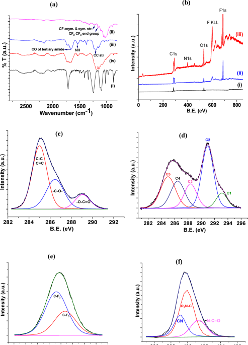
The PET control film (Fig. 1a(i)) displayed characteristic peaks corresponding to aromatic rings (846, 870 and 968 cm− 1), methylene group (bending mode at 1460 cm− 1), vibrations of the ester CO bond (C = O stretching mode at 1715 cm− 1 and C-O stretching at 1245 cm− 1), O-C-C stretching (1100 cm− 1), bending and wagging vibrational modes of the ethylene glycol segment (1339, 1409 and 1458 cm− 1), vibrations of aromatic skeleton with stretching of C = C (1505 and 1580 cm− 1), and the broad axial symmetrical deformation of CO2 (~ 2200 cm− 1)24. The PFDA deposited PET films (Fig. 1a(ii)) were recognizable by the appearance of CF2 and CF3 end group vibrations at 1155 cm− 1, besides the CF symmetric and asymmetric stretching modes at 1200 cm− 1. The weak absorption band at 1338 cm− 1 was assigned to C-F stretching vibration in the -CF2CF3 group. The absence of an ester group peak could be attributed to the fragmentation of the same during the plasma polymerization process. The electrons generated in the plasma can induce cleavage of the ester bonds, either homolytically or heterolytically, to form smaller fragments that finally form the fluorocarbon coating. 2E2O coating on PET (Fig. 1a (iii)) displayed bands corresponding to C = O stretching (1730 cm− 1), CC stretching (1202 cm− 1), NH stretching (1540 cm− 1), and CO of the tertiary amide (1659 cm− 1). These amide groups are probably formed during post deposition reactions in the film24. The bands between 1463 and 1350 cm− 1 are typical of the bending vibrations of CH3 and CH2 groups. The small but broad peak at 2160 cm− 1 arises from the nitrile C ≡ N groups formed during the plasma polymerization process through fragmentation and recombination of the oxazoline monomers in the plasma. 2E2O-PFDA amphiphilic coatings (Fig. 1a(iv)) presented strong CC stretching vibrations at ~ 1200 cm− 1 that overlapped with the CF2 and CF3 end group vibrations of PFDA falling in the same region. Additionally, the main peaks of Poly(2E2O) (C = O stretching at 1735 cm− 1, CC stretching at 1192 cm− 1, NH stretching at 1533 cm− 1, CO of the tertiary amide at 1670 cm− 1 and nitrile CN at 2160 cm− 1) were all visible in the spectra, although the C = O stretching and tertiary amide CO bands were not well resolved and appeared to merge, forming a broader band centered around 1693 cm− 1. This confirmed the presence of 2E2O in the amphiphilic coatings.
The high-resolution XPS survey spectra of untreated PET, PFDA coating on PET and amphiphilic coating on PET are presented in Fig. 1b. The survey spectra of the PFDA coating clearly displays peaks corresponding to F1s and the auger peak of F (F KLL) while the amphiphilic coating shows an additional peak corresponding to N1s introduced by the 2E2O groups. The C1s peak of PET was deconvoluted into three components. Binding energies of 285 eV, 286.47 eV and 289.06 eV were assigned to C-C/C-H, C-O and O-C = O bonds, respectively (Fig. 1c). In the case of the PFDA deposited films, the C1s peak could be deconvoluted to reveal peaks corresponding to the CF3 (C1: 293.54 eV) and CF2 (C2: 291.42 eV) bonds, in addition to CF/O-C = O (C3: 288.40 eV), C-O(CF) (C4: 286.46 eV) and C-CF/C-O (C5: 285.04 eV) (Fig. 1d). The well resolved XPS peaks of CF2 and CF3 groups of plasma polymerized PFDA coatings signified lower degree of fragmentation and rearrangement of the perfluorocarbon chain, notwithstanding the cleavage of the ester bond as revealed from the FTIR analysis. Additionally, the ratio of the areas of the C1 and C2 peaks did not deviate significantly from the theoretical ratio of 1:7. A F1s peak corresponding to organic fluorine was also visible and deconvoluted into two peaks at 687.61 eV and 688.36 eV, assigned to CF2 and CF3 respectively (Fig. 1e). This was a clear confirmation of incorporation of PFDA onto the PET surface. The 2E2O-PFDA amphiphilic coatings were distinguishable by the appearance of an additional N1s band visible at 399.12 eV, introduced by the oxazoline groups of 2E2O. The N1s peak was deconvoluted into binding energies of 399.1 eV, 399.9 eV and 401.3 eV (Fig. 1f), attributable to CN, R2N-C and N-C = O bonds, respectively. Although it is reported that the C1s peak of polyoxazoline films can also provide information about the existence of new chemical bonds containing nitrogen (C-N, C ≡ N, N-C = O) and oxygen (C = O)25, the peaks could not be specifically identified in this case probably because of overlap with the existing peaks arising from PET and PFDA. The atomic % values, as obtained from the peak areas of the respective elements after correction with their relative sensitivity factors (RSF), are presented in Table 1. The F content in PFDA coated sample was as high as ~ 40%, which expectedly dropped to ~ 13% in the amphiphilic coating wherein the more volatile 2E2O was being injected simultaneously. This value has been corroborated by the elemental analysis data reported in Table 2. The N/O ratio in both 2E2O coated and amphiphilic samples was slightly higher than that of 2E2O itself (1 : 1), which implied that the plasma treatment did not cause significant oxidation of the films but instead led to the formation of volatile oxygenated species that were not retained in the final film. The fragmentation of the PFDA ester group also contributes to the loss of O in the final amphiphilic films.

(a) FTIR spectra of (i) untreated PET (ii) PFDA coated PET (iii) 2E2O coated PET and (iv) 2E2O: PFDA (1:1, 30 min) coated PET. (b) High resolution XPS survey spectra of (i) untreated PET (ii) PFDA coated PET (iii) 2E2O: PFDA (1:1, 30 min) coated PET. Deconvoluted spectra of (c) C1s in untreated PET (d) C1s in PFDA coated PET (e) F1s in PFDA coated PET and (f) N1s in 2E2O: PFDA (1:1, 30 min) coated PET.
The mechanisms of polymerization of PFDA and 2E2O are presented in Fig. 2. In the case of PFDA (Fig. 2a), application of RF energy to the gas mixture results in the disassociation of the PFDA molecules, generating reactive species such as radicals and ions. As evidenced from the FTIR and XPS analysis, the disassociation occurs in the ester linkages while the fluorocarbon chain remains more or less intact. These reactive species migrate to the substrate surface and undergo polymerization and crosslinking, with each new PFDA molecule adding to the growing polymer chain to form a thin polymeric film whose properties are controlled by the applied plasma power and mode, pressure, gas flow rate and substrate temperature.
The polymerization of 2E2O (Fig. 2b), on the other hand, proceeds either through retention of the oxazoline ring structure or via a ring opening polymerization mechanism. The ring retention is generally favoured under gentle deposition conditions (low power and longer duration), whereas ring opening polymerization is the dominant route under harsher reaction conditions (high power and shorter duration) The ring opening typically occurs through an electrophilic attack initiated by a radical generated in the plasma, the most favoured position being the C atom adjacent to the N atom in the ring. More oxazoline molecules are subsequently added with the growing polymer chain, finally depositing as a thin film on the substrate surface. While an intact oxazoline ring is desired to initiate further functionalization through covalent bond formation with other ligands or biomolecules, the stability of the film gets compromised in the process due to the mild deposition conditions required. In contrast, high powers favour formation of sturdier films with greater extent of polymerization and crosslinking20. Therefore, in order to achieve stable films with better biocidal activity retention capacity, all depositions in the present work were carried out at 40 W RF power, with durations varied between 10 and 30 min. The most likely mechanism for polymerization in this case was therefore through the ring opening route, which could also be confirmed through FTIR analysis, where the appearance of a peak corresponding to nitrile C ≡ N groups at 2160 cm− 1 could only be explained by the fragmentation and recombination of the oxazoline monomers in the plasma. Moreover, the absence of the characteristic etheric oxygen band of the oxazoline ring, that typically appears at ~ 1072 cm− 1, and the presence of an amide carbonyl stretch band at 1659 cm− 1 (formed during ring opening polymerization) clearly indicate disintegration of the oxazoline ring structure. In tune with the FTIR results, XPS analysis as well revealed the presence of CN (399.1 eV), N-C = O (amide linkage, 401.3 eV) bonds confirming the ring opening process. The appearance of an XPS peak corresponding to C-NR2 (399.9 eV) could result from the reduction of the amide groups by the strong reducing environment provided by the plasma system, possibly triggered by a hydride ion induced nucleophilic attack.
The formation of radical species in the plasma was also confirmed through optical emission spectroscopic measurements. The OES of pure Ar and Ar/2E2O-PFDA plasmas under similar experimental conditions (P = 40 W Continuous Wave (CW), Ar flow rate = 20 sccm, pressure = ~ 0.5 mbar) are presented in Fig. 1c. The OES of pure Ar displayed characteristic peaks in the visible region, mainly six dominant lines at 698.02 nm, 709.51 nm, 753.01 nm, 765.53 nm, 803.28 nm and 813.54 nm corresponding to different Ar I transitions. Simultaneously, some low intensity lines originating from oxygen plasma were also visible at ca. 774.76 nm, 844.53 nm, and a small peak at 926.81 nm. Some minor lines, probably attributable to the N2 second positive system, could also be seen in the 360–454 nm region. In contrast, the Ar/2E2O-PFDA plasma demonstrated a significant increase in intensity (~ 7 fold) in the low wavelength region i.e. 341–437 nm. The peaks that could be distinctly identified were the CHO emission line (359.43 nm), CN emission lines (383.65 nm, 422.43 nm) and CH* excited state lines (430.44 nm), all of which could be traced back to the inclusion of 2E2O in the plasma system and the subsequent disruption of its ring structure in the plasma medium. A prominent band corresponding to H2 emission lines, interspersed with various minor Ar emission lines, could also be observed in the 654–680 nm region, probably arising from the fragmentation of both precursor molecules. This was accompanied by the complete disappearance of two minor intensity Ar plasma lines initially detected at 698.02 nm and 709.51 nm. The spectral lines corresponding to atomic F, which are expected to form upon fragmentation of PFDA, normally lie in the 600–750 nm region but could not be specifically assigned due to overlap with multiple Ar I and H2 emission lines in the system. Nevertheless, the OES analysis shed significant light on the qualitative composition of the plasma generated in the presence of both precursors.
Electron temperature (Te) for both plasma systems were determined using the Boltzman plot method, with relative line intensities of various emission lines of Ar atom in both plasmas being used to generate the Boltzmann plots. Based on the assumption that the Boltzmann distribution accurately describes the excited state populations of different Ar atoms in the plasma, the following relation (1) was used:
$$:lnleft(frac{lambda:I}{{Ag}_{u}}right)=-frac{1}{{k}_{B}{T}_{e}}{E}_{u}+:C$$
(1)
Where, kB is the Boltzmann constant and A, the transition probability. λ and I are wavelength and relative intensity of the emitted line, respectively, Eu is energy of upper level of transition, and gu is the statistical weight of the upper level. The values of A, Eu and gu are available in NIST standard database. The Te value could be determined from the slope of the plot (1/kBTe).
The Te values hence obtained were 5292 K (0.46 eV) for Ar plasma and 3369 K (0.29 eV) for the Ar/2E2O-PFDA plasma. The decrease in Te is attributed to the loss in energy caused by inelastic collisions between electrons and the precursor molecules, which trigger the ionization, excitation and dissociation of 2E2O and PFDA molecules. The initial Te plays a crucial role in determining the film characteristics. Typically, high Te values lead to increased dissociation of precursor gases, resulting in a higher density of reactive species that participate in film growth. Although this can enhance deposition rates, there is also a possibility of introducing defects into the film due to higher ion bombardment, increased film stress or roughness, unless process parameters are carefully controlled. On the other hand, low Te reduces ion bombardment energy at the growing film surface, which can ensure minimal defect density and improve film uniformity. This is because the ion bombardment energy is determined by the sheath potential, which is a function of Te. But, on the flip side, it may retard the deposition rate and influence the incorporation of specific chemical groups. All these factors can be precisely controlled by fine-tuning parameters such as RF power, pressure, and excitation frequency.
With Ar/2E2O-PFDA plasma being a complex system, the electron density (ne) for only the Ar plasma was determined by the Stark broadening method, using the stark broadened profile of Ar atomic emission line at 430 nm. The total broadening profile of the emission line, obtained from the fitted Voigt profile was deconvoluted into Gaussian ((:{varDelta:lambda:}_{1/2}^{G})) and Lorentzian ((:{varDelta:lambda:}_{1/2}^{L})) components using Eq. (2).
$$:{varDelta:lambda:}_{voigt}=frac{1}{2}varDelta:{lambda:}_{1/2}^{L}:+:sqrt{{left(frac{1}{2}{varDelta:lambda:}_{1/2}^{L}right)}^{2}+{left({varDelta:lambda:}_{1/2}^{G}right)}^{2}}$$
(2)
While the Gaussian profile contains contributions from instrumental and Doppler broadenings, the Lorentzian profile includes stark, Van der Waal and resonance broadenings. Since the stark ((:{varDelta:lambda:}_{S:})) and instrumental ((:{varDelta:lambda:}_{I:})) broadenings are the only significant sources under moderate plasma temperatures and low pressure conditions, all other contributions were neglected, and (:{varDelta:lambda:}_{S:})(≈(:{varDelta:lambda:}_{1/2}^{L})) was calculated using measured values of (:{varDelta:lambda:}_{voigt}) and (:{varDelta:lambda:}_{I:})(≈(:{varDelta:lambda:}_{1/2}^{G})). Subsequently, electron density was estimated using the approximation (3):
$$:varDelta:{lambda:}_{S}=2left[1+1.75times:{10}^{-4}{n}_{e}^{1/4}alpha:left(1-0.068{n}_{e}^{1/6}{T}_{e}^{-1/2}right)right]w{n}_{e}times:{10}^{-16}$$
(3)
Where, Te is the electron temperature in K, α is the static ion broadening parameter and ѡ represents electron impact half width. Both α and ѡ were estimated from Table 1 in Ref6. Using the parameters presented in Supplementary Table 1, the Ar plasma electron density was calculated to be 2 × 1015 cm− 3.

Mechanism of plasma polymerization of (a) PFDA and (b) 2E2O (c) OES of (i) Ar plasma and (ii) Ar/2E2O-PFDA plasma with identified spectral lines.
Surface characterization
SEM-EDX and AFM analysis
The plasma polymerized 2E2O coatings (Fig. 3a) deposited under CW plasma mode exhibited a smooth, featureless morphology devoid of any distinct characteristics at a magnification of ~ 50 kx. With the simultaneous introduction of PFDA, the surface roughness, under same extent of magnification, was observed to increase (Fig. 3b). With increase in the PFDA content (2E2O: PFDA = 1:2), the surface roughness increased further, as evident from Fig. 3c, due to the introduction of CF2 and CF3 groups through PFDA deposition26. Elemental analysis of the amphiphilic coatings revealed an increase in the F content from 10.83 to 18.87 atomic % (Table 2) concomitant with the increase in feed ratio of 2E2O and PFDA monomers. The lower O content in the deposited films compared to the control PET film, as well as the further lowering with increase in PFDA content, is indicative of O loss through formation of volatile oxygenated species and replacement by F subsequent to deposition.

SEM micrographs at 50kx for (a) 2E2O coated PET (b) 2E2O: PFDA (1:1, 30 min) coated and (c) 2E2O: PFDA (1:2, 30 min) coated PET with respective EDX elemental mapping for C Kɑ, O Kɑ and F Kɑ lines.
The 3D AFM images of PET, PFDA, 2E2O and amphiphilic coatings on PET are shown in Fig. 5. In correlation to the information derived from SEM analysis, the coating deposited from 2E2O alone (Fig. 4b) has a lower roughness compared to bare PET (Fig. 4a), with the average roughness decreasing by ~ 34 nm. The roughness increased again with the introduction of PFDA (Fig. 4c), with maximum roughness obtained for the 2E2O: PFDA = 1:2 ratio (Fig. 4d). It can be speculated that the roughness is introduced by the randomly oriented crystalline segments of PFDA formed by packing of adjacent fluorinated side chains15 Therefore, the final roughness in the amphiphilic samples is a result of surface instability caused by the molecular-level mixing of the hydrophilic and hydrophobic moieties. The root mean square as well as average roughness values of deposited films and untreated PET are provided in Table 3. The average roughness values were determined from measurements at five different places with dimensions 10 μm × 10 μm for each sample.

3D AFM images of (a) untreated PET (b) 2E2O coated PET (c) 2E2O: PFDA (1:1, 30 min) coated and (d) 2E2O: PFDA (1:2, 30 min) coated PET.
Contact angle studies
The 2E2O films deposited on PET were hydrophilic in nature, with water contact angles in the range of 23–290 (Fig. 6) (Table 4) and surface free energy in the range of 52–64 mJ/m2. On the other hand, the PFDA coatings were highly hydrophobic in nature, as reflected in their WCA values which all ranged between 125 and 1420 (Fig. 5). For these coatings, surface energies were ~ 8–15 mJ/m2. The low surface energy, significantly lower than that reported for untreated PET films, arises due to the presence of CF2 and CF3 end groups, as confirmed by XPS and FTIR, which are distinguished by their very weak intermolecular interactions compared to relatively more polar groups. The decreased surface tension is manifested through an incompatibility with water molecules. In fact, it has been reported that CF2 and CF3 groups are capable of reducing surface energies to values as low as 7 mJ/m227.
Expectedly, the amphiphilic coatings, deposited using different proportions of 2E2O and PFDA and for different durations, all demonstrated WCAs in the intermediate region (Fig. 6). The 1:1 coatings showed enhanced WCA values with increase in the deposition duration, indicating a preferential deposition of PFDA as the deposition time is increased. It is speculated that this can be due to the relatively higher vapour pressure of 2E2O (~ 13 mm [Hg] at 25 °C) which facilitates its early deposition in the initial stages of the process, resulting in an amphiphilic behaviour tilted towards higher hydrophilicity, with WCA values slightly lower than that of control PET. Thereafter, PFDA (Vapour pressure = ~ 0.5 mm [Hg] at 25 °C), which is maintained at 60 °C, also generates sufficient vapour pressure and an equilibrium is attained between the two monomers in the system, resulting in deposition of both monomers with equal preference. This yields surfaces with WCAs midway between control PET and purely hydrophobic PFDA coatings. These WCA values were more or less retained after five mild washing cycles, indicating the stability of the coatings prepared under optimized plasma deposition conditions.

Water contact angle (WCA) values of coatings deposited under various 2E2O: PFDA ratios and deposition durations (10–30 min).
Antibacterial and antibiofouling performance evaluation
The antibacterial tests were performed according to ISO 22196:201126 using S. aureus (CCM 4516) and E. coli (CCM 4517) bacteria. Table 5 presents the results of the number of viable bacteria per cm2 of a sample (Log cfu/mL) for 2E2O, PFDA and the amphiphilic coatings, reported as the arithmetic mean for three samples. All the samples, to some extent or the other, demonstrated antibacterial efficacy towards both bacterial strains within a 24 h observation period, although the efficiency against S. aureus appeared to be superior compared to that for E. coli. For instance, both 2E2O and PFDA coatings could achieve only around 50% killing of E. coli after 24 h, whereas the corresponding figures for S. Aureus were ~ 77%. The amphiphilic coatings, in contrast, recorded much better activities, with complete killing observed for all samples except those prepared using a 2E2O: PFDA ratio of 1:1 for 10 min deposition duration. Although it is generally observed that low WCAs (hydrophilic surfaces) or superhydrophobic surfaces favour reduced bacterial adhesion, in the case of amphiphilic surfaces, surfaces with intermediate WCAs (51–1140) were observed to be the best performers, hinting that there was no specific correlation between WCA and antibacterial efficacy for amphiphilic surfaces. As far as surface roughness was concerned, amphiphilic surfaces with average roughness greater than that of the control PET demonstrated the best antibacterial activities. This is because in the nanoscale regime, rougher surfaces offer more contact points for bacteria by expanding the surface area28. This facilitates physical damage of cell membranes or enhances the chemical interactions with the antimicrobial moieties present on the surface.
In order to assess the antibacterial activity retention capacity, amphiphilic coating deposited under the conditions: 2E2O: PFDA = 1:1, 30 min deposition, was also subjected to five mild washing cycles using tap water under room temperature conditions. The antibacterial assay performed using E. coli and S. aureus demonstrated that the sample retained its activity with near complete killing in 24 h even after the multiple washing cycles, indicating excellent stability of the deposited films. The stability of the films is on expected lines due to the use of high power (40 W) for the deposition, which favours greater radical density generation in the plasma and thereby, a greater extent of polymerization and crosslinking in the deposited films. The results are highlighted in Fig. 6a and b.
Although the exact antibacterial mechanism of action of oxazolines on bacteria is still unclear, a reasonable explanation has been provided by Concilioet al.29, who reported that poly(2-oxazoline)s bind to intracellular targets such as DNA, RNA and proteins to disrupt the cellular processes and metabolic pathways and of bacteria. This also hinders the bacteria intercommunication essential for biofilm formation. Another plausible hypothesis is that the coatings hydrate in aqueous environments leading to softening of the film and subsequent weaker bacterial adhesion30. In the case of amphiphilic coatings, the synergistic effect of electrostatic attraction and hydrophobic insertion can disrupt membrane structure leading to cell rupture. While the charged domains of such systems promote attachment to the bacterial surface, which typically carry a net negative charge, the hydrophobic portions subsequently insert into the lipid bilayer to trigger the bactericidal activity31.
In addition to antibacterial properties, amphiphilic coatings have also been reported to display fouling resistance. The amphiphilic coatings prepared with a monomer ratio of 1:1 for 20 and 30 min durations were subjected to antibiofouling studies using the standard Viable cell enumeration method. As can be seen from Fig. 6c, both coatings demonstrated excellent antibiofouling behaviour against E.coli and S. aureus bacteria, indicating that the coatings effectively inhibited the process of biofilm formation. This behaviour can be attributed to molecular-scale heterogeneities over and above the switchable surface energy states in response to wetting. The combination of random hydrophilic and hydrophobic nanopatterns in amphiphilic coatings can inhibit surface adhesion of biomolecules at the solid–liquid–air interface. The coating efficiency relies on the huge contrast in surface energy introduced by the hydrophilic and hydrophobic monomers, which facilitate rapid surface chain reorientation. It has been reported that the amphiphilic copolymers preferentially presents zwitterionic groups at the water–solid interface and fluorinated side chains at the air–solid interface15.

Antibacterial assay of amphiphilic coatings [2E2O: PFDA = 1:1, 30 min deposition] after five washing cycles against (a) E. coli and (b) S. aureus (c) Antibiofouling assay of amphiphilic coatings [2E2O: PFDA = 1:1, 20 and 30 min deposition duration] against E. coli and S. Aureus.
The antibiofouling properties of oxazoline films alone have been reported previously. For instance, Cavallaro et al.22 and Macgregor-Ramiasa et al.32 have deposited 2-methyl-2-oxazoline and 2-ethyl-2-oxazoline based thin films and demonstrated their ability to resist biofilm formation by more than 90%, although they have not commented on their antibacterial properties. On similar lines, plasma-polymerized poly(ethylene glycol)-like coatings have been developed for antibacterial applications33. At the other end of the spectrum, superhydrophobic34 and superamphiphobic35 surfaces based on fluoride containing low surface energy monomers have also garnered sizeable attention for their antibacterial properties. Bhatt et al.26 and Kumar et al.36 developed nanostructure protein repellant amphiphilic copolymer coatings using PFDA and DEGVE/DEGDME which were analyzed for their antibiofouling properties. But long term application of DEGVE, an unsaturated form of PEG, is limited by stability issues. More recently, photochemical thiol–ene click reaction between mercapto functional group (trimethylolpropane tris(3-mercaptopropionate)) and vinyl functionalized silica precursor (3-(trimethoxysilyl)propyl methacrylate) has been adopted to tailor amphiphilic coatings with bactericidal properties37. The film fabrication process involves two steps and spin coating, which is generally not considered a reliable method to achieve durable films. However, plasma mediated co-deposition PFDA and 2E2O based amphiphilic coatings are yet to be investigated. With the antibacterial and antibiofouling properties reported in this work observed to be at par with those in the literature, the green credentials of the fabrication process (solvent free, ambient condition PECVD method) and reusability of the films extends an additional benefit that can facilitate potential upscaling for practical applications.
Conclusion
Fluorocarbon based coatings, fabricated using monomers such as PFDA and bearing low surface energies, are not the only surfaces that demonstrate appropriate protein repellent behaviour, while hydrophilic polymers like polyoxazolines also cannot be considered as universal protein repellent agents. Amphiphilic coatings derived through copolymerization of incompatible precursors, however, offer a middle ground by creating surfaces with switchable surface energies and superior antibacterial and antibiofouling behaviour. Coatings prepared under optimized reaction conditions were observed to exhibit surface roughness and water contact angle values that could be fine tuned by varying the 2E2O: PFDA ratios as well as deposition duration. Antibacterial studies revealed the amphiphillic coatings to be superior to both the purely hydrophilic and hydrophobic coatings, barring the one prepared under 10 min deposition duration, with complete killing achieved over a 24 h duration. Additionally, samples were observed to retain their antibacterial activity after five washing cycles, indicating high system stability. The 1:1 samples prepared under deposition durations of 20 and 30 min were also observed to display excellent antibiofouling properties, as confirmed through the viable cell enumeration studies. These results open a vista of opportunities for potential application of these amphiphilic coatings in the healthcare domain.
Experimental
Materials
2-Ethyl-2-oxazoline (2E2O) (C5H9NO, 99% purity, mol. wt.:99.13, BP:128.4 °C, density:0.982 g/mL at 25 °C) and 1 H,1 H,2 H,2 H-perfluorodecyl acrylate (PFDA) (H2C = CHCO2CH2-CH2(CF2)7CF3, 97% purity, mol. wt.:518.17, BP:90 °C, density:1.637 g/mL at 25 °C) were purchased from Sigma Aldrich, India. Polyethylene terephthalate (PET) sheets of commercial grade were purchased from a local supplier and ultrasonically cleaned in a water-ethanol (1:1) bath for 30 min. PET sheets were air dried to a constant weight and cut into substrates of size 6 × 6 cm prior to use. The 2E2O-PFDA copolymer coatings were deposited using an RF powered capacitively coupled low-pressure Plasma Enhanced Chenical Vapor Deposition (PECVD) set up developed indigenously. The details of the setup have been discussed elsewhere in our earlier work38. (Supplementary Fig. 1). The PFDA line was maintained at 60 °C, while the 2E2O line was kept at room temperature. High purity Argon was used as the carrier gas, with gas flow rates controlled using digital mass flow controllers (MKS instruments). The partial pressures of precursor molecules were controlled and calculated using Ar flow rate (sccm) to prepare copolymer coatings with varying chemical compositions. For individual coatings, i.e. of 2E2O or PFDA alone, only the 2E2O or PFDA lines were run after isolating the undesired monomer line. Before each run, the PECVD chamber was cleaned thoroughly with isopropanol and dried. The set-up was reassembled and further cleaned using a 20 W Ar plasma discharge operated at ~ 0.1 mbar pressure for 30 min.
The 2E2O-PFDA amphiphilic coatings were deposited on PET by simultaneously introducing 2E2O and PFDA precursor vapors. A CW power source (40 W) and Ar flow rate of 20 sccm was used for all deposition experiments and the duration varied between 10 and 30 min.
Characterization
A Contact Angle Goniometry System (Drop Shape Analyzer, Model No: DSA100S) was used to measure sessile drop contact angle values. Measurements were captured by dispensing a 6-µL deionized water droplet on the coating surface and recording the droplet images continuously for time-resolved water contact angle values. For evaluating surface morphology, Scanning electron microscopy (SEM) was performed using a Field Emission Scanning Electron Microscope (Carl Zeiss, Model: Auriga 4553) operated at 20 kV under high vacuum and equipped with an energy-dispersive X-ray microanalyzer (OXFORD Instruments, Model: X-Max). Sample preparation prior to imaging involved sputter-coating with a thin gold film using a MCM-100P gold coating unit delivering an average 10 nm coating thickness under 8–10 Pa vacuum. For structure elucidation and bond identification, XPS analysis was carried out on a DESA-150 electron analyser (M/s. Staib Instruments, Germany) operated with a Mg Kɑ X-ray source (hν = 1253.6 eV). Binding energy positions were calibrated from the C-C/C-H C1s peak position assigned at 285 eV. Spectrum deconvolution was conducted using a Gaussian curve-fitting technique, while background correction was done via the Shirley method. For FTIR analysis, an IR Affinity-1 spectrometer (Shimadzu, Japan) operated in ATR mode was used, with spectra recorded under 4 cm–1 resolution, wavenumber range of 400–4000 cm–1, and averaged over 50 scans. Surface roughness of the samples was determined via tapping mode AFM, using a Nanosurf easy scan 2 atomic force microscope (Switzerland) operated with a dynamic AFM cantilever tip Tap190Al-G (~ 160 kHz, 48 N/m force constant). To identify the radical species generated in the plasma, Optical Emission Spectra (OES) of the plasma were recorded using an OCEAN SR (Ocean optics) compact spectrophotometer.
Antibacterial and antibiofouling studies
The standard colony count method was deployed to evaluate antibacterial properties of the coated samples. Bacterial cultures of S. aureus and E. coli were grown in a nutrient broth for 18 h and the cells harvested by centrifugation for 10 min at 6000 rpm. Cells were double washed using 0.1 M phosphate buffered saline (PBS) (pH = 7.0) before re-suspension in the buffer medium. Initial counts were recorded after spread plating sample aliquots on plate count agar. Subsequently, the control (uncoated PET) and coated samples were added individually to the suspension and stirred using a rotary shaker at 37 °C. Sample aliquots were collected at predetermined time intervals and spread plated for colony counting. Antibacterial activity retention capacity was determined by adopting the same protocol after subjecting the samples to multiple washing cycles.
The antibiofouling property of the samples was investigated using the standard viable cell enumeration method, also known as cfu/mL assay or aerobic plate count method. For this, a mature biofilm was first suspended in a liquid medium and sonicated for homogenization, followed by aseptic removal of aliquots of the suspended biofilm. These aliquots were subjected to serial dilution and plated onto agar containing nutrient media. Samples were incubated for 24 h and colonies counted on the plates. The number of cells per milliliter (cfu/mL) in the original culture was calculated from the mean colony counts, volume of culture plated, and the dilution factor from the suspended biofilm to the plate. Antifouling performance was evaluated by comparing the bacterial counts on treated and untreated surfaces.
Data availability
All data generated or analysed during this study are included in this published article and its supplementary information files.
References
-
Francolini, I., Vuotto, C., Piozzi, A. & Donelli, G. Antifouling and antimicrobial biomaterials: an overview. APMIS 125 (4), 392. https://doi.org/10.1111/apm.12675 (2017).
-
Ahmed, S. & Darouiche, R. O. Anti-biofilm agents in control of Device-Related infections. Biofilm-based Healthcare-associated Infections: II. 137 https://doi.org/10.1007/978-3-319-09782-4_9 (2015).
-
Weber, D. J., Rutala, W. A., Anderson, D. J. & Sickbert-Bennett, E. E. Biofilms on medical instruments and surfaces: do they interfere with instrument reprocessing and surface disinfection. Am. J. Infect. Control. 51 (11), A114. https://doi.org/10.1016/j.ajic.2023.04.158 (2023).
-
Szabó, S. et al. An overview of healthcare associated infections and their detection methods caused by pathogen bacteria in Romania and Europe. J. Clin. Med. 11 (11). https://doi.org/10.3390/jcm11113204 (2022).
-
Maan, A. M. C., Hofman, A. H., de Vos, W. M. & Kamperman, M. Recent developments and practical feasibility of Polymer-Based antifouling coatings. Adv. Funct. Mater. 30 (32), 2000936. https://doi.org/10.1002/adfm.202000936 (2020).
-
Ielo, I. et al. Development of antibacterial and antifouling innovative and Eco-Sustainable Sol–Gel based materials: from marine areas protection to healthcare applications. Gels 8 (1). https://doi.org/10.3390/gels8010026 (2022).
-
Nir, S. & Reches, M. Bio-inspired antifouling approaches: the quest towards non-toxic and non-biocidal materials. Curr. Opin. Biotechnol. 39, 48. https://doi.org/10.1016/j.copbio.2015.12.012 (2016).
-
Wancura, M. et al. PEG-Based hydrogel coatings: design tools for biomedical applications. Ann. Biomed. Eng. 52 (7), 1804. https://doi.org/10.1007/s10439-023-03154-9 (2024).
-
Ozcelik, B. et al. Poly(ethylene glycol)-Based coatings combining Low-Biofouling and Quorum-Sensing inhibiting properties to reduce bacterial colonization. ACS Biomaterials Sci. Eng. 3 (1), 78. https://doi.org/10.1021/acsbiomaterials.6b00579 (2017).
-
Pidhatika, B. et al. Comparative stability studies of Poly(2-methyl-2-oxazoline) and Poly(ethylene glycol) brush coatings. Biointerphases 7, 1. https://doi.org/10.1007/s13758-011-0001-y (2012).
-
Chen, Y. et al. Comparative assessment of the stability of nonfouling poly(2-methyl-2-oxazoline) and poly(ethylene glycol) surface films: an in vitro cell culture study. Biointerphases 9, 031003. https://doi.org/10.1116/1.4878461 (2014).
-
Christova, D. et al. Amphiphilic segmented polymer networks based on poly(2-alkyl-2-oxazoline) and poly(methyl methacrylate). Polymer 43 (17), 4585. https://doi.org/10.1016/S0032-3861(02)00313-0 (2002).
-
Kumar, V. et al. Fluorocarbon coatings via plasma enhanced chemical vapor deposition of 1H,1H,2H,2H-perfluorodecyl acrylate – 2, Morphology, wettability and antifouling characterization. Plasma Processes Polym. 7 (11), 926. https://doi.org/10.1002/ppap.201000038 (2010).
-
Snyders, R. et al. Foundations of plasma enhanced chemical vapor deposition of functional coatings. Plasma Sources Sci. Technol. 32 (7), 074001. https://doi.org/10.1088/1361-6595/acdabc (2023).
-
Donadt, T. B. & Yang, R. Amphiphilic polymer thin films with enhanced resistance to biofilm formation at the Solid–Liquid–Air interface. Adv. Mater. Interfaces. 8 (5), 2001791. https://doi.org/10.1002/admi.202001791 (2021).
-
Shu, H., Chen, P. & Yang, R. Advances in antibacterial polymer coatings synthesized via chemical vapor deposition. Chem. Bio Eng. 1 (6), 516. https://doi.org/10.1021/cbe.4c00043 (2024).
-
Guo, H. et al. Direct formation of amphiphilic crosslinked networks based on PVP as a marine anti-biofouling coating. Chem. Eng. J. 374, 1353. https://doi.org/10.1016/j.cej.2019.06.025 (2019).
-
Pidhatika, B. et al. The role of the interplay between polymer architecture and bacterial surface properties on the microbial adhesion to polyoxazoline-based ultrathin films. Biomaterials 31 (36), 9462. https://doi.org/10.1016/j.biomaterials.2010.08.033 (2010).
-
Arsenie, L. V. & Lapinte, V. A recent state of Art on polyoxazoline-containing antifouling coatings for biological and environmental applications. Prog. Org. Coat. 186, 107993. https://doi.org/10.1016/j.porgcoat.2023.107993 (2024).
-
Macgregor, M. & Vasilev, K. Perspective on plasma polymers for applied biomaterials nanoengineering and the recent rise of oxazolines. Materials 12 (1). https://doi.org/10.3390/ma12010191 (2019).
-
Sťahel, P. et al. Atmospheric pressure plasma polymerized Oxazoline-Based thin Films—Antibacterial properties and cytocompatibility performance. Polymers 11 (12). https://doi.org/10.3390/polym11122069 (2019).
-
Cavallaro, A. A., Macgregor-Ramiasa, M. N. & Vasilev, K. Antibiofouling properties of Plasma-Deposited Oxazoline-Based thin films. ACS Appl. Mater. Interfaces. 8 (10), 6354. https://doi.org/10.1021/acsami.6b00330 (2016).
-
Pidhatika, B. et al. Plasma-assisted surface engineering for value added in starch bioplastics: A study on enhanced surface properties and natural dye immobilization. J. Appl. Polym. Sci. 141 (12), e55130. https://doi.org/10.1002/app.55130 (2024).
-
Pereira, A. P. S. et al. Processing and characterization of PET composites reinforced with geopolymer concrete waste. Mater. Res. 20 (suppl 2), 411. https://doi.org/10.1590/1980-5373-mr-2017-0734 (2017).
-
St’ahel, P. et al. Comparison of Plasma-Polymerized thin films deposited from 2-Methyl-2-oxazoline and 2-Ethyl-2-oxazoline: I film properties. Int. J. Mol. Sci. 24 (24). https://doi.org/10.3390/ijms242417455 (2023).
-
Bhatt, S. et al. Nanostructure protein repellant amphiphilic copolymer coatings with optimized surface energy by inductively excited low pressure plasma. Langmuir 27 (23), 14570. https://doi.org/10.1021/la203256w (2011).
-
Yang, S., Wang, J., Ogino, K., Valiyaveettil, S. & Ober, C. K. Low-Surface-Energy fluoromethacrylate block copolymers with patternable elements. Chem. Mater. 12 (1), 33. https://doi.org/10.1021/cm990241+ (2000).
-
Georgakopoulos-Soares, I., Papazoglou, E. L., Karmiris-Obratański, P., Karkalos, N. E. & Markopoulos, A. P. Surface antibacterial properties enhanced through engineered textures and surface roughness: A review. Colloids Surf., B. 231, 113584. https://doi.org/10.1016/j.colsurfb.2023.113584 (2023).
-
Concilio, M. et al. Mechanism of action of Oxazoline-Based antimicrobial polymers against Staphylococcus aureus: in vivo antimicrobial activity evaluation. Adv. Healthc. Mater. 12 (29), 2301961. https://doi.org/10.1002/adhm.202301961 (2023).
-
Ramiasa, M. N. et al. Plasma polymerised polyoxazoline thin films for biomedical applications. Chem. Commun. 51 (20), 4279. https://doi.org/10.1039/C5CC00260E (2015).
-
Cheng, Z. & Raffa, P. From design to application: amphiphilic copolymers as antimicrobial materials. Mater. Adv. 6 (15), 4939. https://doi.org/10.1039/d5ma00335k (2025).
-
Macgregor-Ramiasa, M. N., Cavallaroa, A. A. & Vasilev, K. Properties and reactivity of polyoxazoline plasma polymer films. J. Mater. Chem. B. 3, 6327. https://doi.org/10.1039/C5TB00901D (2015).
-
Khajavi, M. Z. et al. Sustainable plasma-polymerized poly(ethylene glycol)-like coatings with nitrogen-rich functionalities for antibacterial food packaging applications. Food Packaging Shelf Life. 46, 101371. https://doi.org/10.1016/j.fpsl.2024.101371 (2024).
-
Castaneda-Montes, I., Ritchie, A. W. & Badyal, J. P. S. Atomised spray plasma deposition of hierarchical superhydrophobic nanocomposite surfaces. Colloids Surf., A. 558, 192. https://doi.org/10.1016/j.colsurfa.2018.08.054 (2018).
-
Jarad, N. A., Imran, H., Imani, S. M., Didar, T. F. & Soleymani, L. Fabrication of superamphiphobic surfaces via spray Coating; a review. Adv. Mater. Technol. 7 (10), 2101702. https://doi.org/10.1002/admt.202101702 (2022).
-
Kumar, V. et al. Amphiphilic copolymer coatings via plasma polymerization process: switching and anti-biofouling characteristics. Plasma Processes Polym. 8, 373. https://doi.org/10.1002/ppap.201000109 (2011).
-
Jeon, Y. et al. Highly Transparent, robust Hydrophobic, and amphiphilic Organic–Inorganic hybrid coatings for antifogging and antibacterial applications. ACS Appl. Mater. Interfaces. 13 (5), 6615. https://doi.org/10.1021/acsami.0c20401 (2021).
-
Misra, N. et al. Mitigation of Cr(VI) toxicity using Pd-nanoparticles immobilized catalytic reactor (Pd-NICaR) fabricated via plasma and gamma radiation. Environ. Sci. Pollut. Res. 25 (16), 16101. https://doi.org/10.1007/s11356-018-1709-8 (2018).
Acknowledgements
Authors acknowledge Dr. A.K. Satpati, Analytical Chemistry Division, BARC for carrying out the AFM analysis and Dr. N.K. Goel, BARC for auxiliary support.
Funding
Open access funding provided by Department of Atomic Energy.
Ethics declarations
Competing interests
The authors declare no competing interests.
Ethical approval
Ethical approval is not applicable since no human or animal studies are involved.
Additional information
Publisher’s note
Springer Nature remains neutral with regard to jurisdictional claims in published maps and institutional affiliations.
Supplementary Information
Rights and permissions
Open Access This article is licensed under a Creative Commons Attribution-NonCommercial-NoDerivatives 4.0 International License, which permits any non-commercial use, sharing, distribution and reproduction in any medium or format, as long as you give appropriate credit to the original author(s) and the source, provide a link to the Creative Commons licence, and indicate if you modified the licensed material. You do not have permission under this licence to share adapted material derived from this article or parts of it. The images or other third party material in this article are included in the article’s Creative Commons licence, unless indicated otherwise in a credit line to the material. If material is not included in the article’s Creative Commons licence and your intended use is not permitted by statutory regulation or exceeds the permitted use, you will need to obtain permission directly from the copyright holder. To view a copy of this licence, visit http://creativecommons.org/licenses/by-nc-nd/4.0/.
About this article

Cite this article
Misra, N., Rawat, S., Kanatt, S.R. et al. Plasma copolymerized oxazoline and fluoro monomer based amphiphilic coatings for antibacterial and antifouling applications. Sci Rep 15, 42998 (2025). https://doi.org/10.1038/s41598-025-27048-z
-
Received:
-
Accepted:
-
Published:
-
Version of record:
-
DOI: https://doi.org/10.1038/s41598-025-27048-z