
- Article
- Open access
- Published:
- Amina Hayat1,
- Asma Irshad1,
- Uzair Ishtiaq2,3,
- Qudsia Mushtaq4,
- Alexis Spalletta5,
- Patrick Martin5,
- Rabbia Jawad6 &
- …
- Tahira Batool7
Scientific Reports volume 15, Article number: 27889 (2025) Cite this article
Subjects
Abstract
The leather industry is a key contributor to the country’s economy but faces serious concerns about surface protection from microbial contamination. Various chemical methods are being applied to leather surface processing but they often release topic compounds dangerous for human body. Nanoparticles endowed with antimicrobial properties are proved to be an efficient approach for leather protection. The current study provides eco-friendly approach for synthesis and characterization of citric acid-coated magnetite nanoparticles, examining their potential antimicrobial agent within the leather industry. Magnetite nanoparticles (Fe3O4) were synthesized via aqueous co-precipitation method, subsequently functionalized with citric acid and characterized through UV-visible spectroscopy, FTIR, SEM-EDAX, and XRD. The antimicrobial activity against pathogenic bacteria and fungi was evaluated through agar well-diffusion method, minimum inhibitory concentration (MIC), and biofilm inhibition. All the results were statistically calculated through one-way ANOVA. UV-visible spectroscopy showed peak for Fe3O4 NPs at 280 nm while for Fe3O4@CA at 310 nm. The FTIR spectrum showed various distinct peaks at 3211.48, 1579.70, 1409.96, 1344.38, 1018.41, and 675.09 cm−1 and SEM-EDAX revealed semi-spherical morphology of nanoparticles with average particle size 40 nm. The XRD graph showed peaks at 2Ɵ of 27.2o, 35.7o, 47.1o, 57.0o and 60.8o which intimated to the crystal plane of (220), (311), (400), (511) and (440), respectively. The distinct zones of inhibition were observed against these pathogenic strains i.e. and Escherichia coli (ATCC 15597) (27 ± 0.9 mm), followed by Aspergillus niger (23 ± 0.2 mm) and Staphylococcus aureus (ATCC 25923) (22 ± 0.7 mm). Results of MIC i.e. 0.3 mg/mL for bacterial strains and 0.625 mg/mL for fungal strains were the least concentration of inhibition while biofilm inhibition with no visible growth in Fe3O4@CA containing samples, revealed the excellent antimicrobial potential of Fe3O4@CA nanoparticles. These findings suggest an effective method for synthesizing Fe3O4@CA nanoparticles, whose antimicrobial properties will be advantageous for protecting leather material from various microbial contaminations.
Introduction
Leather industry is a keystone for the global economy as it significantly contributes to various sectors including fashion, furniture, bags and automotive engineering1. Despite economic importance, the safe and eco-friendly leather production and protection remains a pressing concern. Leather processing has various stages, which causes contamination issues, for environment and leather consumers, comprising different factors i.e. harmful chemical residues, pollutant absorption, and microbial proliferation and penetration2. Hence these problems imposed substantial challenges in the quality and durability of leather and leather-based accessories3. Moreover, after leather processing, improper disposal of excessive waste material from leather factories introduces environmental hazards due to integration of toxic chemicals and reagents from leather manufacturing into biological life cycle4. Despite all these artificial chemical-based treatments, pathogenic microbial strains are still a serious challenge for leather industry as various fungal and bacterial strains i.e. Aspergillus niger and Escherichia coli, respectively, continuously grow on the leather surface leads to reduced quality and life span of leather products5. Addressing these issues, need an innovative, sustainable and eco-friendly approach, which aligns with the global requirements in order to reduce microbial pollution as well as human health hazards. In this regard, nanotechnology has played remarkable role in the field of scientific research where wide range of nanoparticles, with their excellent and unique physio-chemical properties6, are extensively used to treat leather and leather based-products7,8.
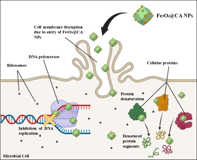
Recent advancements in nanotechnology have led to the manufacturing of multifunctional nanomaterials with wide range of applications in biomedical and environmental sectors. For example, waste water treatment through reduced porphyrin conjugated graphene oxide nanocomposite9, degradation of methylene blue10 and microbial contamination11 in waste water through reduced graphene oxide by using its photocatalytic activity12. Similarly, surface modification of nanoparticles can improve their antimicrobial potential and helps to treat various pathogenic resistant microbial strains. A study has explained the antifungal and antibacterial nature of titanium dioxide (TiO2) doped zinc oxide (ZnO) nanoparticles to protect leather surface and provides a better quality and durable leather products13. They have reported significant antimicrobial effects of TiO2 doped ZnO NPs which highlighted the importance of nanotechnology in leather industry13. Among various nanomaterials, magnetite (Fe3O4) nanoparticles have gained tremendous attention due to their exclusive physio-chemical and magnetic properties, high surface-to-volume ratio, and biologically compatible nature14,15. Based on these properties, Fe3O4 nanoparticles are widely used in water purification16, pollutant remediation17, and biomedical treatments18. The nature of nanoparticles to be manipulated through surface modifications make them particularly attractive for targeted applications19. Fe3O4 nanoparticles exhibited excellent antibacterial potential against broad range of pathogenic microorganisms by producing excessive amount of reactive oxygen species (ROS)20,21. These ROS enters into the bacterial cell where they penetrate into the cellular organelles, damage protein functioning, destruct enzymes molecules and inhibit nucleic acid proliferation and replication as shown in the Fig. 1. By incorporating these mechanisms, Fe3O4 nanoparticles create pores in the cell membrane of bacteria and ultimately leads to the cell death22.

(Self-Drawn on bio render (Online)): Antimicrobial Potential mechanism of citric acid coated magnetite nanoparticles by inhibiting DNA proliferation and replication, protein denaturation and cell membrane destruction.
Literature studies have reported limitations regarding chemical-based methods i.e. high cost, usage of toxic chemicals, and extra time consumption. These methods also prevented nanoparticles to be used for biological applications. Hence, for human use, the synthesis of nanoparticles through organic molecules is gaining importance due to better quality nanoparticles with reduce cytotoxicity. In this context, the functionalization of nanoparticles with biocompatible and eco-friendly agents, i.e. citric acid, further enhances their potential activities. Citric acid, a natural organic acid, have tremendous bioactive compounds with excellent antibacterial properties which can improves the dispersibility, stability, and functional properties of various nanoparticles23,24. Selenium nanoparticles have synthesized in the presence of citric acid using a green synthesis approach with excellent antibacterial, antifungal, antioxidant and anticoagulant potential activities25. Organic compounds are crucial to synthesize high-quality and biocompatible nanoparticles, hence offering a sustainable and bio-friendly alternative to traditional chemical and physical methods. Rich in varied bioactive components including flavonoids, polyphenols, and alkaloids, organic compounds serve as natural capping, stabilizing, and reducing agents during nanoparticle synthesis. This green synthesis approach has eradicated the use of toxic chemicals in comparison to conventional chemical methods.
While use of citric acid, to synthesize nanoparticles, has been expansively employed in various applications, its use in leather processing represents a novel and propitious frontier. Incorporating green-synthesized nanoparticles into leather processing can provides an innovative approach to leather industries to revolutionize traditional methods with clean, safe, and efficient substitute to traditional chemical treatments in order to get better quality and durable leather products. This study has focused on the synthesis, characterization and biological applications of Fe3O4 nanoparticles coated with citric acid encountering adverse effects of chemical residues and microbial growth on leather surface. The structural, chemical, functional and antimicrobial properties of Fe3O4 nanoparticles have investigated in addressing leather contamination challenges. This work aims to provide a comprehensive background for the excellent antimicrobial potential of Fe3O4 nanoparticles against resistive microorganisms hence providing better leather processing, contributing to both industrial innovation and environmental conservation.
Materials and methods
The leather samples were randomly collected from various leather industries in Sialkot, Pakistan. The microorganisms i.e. Staphylococcus aureus, Escherichia coli and Aspergillus Niger were isolated from collected leather samples. The citrus lemon was purchased from local market in Lahore, Pakistan.
Chemicals
All the chemicals and reagents of analytical grade were used without further purification. Iron (II) chloride tetrahydrate (FeCl2. 4H2O > 99% purity), Iron (III) chloride hexahydrate (FeCl3. 6H2O > 99% purity), ammonia (NH4OH), and sodium hydroxide were purchased from Sigma Aldrich while citric acid was obtained from Merck. De-ionized water was used as a solvent.
Isolation and incubation of fungal strain
The obtained leather samples were placed inside the desiccator as a source of tropical chamber at temperature 28 °C. Continuous sprinkling of autoclaved water was applied to maintain humidity required for the optimal growth of fungal strains. This was kept for 7 days and cultured on Potato Dextrose Agar (PDA) in the presence of 10 mg of streptomycin and 1 mL of 10% tartaric acid and incubated at 28 °C for two days. This protocol was followed by the guidelines of Asma Irshad et al., 202413. The isolated fungus was purified on yeast peptone dextrose agar, identified through Olympus CX23 microscope and characterized by Fungal Bank of University of the Punjab, Lahore, Pakistan.
Synthesis of citric acid coated Fe 3 O 4 nanoparticles
The magnetite nanoparticles were prepared through chemical co-precipitation method by reacting different concentration of ferrous chloride and ferric chloride i.e. 1 molar concentration of Fe2+ (FeCl2. 4H2O) and 2 molar concentration of Fe3+ (FeCl3. 6H2O), respectively. The mixture was homogenized by adding 25% ammonia (NH4OH) dropwise, with continuous stirring for 10 min at 80 °C. The pH was maintained at 8 to 12 and black precipitates were obtained, washed with de-ionized water and subjected to magnetic field. The protocol was followed by the guidelines of26 with slight modifications. The purified magnetic nanoparticles were dried, ground to fine powder and further processed for citric acid coating. For this purpose, 1 g citric acid and 1 g nanoparticles with 2 mL water were dissolved, stirred for 30 min at 90 °C and reduced the temperature as coating process was went to completion. The citric acid coated magnetite nanoparticles (Fe3O4@CA) were washed with de-ionized water, dried and ground to fine powder.
Characterization of citric acid coated magnetite nanoparticles
UV-Vis spectroscopy
The concentration of synthesized nanoparticles was determined through UV-Vis spectroscopy. For this, solution of Fe3O4@CA nanoparticles was prepared in de-ionized water and UV-Vis analysis was done with the instrument UV-Vis spectrophotometer Shimadzu UV-1900i (Shimadzu Corporation, Kyoto, Japan). The spectrum was obtained in the wavelength ranges from 200 to 500 nm by keeping de-ionized water as blank13.
Fourier transform infrared (FT-IR) spectroscopy
The functional groups and chemical bonds present between citric acid components and magnetite nanoparticles were investigated through FT-IR in dry air at room temperature (26 °C). The spectrum for Fe3O4@CA NPs was obtained in the presence of potassium bromide pellet by using Fourier transform infrared spectroscopy type ThermoFisher-Scientific Nicolet iS50 (ThermoFisher Scientific, USA) in the wavenumber ranges from 400 to 4000 cm−127.
Scanning electron Microscopy-Energy dispersive X-Ray (SEM-EDX)
The surface morphology and size distribution of Fe3O4@CA NPs was determined via scanning electron microscopy by using Nova NanoSEM 450 analyzer (ThermoFisher Scientific, USA), and Image J software was used to get the size of nanoparticles. Furthermore, SEM associated EDX profile was obtained to determine the elemental composition of Fe3O4@CA nanoparticles27.
X-Ray diffraction (XRD)
The crystalline structure and phase purity of Fe3O4@CA nanoparticles were determined by using Diffractometer-Bruker AXS D8 Advance (Bruker Corporation, Karlsruhe, Germany) using copper anticathode (Cu K = 1.5406 Å) under maintained constant conditions over a 2θ range from 10° to 80° for the step of 0.0101/minute13. The obtained diffractograms have been analyzed via system, based on the data sheets ASTM (American Society for Testing and Materials) with interplanar spacing (d) 2θ recorded software.
The average crystalline size of Fe3O4@CA nanoparticles from FWHM (full width at half maximum) was calculated using Scherrer Eq. 1 given below;
$${rm D = Klambda/beta:cos:theta}$$
(1)
, where D is the average crystalline size, K is constant (shape factor, commonly 0.9), λ is the wavelength (1.5406 Å) of X-rays, β is the full width at half maximum (FWHM) of diffraction peak, and θ is the Bragg angle.
Antimicrobial potential of citric acid coated magnetite nanoparticles
Agar well-diffusion method
Antimicrobial activities of Fe3O4@CA nanoparticles were estimated by agar well diffusion method against bacterial strains i.e. gram-positive strain Staphylococcus aureus, gram-negative strain Escherichia coli and fungal strain fungal strain Aspergillus niger. Potato dextrose and Luria Bertani agar were used to cultured fungal and bacterial strains, respectively. Wells were formed using cork-borer, each filling 100 µl of 1 mg/mL Fe3O4@CA nanoparticles solution. Streptomycin was used as a positive control for bacterial species while Fluconazole for fungal species by keeping water as a negative control. Plates were incubated at 37 °C (bacteria) or 28°C fungi for 24–48 h13,28.
Minimum inhibitory concentration (MIC)
The antimicrobial efficacy of Fe3O4@CA nanoparticles was also investigated through standard broth dilution method with reference to minimum inhibitory concentration (MIC). The MIC is the lowest nanoparticle concentration, which has ability to inhibit microbial growth. Serial two-fold dilutions of Fe3O4@CA nanoparticles were prepared in the concentrations 5, 2.5, 1.25, 0.625, and 0.312 mg/mL. The microbial concentration was arranged to 108 CFU/mL (0.5 McFarland’s standard) and both control and experimental samples were incubated at 37 °C for 24 h. Control samples were Streptomycin (for bacteria) and Fluconazole (for fungus)28.
Anti-biofilm potential
The crystal violet dye assay was performed to determine the biofilm inhibition potential of Fe3O4@CA nanoparticles. The CV dye has ability to binds with the extracellular polymeric substances of bacterial cells. The total biofilm mass formation was measured through this assay. For this, bacterial and fungal cultures were prepared and incubated at 37 °C for 48 h. Streptomycin and Fluconazole were used as reference for bacteria and fungi, respectively. The MIC Fe3O4@CA nanoparticles were added in the test sample tubes and after incubation, this material was discarded, tubes were stained with 2% CV dye, and washed with phosphate buffer saline (PBS). Then test-tube material was dissolved in 30% glacial acetic acid and absorbance was measured at 570 nm to obtain biofilm inhibition activity13.
Statistical analysis
The obtained values were calculated through Minitab Statistical Software version 22.1 and results were expressed as mean ± SD. The p < 0.05 was considered statistically significant. The experiments were conducted in triplicates and these results are mentioned as mean with standard deviation.
Results and discussion
Microscopic analysis of isolated fungus
The microscopic examination of isolated fungal samples has shown the presence of Aspergillus niger. The physical characteristics and morphological features of Aspergillus niger i.e. tiny, upright conidiophores, phialides growing from the center of spore and small globule swelling at the ends have observed under microscope. The conidia were light to dark brown, single celled and globule shaped.
Citric acid coated magnetite (Fe 3 O 4 @CA) nanoparticles
The fine powder of citric acid coated Fe3O4 NPs has obtained by co-precipitation of Iron (III) chloride hexahydrate (FeCl3. 6H2O) and Iron (II) chloride tetrahydrate (FeCl2. 4H2O) with 25% ammonium hydroxide in the presence of citric acid. The particles have been observed in the reaction mixture via color shifting from orange to black, indicated Fe3O4 NPs formation through magnetic decantation. The fine black powder of Fe3O4@CA has collected from bottom of beaker and confirmed through characterization methods.
Characterization of Fe 3 O 4 @CA NPs
UV-Visible spectroscopic analysis
The optical properties of Fe3O4 NPs and citric acid coated Fe3O4 nanoparticles have been studied through UV-Vis spectroscopy under the wavelength ranges from 200 to 1100 nm. The obtained UV-Vis spectrum for Fe3O4 NPs, as shown in the Fig. 2, has represented long and broad peak around 200–350 nm before and after coating of citric acid. Figure 2a has indicated the presence of Fe3O4 NPs with the spectrum peak around 250–300 nm. This peak has also confirmed through a literature study, in which Fe3O4 NPs were prepared and characterized through different techniques29. These results have also coincided with different existing studies30,31. In Fig. 2b, shifted peak has observed around 300–320 nm due to the coating of citric acid with Fe3O4 NPs. This shifting is due to conjugation of different biologically active components of citric acid with Fe3O4 NPs. A study was synthesized Fe3O4 NPs using papaya fruit extract coating and biologically characterized through UV-Vis spectroscopy. Their results were showed spectral peaks at 295 nm for FeCl3 NPs while 346 nm for Fe2O3 NPs32. These findings have shown the concept of shifting of wavelength due to the presence of citric acid.

UV–Visible spectra of (a) Fe3O4 nanoparticles and (b) citric acid coated Fe3O4 NPs.
Fourier transform infrared spectroscopy (FTIR)
The functional groups and chemical bonding between citric acid (C6H8O7) and Fe3O4 NPs have determined through FTIR spectroscopy. The spectrum was obtained between 4000–400 cm−1, with various broad and short peaks for correct formation of Fe3O4 nanoparticles (Fig. 3a) and citric acid coated Fe3O4 nanoparticles (Fig. 3b). Figure 3b has represented short peaks around 3211.48 cm−1 for O-H (hydroxyl) stretching vibrations of citric acid. A sharp prominent peak at 1579.70 cm−1 has revealed the binding of citric acid with the surface of nanoparticles through chemisorption of citrate ion33. The peak at 1409.96 cm−1 has confirmed the conjugation between citric acid and Fe3O4 NPs while stretching of the CO bond within the COOH group has recognized through the demonstration of an intense band at 1344.38 cm−1. At 447.49 cm−1, an absorption band has signified Fe-O bonding as well as verified Fe3O4 phase formation. Hence, these FTIR peaks have confirmed the citric acid coating over magnetite core as described by another literature study. Dheyab et al. 2020 synthesized citric acid functionalized iron oxide nanoparticles in colloidal solution. Their FTIR spectrum showed similar peaks which provided clear evidence to the current research work34.

FTIR spectrum of (a) Fe3O4 nanoparticles (b) Fe3O4@CA Nanoparticles.
Scanning electron microscopy-energy dispersive X-Ray (SEM-EDAX)
Scanning electron microscopy was performed to determine the high-resolution structural morphology and characteristics features of Fe3O4@CA NPs. Three-dimensional structure of Fe3O4@CA was produced when electron in SEM interacted with the surface of nanoparticles. Figure has represented particles with average size of obtained nanoparticles approximately 40 nm (Fig. 4a) which indicating the crystalline shape forming less agglomerates linked to active sites on the nanoparticle’s surface, including steric effects through interaction. Figure 4b, has shown the elemental composition of citric acid coated magnetite nanoparticles as shown in the EDX profile. The iron (Fe) has shown the predominant element followed by carbon (C) and oxygen (O) due to the coating of citric acid over the surface of magnetite nanoparticles.

Representative image of citric acid Fe3O4 nanoparticles showing (a) scanning electron microscopy (SEM) (b) energy dispersive X-ray profile (EDAX).
X-ray diffraction (XRD)
The crystalline structure and phase purity of Fe3O4@CA NPs has been determined through X-ray diffraction (Table 1). Figure 5 represented well-defined peaks at 2Ɵ of 27.2o, 35.7o, and 47.1o which intimated to the crystal plane of (220), (311), and (400), respectively33, while some short peaks were observed at 2Ɵ of 57.0o and 60.8o for (511) and (440), respectively35. The resulted peaks were narrow and well-defined which confirmed the high crystallinity of Fe3O4@CA NPs. These peaks were consistent to the diffraction pattern of Fe3O4 (Ref. Code 01-075-0033). A study was conducted in which magnetite nanoparticles were synthesized via co-precipitation method and coated with citric acid. Their XRD results are coincided with the obtained spectrum of Fe3O4@CA NPs34.

Representative image showing XRD spectrum for citric acid magnetite nanoparticles.
Antimicrobial potential analysis of Fe 3 O 4 @CA NPs
Agar well-diffusion
The antimicrobial potential of Fe3O4@CA NPs has determined through agar well-diffusion method against both gram-positive Staphylococcus aureus and gram-negative Escherichia coli bacterial strains and fungal specie Aspergillus Niger. The synthesized nanoparticles have shown excellent antimicrobial efficacy against both bacterial and fungal strains. Figure 6 have represented diameter of clear zones of inhibition which were recorded and documented as shown in the Table 2. Fe3O4 nanoparticles produced greater number of reactive oxygen species (ROS) which incorporated into the microbial cell and invade cellular organelles along with genetic material36. The maximum zone of inhibition, i.e. 27 ± 0.9 mm (Fig. 6a) has been observed against Escherichia coli followed by 23 ± 0.2 mm (Fig. 6c), fungal specie Aspergillus Niger and Staphylococcus aureus (22 ± 0.7 mm) (Fig. 6b) and. the obtained ZOI have demonstrated that these nanoparticles can provide excellent antimicrobial efficacy against Aspergillus niger as compared to bacterial strains, hence beneficial for preventing resistant fungal growth on the surface of leather and related products. Streptomycin and Fluconazole were applied as reference against bacterial and fungal strains, respectively. The synthesized nanoparticles have shown excellent antimicrobial properties in comparison to standard antibiotics. A study was designed to synthesize magnetite nanoparticles by busing Calotropis procera aqueous leaf extract through green synthesis approach. They revealed the excellent antimicrobial activity of magnetite nanoparticles with notable zones of inhibition i.e. 7.1 mm to 22.5 mm against S. aureus, K. pneumonia, (A) niger, F. oxysporum and (B) subtilis, through agar well-diffusion method37. Various other studies have also provided evidence for the antimicrobial potential of magnetite nanoparticles38,39. These results have revealed the antimicrobial potential of synthesized Fe3O4@CA NPs.

Zones of inhibition of Fe3O4@CA nanoparticles showing antimicrobial potential against (a) gram-negative strain Escherichia coli, (b) gram-positive strain Staphylococcus aureus, (c) fungal strain Aspergillus niger.
Minimum inhibitory concentration (MIC)
Due to excellent antimicrobial activity of Fe3O4@CA NPs against both bacterial and fungal species, they have further assessed for minimum inhibitory concentration (MIC) through micro-dilution technique, with dilutions ranges 5-0.156 mg/mL. MIC is the minimum concentration of nanoparticles able to degrade microbial cells13. Table 3 has demonstrated the MIC values of Fe3O4@CA NPs against selected microbial strains. The minimum the value of MIC, greater the inhibitory mechanism. For bacterial strains, the results have shown MIC value 0.3 mg/mL while 0.625 mg/mL for Aspergillus niger. These values have depicted that bacterial strains are more susceptible to synthesized Fe3O4@CA NPs as compared to fungal strain. The obtained results have coincided with the literature findings of similar studies performed by Lathakumari, R. H. et al., 2022. They reported similar MIC values of bare and citrate coated magnetite nanoparticles i.e. 0.0375-0.3 mg/mL against broad range of microorganisms40. Al-Rawi M et al., 2021 conducted experiment with Fe3O4 nanoparticles to determine minimum inhibitory concentration against local virulent bacterial isolates in urine samples. Their results represented MIC 550 µg/mL against gram-negative bacterium E. coli. The value of MIC was different because of different method of production and precursors for Fe3O4 nanoparticles41.
Antibiofilm Potential
Various groups of microorganisms exhibit network of complex polysaccharides in the form of biofilm for their growth, survival and protection. The synthesized Fe3O4@CA NPs have subjected to investigate the antibiofilm potential against both bacterial and fungal strains. The optical density of each sample has recorded to depict the biofilm production. Table 4 has shown no optical density for Fe3O4@CA NPs containing samples against each selected microbial strain while non-Fe3O4@CA NPs samples have different optical densities (OD) against Staphylococcus aureus (1.79 ± 0.05), Escherichia coli (1.58 ± 0.06), and Aspergillus Niger (1.86 ± 0.07). This experiment has clearly demonstrated the antibacterial and antifungal potential of Fe3O4@CA NPs. Similar study was conducted in 2020, where magnetite nanoparticles were synthesized by using ferrous and ferric salts through co-precipitation method and further coated with polyethylene imine. They performed biofilm inhibition analysis of magnetite and nickel ferrite nanoparticles, separately. Their results showed excellent anti-biofilm potential against pathogenic strains. The highest biofilm inhibition concentration of synthesized nanoparticles was 10 µg/mL against E. coli followed by other strains42. The difference in results values may be due to different coating material and concentration of nanoparticles. These studies have provided clear evidence about the antimicrobial nature of magnetite nanoparticles26,43.
Conclusion
Magnetite nanoparticles have prepared by co-precipitation method and further coated with citric acid. These nanoparticles have characterized through various techniques i.e. UV-Vis spectrometry, FTIR, SEM-EDAX, and XRD, which have demonstrated favorable outcomes indicating precise and accurate synthesis of these magnetite nanoparticles. These nanoparticles have prepared devoid of impurities, and their antimicrobial efficacy has assessed against various pathogenic bacterial and fungal strains. The results have shown that citric acid coated Fe3O4 formation can able to reduce the leather damaging caused by microorganisms. Hence, green synthesized magnetite nanoparticles can be a promising and innovative solution for leather industries to process leather materials against pathogenic microorganisms. Although these nanoparticles have proved their antimicrobial efficiency in the current study but further prospects are required to investigate about biosafety potential and their exact use in leather processing.
Data availability
All the data used for this study are present in this manuscript.
References
-
Mendanha, D. Driving sustainability in the automotive industry: bio-coated materials and modern strategies. Acad. Mater. Sci. 1 (2). https://doi.org/10.20935/AcadMatSci6188 (2024).
-
Sivakumar, V. Towards environmental protection and process safety in leather processing–a comprehensive analysis and review. Process. Saf. Environ. Prot. 163, 703–726. https://doi.org/10.1016/j.psep.2022.05.062 (2022).
-
Lasoń-Rydel, M., Sieczyńska, K., Gendaszewska, D., Ławińska, K. & Olejnik, T. P. Use of enzymatic processes in the tanning of leather materials. Autex Res. J. 24 (1), 20230012. https://doi.org/10.1515/aut-2023-0012 (2024).
-
Merlyn, S. R., Saranya, R., Jayapriya, J., Aravindhan, R. & Tamilselvi, A. Biological Treatment of Leather Industry Solid Waste Current Status and Future Perspectives. Solid Waste Management: pp. 103–118.
-
Abdulhusein, H. S. & Kadim, B. M. Antimicrobial substances and strategies to avoid bacterial and fungal effects in leather manufacturing. Kafkas Üniversitesi Fen Bilimleri Enstitüsü Dergisi. 17 (2), 81–91. https://doi.org/10.58688/kujs.1467530 (2024).
-
Devi, L., Kushwaha, P., Ansari, T. M., Kumar, A. & Rao, A. Recent trends in biologically synthesized metal nanoparticles and their biomedical applications: a review. Biol. Trace Elem. Res. 202 (7), 3383–3399. https://doi.org/10.1007/s12011-023-03920-9 (2024).
-
Sahu, B., Ramesh, A. & Zameer, F. Impact of nanomaterials on leather: a nano-Saga from processing to application. Clean. Technol. Environ. Policy. 1–28. https://doi.org/10.1007/s10098-024-02912-0 (2024).
-
Hasanah, U., Miran, M. S. & Islam, M. M. Nanomaterials for leather production. Appl. Emerg. Nanomaterials Nanatechnol. 148, 170–199. https://doi.org/10.21741/9781644902554-6 (2023).
-
El-Khawaga, A. M., Tantawy, H., Elsayed, M. A. & Abd El-Mageed, A. I. Synthesis and applicability of reduced graphene oxide/porphyrin nanocomposite as photocatalyst for waste water treatment and medical applications. Sci. Rep. 12 (1), 17075. https://doi.org/10.1038/s41598-022-21360-8 (2022).
-
Abuzeyad, O. H., El-Khawaga, A. M., Tantawy, H., Gobara, M. & Elsayed, M. A. Merits photocatalytic activity of rGO/zinc copper ferrite magnetic nanocatalyst for photodegradation of methylene blue (MB) dye. Discover Nano. 20 (1), 1–16. https://doi.org/10.1186/s11671-024-04162-x (2025).
-
Abuzeyad, O. H., El-Khawaga, A. M., Tantawy, H. & Elsayed, M. A. An evaluation of the improved catalytic performance of rGO/GO-hybrid-nanomaterials in photocatalytic degradation and antibacterial activity processes for wastewater treatment: A review. J. Mol. Struct. 1288, 135787. https://doi.org/10.1016/j.molstruc.2023.135787 (2023).
-
Abuzeyad, O. H., El-Khawaga, A. M., Tantawy, H., Gobara, M. & Elsayed, M. A. Reduced graphene oxide loaded with ZCF magnetic nanoparticles as a promising photocatalyst and antibacterial agent. J. Cluster Sci. 36 (1), 1. https://doi.org/10.1007/s10876-024-02718-6 (2025).
-
Irshad, A. et al. Unveiling the power of TiO2 doped ZnO nanomaterial as an effective antimicrobial solution in the leather industry. Heliyon 10 https://doi.org/10.1016/j.heliyon.2024.e38414 (2024).
-
Piro, N. S., Hamad, S. M., Mohammed, A. S. & Barzinjy, A. A. Green synthesis magnetite (Fe₃O₄) nanoparticles from Rhus coriaria extract: a characteristic comparison with a conventional chemical method. IEEE Trans. Nanobiosci. 22 (2), 308–317. https://doi.org/10.1109/TNB.2022.3187344 (2022).
-
Jafari, N., Mohammadpourfard, M. & Hamishehkar, H. A comprehensive study on doxorubicin-loaded aspartic acid-coated magnetic Fe3O4 nanoparticles: synthesis, characterization and in vitro anticancer investigations. J. Drug Delivery Sci. Technol. 100, 106133. https://doi.org/10.1016/j.jddst.2024.106133 (2024).
-
Liang, Y. et al. Synthesis and characterization of Fe3O4 nanoparticles prepared by solvothermal method. J. Mater. Eng. Perform. 33 (13), 6804–6815. https://doi.org/10.1007/s11665-023-08431-1 (2024).
-
Rouhani, M., Ashrafi, S. D., Taghavi, K., Joubani, M. N. & Jaafari, J. Evaluation of Tetracycline removal by adsorption method using magnetic iron oxide nanoparticles (Fe3O4) and clinoptilolite from aqueous solutions. J. Mol. Liq. 356, 119040. https://doi.org/10.1016/j.molliq.2022.119040 (2022).
-
Montiel Schneider, M. G. et al. Biomedical applications of iron oxide nanoparticles: current insights progress and perspectives. Pharmaceutics 14 (1), 204. https://doi.org/10.3390/pharmaceutics14010204 (2022).
-
Largani, S. P. H., Salimi-Kenari, H., Nabavi, S. R. & Darzi, A. A. R. Manipulation of the thermo-rheological properties of stable Fe3O4 nanoparticles-embedded PCM nanoemulsions. J. Energy Storage. 80, 110351. https://doi.org/10.1016/j.est.2023.110351 (2024).
-
Ghorbanizadeh, S. et al. Antibacterial effects and cellular mechanisms of iron oxide magnetic nanoparticles coated by piroctone olamine against some cariogenic bacteria. Annals Med. Surg. https://doi.org/10.1016/j.amsu.2022.104291 (2022).
-
Allogmani, A. S., Mohamed, R. M. & Hasanin, M. S. Green, eco-friendly, highly biocompatible and bioactive nanocomposite-based biopolymers loaded with zno@ Fe3O4 nanoparticles. Polymers 15 (17), 3641. https://doi.org/10.3390/polym15173641 (2023).
-
Rukhsar, M. et al. An overview of iron oxide (Fe3O4) nanoparticles: from synthetic strategies, characterization to antibacterial and anticancer applications. Crystals 12 (12), 1809. https://doi.org/10.3390/cryst12121809 (2022).
-
Mikelashvili, V. et al. Synthesis and characterization of citric acid-modified iron oxide nanoparticles prepared with electrohydraulic discharge treatment. Materials 16 (2), 746. https://doi.org/10.3390/ma16020746 (2023).
-
Singh, S., Maurya, I. C., Tiwari, A., Srivastava, P. & Bahadur, L. Green synthesis of TiO2 nanoparticles using citrus Limon juice extract as a bio-capping agent for enhanced performance of dye-sensitized solar cells. Surf. Interfaces. 28, 101652. https://doi.org/10.1016/j.surfin.2021.101652 (2022).
-
Alhawiti, A. S. Citric acid-mediated green synthesis of selenium nanoparticles: antioxidant, antimicrobial, and anticoagulant potential applications. Biomass Convers. Biorefinery. 14 (5), 6581–6590. https://doi.org/10.1007/s13399-022-02798-2 (2024).
-
Esfahani, M. B., Khodavandi, A., Alizadeh, F. & Bahador, N. Antibacterial and anti-biofilm activities of microbial synthesized silver and magnetic iron oxide nanoparticles against Pseudomonas aeruginosa. IEEE Trans. Nanobiosci. 22 (4), 956–966. https://doi.org/10.1109/TNB.2023.3268138 (2023).
-
Iram, Z. et al. Nature-Inspired Antimicrobial Agents: Cinnamon-Derived Copper Oxide Nanoparticles for Effective Aspergillus Niger Control. Current Microbiology, 82(1): p. 19 (2025). https://doi.org/10.1007/s00284-024-04000-4
-
Irshad, A. et al. Determination of antibacterial and antioxidant potential of organic crude extracts from Malus domestica, cinnamomum verum and trachyspermum Ammi. Sci. Rep. 15 (1), 976. https://doi.org/10.1038/s41598-024-83506-0 (2025).
-
Nasr, G. M., Thawabieh, O. M., Talaat, R. M., Moawad, M. & Hamshary, M. O. E. Assessment of the in vitro effects of folate Core–Shell conjugated iron oxide nanoparticles as a potential agent for acute leukemia treatment. Front. Bioscience-Landmark. 29 (4), 162. https://doi.org/10.31083/j.fbl2904162 (2024).
-
Buarki, F., AbuHassan, H., Hannan, F. A. & Henari, F. Green synthesis of iron oxide nanoparticles using hibiscus Rosa sinensis flowers and their antibacterial activity. J. Nanatechnol. 2022 (1), 5474645. https://doi.org/10.1155/2022/5474645 (2022).
-
Taherkhani, A., Fazli, H. & Taherkhani, F. Application of Janus magnetic nanoparticle Fe3O4@ sin functionalized with beta-cyclodextrin in thymol drug delivery procedure: an in vitro study. Appl. Organomet. Chem. 35 (11), e. https://doi.org/10.1002/aoc.6399 (2021).
-
Malaikozhundan, B., Krishnamoorthi, R., Vinodhini, J., Nambi, K. S. N. & Palanisamy, S. Multifunctional iron oxide nanoparticles using carica Papaya fruit extract as antibacterial, antioxidant and photocatalytic agent to remove industrial dyes. Inorg. Chem. Commun. 144, 109843. https://doi.org/10.1016/j.inoche.2022.109843 (2022).
-
Khan, S. et al. Antimicrobial activity of citric acid functionalized iron oxide nanoparticles–Superparamagnetic effect. Ceram. Int. 46 (8), 10942–10951. https://doi.org/10.1016/j.ceramint.2020.01.109 (2020).
-
Dheyab, M. A. et al. Simple rapid stabilization method through citric acid modification for magnetite nanoparticles. Sci. Rep. 10 (1), 10793. https://doi.org/10.1038/s41598-020-67869-8 (2020).
-
El-Khawaga, A. M., Farrag, A. A., El-Batal, A. I. & Elsayed, M. A. Evaluation of the antimicrobial activity of citric acid functionalized magnetite nanoparticles. Egypt. J. Med. Microbiol. 29 (4), 143–149. https://doi.org/10.51429/EJMM29418 (2020).
-
Hussain, A. et al. Synthesis, characterization, and applications of iron oxide nanoparticles. Int. J. Health Sci. 17 (4), 3 (2023).
-
Kalu, A., Egwim, E., Jigam, A. & Muhammad, H. Green synthesis of magnetite nanoparticles using Calotropis procera leaf extract and evaluation of its antimicrobial activity. Nano Express. 3 (4), 045004. https://doi.org/10.1088/2632-959X/aca925 (2022).
-
El-Sesy, M. E. & Othman, S. A. Promising antibacterial activities of anethole and green-synthesized magnetite nanoparticles against multiple antibiotic-resistant bacteria. Water Sci. Technol. 87 (3), 729–747. https://doi.org/10.2166/wst.2023.012 (2023).
-
Rajendrachari, S., Karaoglanli, A. C., Ceylan, Y. & Uzun, O. A fast and robust approach for the green synthesis of spherical magnetite (Fe 3 O 4) nanoparticles by tilia tomentosa (Ihlamur) leaves and its antibacterial studies. Pharm. Sci. 26 (2), 175–183. https://doi.org/10.34172/PS.2020.5 (2020).
-
LATHAKUMARI, R. H., RAVI, S., TRISAL, S., VAJRAVELU, L. K. & VISHNUDASAN, D. Efficacy of green synthesised iron oxide nanoparticles against various uropathogens: A Cross-sectional study. J. Clin. Diagn. Res. 16 (10). https://doi.org/10.7860/JCDR/2022/58018.17065 (2022).
-
Al-Rawi, M., Al-Mudallal, N. & Taha, A. Determination of ferrous oxide nanoparticles minimum inhibitory concentration against local virulent bacterial isolates. Arch. Razi Inst. 76 (4), 795. https://doi.org/10.22092/ari.2021.355997.1758 (2021).
-
Ceylan, O., Tamfu, A. N., Doğaç, Y. İ. & Teke, M. Antibiofilm and anti-quorum sensing activities of polyethylene Imine coated magnetite and nickel ferrite nanoparticles. 3 Biotech. 10, 1–12. https://doi.org/10.1007/s13205-020-02509-6 (2020).
-
Li, W., Wei, W., Wu, X., Zhao, Y. & Dai, H. The antibacterial and antibiofilm activities of mesoporous Hollow Fe 3 O 4 nanoparticles in an alternating magnetic field. Biomaterials Sci. 8 (16), 4492–4507. https://doi.org/10.1039/D0BM00673D (2020).
Funding
There was no funding available for this study.
Ethics declarations
Competing interests
The authors declare no competing interests.
Ethics approval and consent to participate
No Ethical concern was raised during this study as no animal or human was used for this study.
Consent for publication
This research work is original, has not been previously published, and is not under consideration for publication elsewhere. Therefore, all authors give their consent for this publication.
Additional information
Publisher’s note
Springer Nature remains neutral with regard to jurisdictional claims in published maps and institutional affiliations.
Rights and permissions
Open Access This article is licensed under a Creative Commons Attribution-NonCommercial-NoDerivatives 4.0 International License, which permits any non-commercial use, sharing, distribution and reproduction in any medium or format, as long as you give appropriate credit to the original author(s) and the source, provide a link to the Creative Commons licence, and indicate if you modified the licensed material. You do not have permission under this licence to share adapted material derived from this article or parts of it. The images or other third party material in this article are included in the article’s Creative Commons licence, unless indicated otherwise in a credit line to the material. If material is not included in the article’s Creative Commons licence and your intended use is not permitted by statutory regulation or exceeds the permitted use, you will need to obtain permission directly from the copyright holder. To view a copy of this licence, visit http://creativecommons.org/licenses/by-nc-nd/4.0/.
About this article

Cite this article
Hayat, A., Irshad, A., Ishtiaq, U. et al. Potent antimicrobial and antibiofilm activity of citric acid coated magnetite nanoparticles for leather preservation. Sci Rep 15, 27889 (2025). https://doi.org/10.1038/s41598-025-14163-0
-
Received:
-
Accepted:
-
Published:
-
DOI: https://doi.org/10.1038/s41598-025-14163-0