
Introduction
Fungal pathogens remain a major cause of morbidity and mortality, particularly among immunocompromised and critically ill patients1,2. Invasive candidiasis, including candidemia, is one of the most common invasive fungal infections and is associated with prolonged hospitalization, high costs, and substantial case-fatality rates3. Recent surveillance reports describe a worrying rise in azole- and echinocandin-resistant Candida isolates and the emergence and global spread of multidrug-resistant species such as Candida auris, emphasizing the need for new antifungal options and better-defined molecular targets4. In Candida spp., resistance is driven by well-characterized mechanisms: mutations and overexpression of ERG11 (encoding lanosterol 14α-demethylase, CYP51), increased expression of ATP-binding cassette and major facilitator efflux pumps, remodeling or bypass of the ergosterol biosynthesis pathway, and biofilm-associated tolerance5,6,7. These adaptations erode the efficacy of current frontline drugs and highlight the importance of natural chemo-types that either act on validated targets in new ways or engage complementary fungal pathways8.
Lanosterol 14α-demethylase (CYP51) is a cytochrome P450 enzyme that catalyzes a key step in ergosterol biosynthesis and is the primary molecular target of azole antifungals9. Inhibition of CYP51 depletes ergosterol and leads to accumulation of aberrant sterols, disrupting membrane function and cell viability10,11. Because CYP51 is essential, druggable, and directly implicated in clinical resistance (via ERG11 mutations and overexpression), it represents a rational focal point for both experimental and computational evaluation of new antifungal candidates12. For this reason, we selected Candida CYP51 as the principal molecular target in our in silico studies to probe the binding behavior and mechanistic plausibility of the metabolite isolated in this work.
Natural products remain a cornerstone of antifungal drug discovery, and fungal secondary metabolites have provided several clinically important agents or lead structures. Endophytic fungi are recognized as a particularly rich source of chemically diverse metabolites with antibacterial, antifungal, antioxidant, antiproliferative, and other bioactivities13,14. Their metabolic profiles may mirror, complement, or diverge from those of their host plants, offering access to unique scaffolds not readily found in free-living fungi15. Rosmarinus officinalis (rosemary) is a well-studied medicinal and culinary plant with documented antimicrobial and antifungal properties, attributed mainly to its essential oils and phenolic constituents16. Extracts and oils of R. officinalis have shown activity against Candida species and various phytopathogenic fungi17. Endophytic fungi associated with R. officinalis therefore represent a logical and underexplored niche for discovering new antifungal metabolites that could reinforce or extend the bioactivity spectrum of the host. Within this context, Aspergillus species are prominent producers of structurally diverse polyketides, terpenoids, and alkaloids18. Aspergillus candidus has previously been reported to yield p-terphenyl derivatives, candidusin-type polyketides, chlorflavonin, and related diphenyl ethers with antibacterial, antifungal, or cytotoxic properties, indicating a versatile biosynthetic capacity18,19. However, the chemical space of endophytic A. candidus, particularly from medicinal hosts such as R. officinalis, remains incompletely characterized.
The present study investigates an endophytic A. candidus isolated from R. officinalis and reports the isolation, structure elucidation, and initial antifungal evaluation of one of its secondary metabolites. Using comprehensive spectroscopic analysis (FTIR, 1H/13C NMR, HSQC), GCMS profiling, and systematic database searches (SciFinder, Reaxys, PubChem, ChemSpider, ChEMBL), we identified a C18 oxygenated polyketide that, to the best of our knowledge, has not been previously described in the literature or major chemical databases. We therefore treat this metabolite as a previously unreported natural product and evaluate its antifungal potential against C. albicans using an agar-based in vitro assay in parallel with CYP51-focused molecular docking and molecular dynamics simulations. By integrating endophyte-derived chemistry, rigorous structure elucidation, and targeted in silico and in vitro assessment, this work aims to introduce a structurally distinct fungal metabolite as a candidate scaffold for future antifungal development.
Methodology
Isolation and preliminary screening of the endophytic fungi
To explore bioactive endophytic fungi, healthy tissues of R. officinalis (leaves, roots, and twigs) were collected from the botanical garden and immediately transferred to the laboratory at the Centre of Biotechnology and Microbiology, University of Peshawar. The samples were stored at -80 °C until further processing. For fungal isolation, plant tissues were surface sterilized to eliminate epiphytic contaminants. This included washing with sterile distilled water, immersion in 75% ethanol for one minute, followed by treatment with 0.1% mercuric chloride for one minute. The sterilized tissues were rinsed thoroughly with sterile distilled water and then aseptically sectioned into approximately 1 cm segments using a sterile scalpel. The sections were placed on Potato Dextrose Agar (PDA) plates supplemented with chloramphenicol (50 µg/mL) to inhibit bacterial growth. Plates were sealed with parafilm and incubated at 25 ± 2 °C for 7–15 days. Fungal growth emerging from the internal plant tissues was carefully monitored, and individual colonies were sub-cultured onto fresh PDA plates to obtain pure cultures. Each purified fungal isolate was subjected to preliminary antibacterial and antifungal screening. Strains that demonstrated promising bioactivity were selected for further identification and secondary metabolite production20.
Identification of the bioactive fungus
The endophytic fungal strain isolated from R. officinalis was identified using a combination of morphological, microscopic, and molecular approaches. For morphological and microscopic characterization, the colony features, hyphal structures, and spore morphology were examined using the slide culture technique. Samples were stained with lactophenol cotton blue and observed under a light microscope at 1000× magnification to document key diagnostic features such as septate hyphae and reproductive structures.
To confirm the identity at the molecular level, genomic DNA was extracted from the pure culture using the Qiagen DNeasy Mini Kit, and its quality was assessed via 1% agarose gel electrophoresis. The Internal Transcribed Spacer (ITS) region of the rDNA was amplified using universal fungal primers ITS1 (5′-TCCGTAGGTGAACCTGCGG-3′) and ITS4 (5′-TCCTCCGCTTATTGATATGC-3′). The amplified products were sequenced by Apical Scientific (Malaysia), and the sequence data were analyzed using the BLAST tool available at NCBI. Phylogenetic relationships were further evaluated using BioEdit and MEGA X software, applying the Fast Minimum Evolution method to construct a distance-based evolutionary tree with a maximum sequence divergence of 0.75. The accession number of the fungal isolate is MH725571.1, and it is available at the following NCBI link: https://www.ncbi.nlm.nih.gov/nuccore/MH725571.1.
Culture conditions and post incubation extraction of fungal secondary metabolites
The producing strain was cultivated in a modified Czapek Yeast Broth (optimized in our laboratory) containing glucose 1% (w/v), peptone 1% (w/v), KCl 0.5% (w/v), MgSO4·7H2O 0.05% (w/v) and 0.001% FeSO4·7H2O(w/v). The initial pH was adjusted to 5.8 ± 0.2 before sterilization. To enhance secondary metabolites production, the base medium was supplemented with an additional 2% (w/v) glucose and 3% (w/v) starch (final concentrations: glucose 3% w/v; starch 3% w/v). Nine 500-mL Erlenmeyer flasks were prepared with 300 mL working volume each (total fermentation volume 2.7 L) and incubated for 14 days at 28 °C on an orbital shaker at 150 rpm. At the end of incubation, whole broth (mycelium plus culture supernatant) was processed for extraction. To facilitate phase separation and precipitation of medium components, 200–250 µL of 40% HCl was added to each flask, followed by liquid–liquid extraction with ethyl acetate (1:1, v/v) performed three times with vigorous mixing. The mycelial biomass was homogenized in an equal volume of ethyl acetate for 30 min with intermittent shaking, then filtered through cheesecloth and Whatman filter paper. Organic phases were combined, dried over anhydrous Na2SO4, and concentrated under reduced pressure at 45 °C on a rotary evaporator to afford the crude extract, which was weighed and subjected to chromatographic purification21.
Purification of fungal metabolites using chromatographic techniques
The fungal crude extract was dissolved in methanol and clarified using sterile 0.22 μm membrane filter (Sigma-Aldrich). The filtrate was mixed with silica gel (60–120 mesh) to form a uniform slurry and gently evaporated to dryness so that the sample was adsorbed onto silica. Normal-phase column chromatography was performed on silica packed and equilibrated in n-hexane. The silica-bound sample was loaded and eluted with a stepwise gradient from n-hexane to ethyl acetate. Fractions were monitored by TLC on silica gel GF254 plates (UV 254/366 nm; anisaldehyde- sulfuric acid visualization as needed). The major bioactive fraction eluted at 65:35 (v/v) ethyl acetate: n-hexane and was carried for further analysis. Preparative TLC was used to sharpen the enriched fraction and the samples were applied as narrow bands to silica gel GF254 plates (20 × 20 cm), developed with 65:35 (v/v) ethyl acetate: n-hexane. The TLC plate was visualized under UV and the target band was excised, eluted with ethyl acetate and then concentrated. Purity was assessed by analytical HPLC on a Waters 2795HT system equipped with a Phenomenex Luna C18 column. The solvent system comprised HPLC-grade ultrapure water (A), HPLC-grade methanol (B), and HPLC-grade acetonitrile (C), each containing 0.055% formic acid. Injections were 20 µL at a flow rate of 1.0 mL min[- [1. The isolated compound eluted as a single, symmetrical peak under these conditions, confirming purity for downstream analyses.
Characterization and structural analysis of the purified compound
The structure of the purified compound was determined using a combination of analytical techniques including Gas Chromatography-Mass Spectrometry (GCMS), Fourier Transform Infrared (FTIR) spectroscopy, and Nuclear Magnetic Resonance (NMR) spectroscopy. Mass analysis was conducted using an Agilent 7890B GC system coupled with an Agilent 5977B MS detector. A 1 µL aliquot of the compound, dissolved in methanol, was injected into the GC system. An Agilent HP 5ms column (0.25 μm film thickness) was used with helium as the carrier gas. The resulting mass spectra were analyzed using Agilent MassHunter software, providing the molecular ion peak and fragmentation pattern essential for initial structural confirmation.
FTIR spectroscopy was performed using a Cary 630 FTIR spectrometer (Agilent Technologies, USA) to identify the functional groups. The powdered sample was analyzed directly, and characteristic absorption bands were recorded, providing a molecular fingerprint of the pure compound. To gain deeper insight into the compound structure, 1D and 2D NMR spectra were recorded using a Varian 500 MHz NMR spectrometer. The sample (6 mg) was dissolved in 0.65 mL of deuterated chloroform (CDCl3), and tetramethylsilane (TMS) was used as the internal reference.
Virtual screening of the pure compound
Molecular docking studies were conducted to evaluate the interaction of the pure fungal compound having nominal mass of 340 with 14α-demethylase, a key enzyme and broad-spectrum antifungal target. The crystal structure of the enzyme (PDB ID: 5FSA) was retrieved from the Protein Data Bank22. Using UCSF Chimera v1.17, the structure was cleaned by removing co-crystallized ligands and water molecules and subjected to energy minimization using steepest descent and conjugate gradient algorithms for 2000 steps to optimize its geometry. The prepared enzyme structure was then imported into PyRx v0.8 for docking analysis. The pure compound, identified as (2E,7E)-6,9-dihydroxy-10-(3-hydroxy-5-oxocyclohexyl)deca-2,7-dien-1-yl acetate, was drawn in ChemDraw 12.0, converted to PDB format, and subsequently prepared as a PDBQT file within PyRx. Docking was performed using AutoDock Vina with the grid box dimensions set to 25 Å in the X, Y, and Z directions, and 100 binding poses were generated. Among the generated docking poses, the one exhibiting the lowest binding energy (kcal/mol) was selected for further interaction analysis. For comparative analysis, posaconazole was used as a control and docked using the same protocol described above. The RMSD between crystal 5FSA–posaconazole and docked 5FSA posaconazole was 0.12 Å, confirming the accuracy of the docking procedure. The docking complex was visualized and analyzed using UCSF Chimera v1.17 and Discovery Studio Visualizer to interpret the molecular interactions within the active site23,24,25.
Molecular dynamics (MD) simulation of the protein-ligand complex
To assess the stability and binding strength of the top-ranked docking pose, Molecular Dynamics (MD) simulations were performed for 100 nanoseconds using the Desmond simulation package. The protein–ligand complex was solvated in an orthorhombic box with a 10 Å buffer on all sides, using the TIP3P water model and periodic boundary conditions. To maintain system neutrality, sodium ions (Na+) were added while ensuring exclusion within 15 Å of the ligand atoms to avoid potential interference with key interactions. The OPLS4 force field was employed for parameterization, and all simulations were conducted under an NPT ensemble maintaining constant pressure, temperature, and particle number. Prior to the production run, the system underwent energy minimization and relaxation using Desmond’s default protocol. A 9 Å cutoff was used for both Lennard–Jones and short-range electrostatic interactions, while long-range electrostatics were handled using the Particle Mesh Ewald (PME) method. The simulation was run at 300 K and 1.01325 bar pressure, regulated using the Nose Hoover chain thermostat and Martyna–Tobias–Klein barostat. A trajectory frame was saved every 200 ps with an integration time step of 2 fs. Post-simulation, trajectory data were analyzed using Desmond’s Simulation Interaction Diagram (SID) and Simulation Event Analysis (SEA) tools. System stability was evaluated through various structural parameters, including Root Mean Square Deviation (RMSD), Root Mean Square Fluctuation (RMSF), Radius of Gyration (Rg), Solvent Accessible Surface Area (SASA), and B-factor. The distance between the electrophilic β-lactam carbonyl carbon and the nucleophilic sulfur of Cys-199 was monitored, with 3.5 Å set as the critical threshold, based on their Vander Waals radii. To ensure the reproducibility of results, simulations were repeated using a different random seed.
Binding energy calculation via MM-PBSA
To estimate the binding free energy of the protein–ligand complex, the Molecular Mechanics Poisson- Boltzmann Surface Area (MM-PBSA) method was applied using the g_mmpbsa tool. This approach calculates the difference in free energy between the bound complex and the sum of the energies of the free protein and ligand. The final 5 ns of the MD simulation trajectory were used for this analysis. Contributions to the total binding energy included van der Waals and electrostatic interactions, along with polar and non-polar solvation energies.
In vitro antifungal activity of the pure compound
Clinical Candida isolates from Lady Reading Hospital (LRH), Peshawar, were sub-cultured to purity. PDA medium was prepared and autoclaved at 121 °C for 15 min in screw-cap culture tubes (10 mL per tube) and cooled to about 50 °C. The purified compound was dissolved in DMSO and added to molten PDA to give final concentrations of 100, 250, 500, and 1000 µg/ mL. Final DMSO was ≤ 1% v/v in all tubes, including solvent controls. Tubes were mixed and set as slants. Each tube was line-inoculated with the test Candida strain using a sterile swab. Fluconazole at the same concentrations served as the positive control; solvent-only tubes were negative controls. The slants were incubated at 37 °C for 72–96 h. Linear growth from the inoculation line to the colony front was measured in millimeters26,27. Experiments were performed in triplicate (n = 3). Data are reported as mean ± SD, and the percent fungal growth inhibition was calculated using the formula:
$${text{Percent}},{text{growth}},{text{inhibition}}:=frac{fungal:growth:in:testleft(mmright):}{fungal:growth:in:control:left(mmright)}times:100$$
Results
Morphological and microscopic identification of the bioactive fungal strain
When cultured on PDA, colonies of A. candidus exhibited rapid growth, attaining 4–5 cm in diameter within 7 days at 25–28 °C. The colonies appeared dense, velvety to floccose in texture, with a bright white to cream surface that became progressively chalky and powdery due to profuse conidiation. The margins were regular and well-defined, and the reverse of the colonies remained pale to light yellow without any distinctive pigmentation. With prolonged incubation, the central region developed a slightly granular appearance, while the overall white coloration persisted, reflecting the species’ typical “candidus” character. On Sabouraud dextrose agar (SDA) colonies also grew rapidly, though they were comparatively less dense than those observed on PDA. The surface was initially cottony and white, later becoming compact and powdery with heavy sporulation. The colonies displayed a granular, chalk-like texture in the central regions, while the periphery remained softer and tufted. The reverse side was unpigmented to faintly yellowish, and no diffusible pigments were produced. Throughout incubation, colonies retained their characteristic pure white appearance, a distinctive trait aiding in the identification of A. candidus.
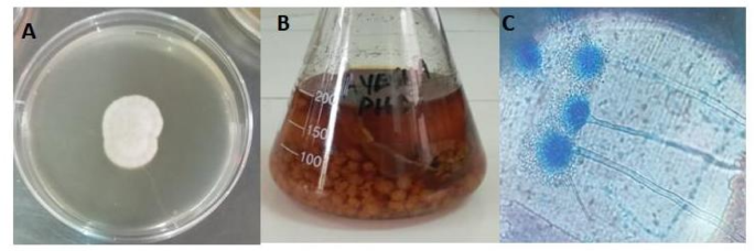
Microscopic examination performed using the slide culture technique, revealed short, smooth, hyaline conidiophores terminating in small globose vesicles (10–20 μm in diameter). The vesicles were typically bi-seriate, bearing flask-shaped metulae that supported cylindrical phialides arranged radially. Conidia were smooth, globose to sub-globose, measuring 2–3 μm in diameter, and produced in small radiating heads that became more compact with age; they appeared bright white rather than pigmented. The hyphae were hyaline, septate, and finely branched, with no sclerotia or cleistothecia observed. These characteristics- particularly the chalky white colonies and smooth, globose, hyaline conidia- distinguish A. candidus from pigmented species such as A. flavus (which forms yellow-green colonies with rough-walled conidia) and A. fumigatus (which produces smoky-green colonies with uniseriate conidial heads). Taken together, the cultural and microscopic traits provided reliable confirmation of A. candidus. The colony morphology, structure of fruiting bodies and growth of the fungus in liquid medium is shown in Fig. 1.

(A) A. candidus grown on PDA: Displays characteristic white and floccose colony growth on solid PDA medium, forming a dense circular mat indicative of healthy sporulation and hyphal expansion. (B) A. candidus cultivated in CYB under agitation: Showing submerged fungal biomass cultured in liquid medium within a shaking incubator. This condition promotes aeration and enhances nutrient absorption, resulting in dispersed hyphal networks and sedimented biomass formation. (C) Fruiting bodies of A. candidus: A microscopic view revealing conidiophores bearing spherical conidia, typical of A. candidus reproductive structures. These bluish structures are interconnected by septate hyphae and represent the asexual reproduction stage, crucial for identification and taxonomy.
Molecular identification of the bioactive fungal strain
Genomic DNA was successfully extracted from the pure culture of the selected endophytic fungus using the DNeasy Mini Tissue DNA Extraction Kit (Qiagen). The quality and integrity of the extracted DNA were confirmed by electrophoresis on a 1% agarose gel, showing distinct PCR amplicons (Fig. 2). The internal transcribed spacer (ITS) region was sequenced and subjected to BLAST analysis, which revealed a 96% identity with Aspergillus candidus, confirming its molecular identity. A phylogenetic tree was constructed to further validate the evolutionary relationship of the isolate, as shown in Fig. 3.

PCR amplicon of the fungal isolate visualized on a 1% agarose gel. Presents the results of agarose gel electrophoresis performed on PCR amplicons of DNA isolated from A. candidus. The electrophoresis was conducted using a 1% agarose gel stained with a DNA-intercalating dye and visualized under ultraviolet light. Lane (1) Shows a DNA ladder, providing reference for determining the approximate size of DNA fragments. Lane (2) Reveals distinct, bright bands corresponding to amplified DNA regions specific to A. candidus. The clarity and intensity of the bands confirm successful PCR amplification, validating the presence and integrity of fungal genomic DNA.

The phylogenetic tree, constructed based on ITS region sequence alignment, illustrates the evolutionary relationship of the tested fungal isolate (highlighted in blue) with reference strains retrieved from the NCBI GenBank database. The isolate clusters closely with A. candidus (accession MH725571.1), indicating a strong genetic match. It also shows close relatedness to A. candidus (NR_077149.1), highlighted in green, further confirming its taxonomic placement within the A. candidus clade. The tree was generated using BLAST pairwise alignments, and the branching pattern reflects high sequence similarity and close evolutionary proximity among the analyzed strains.
Purification of the pure compound from fungal extracts
The fungal crude extract was subjected to chromatographic purification using a column packed with silica gel and eluted with a gradient solvent system of ethyl acetate (EtoAc) and n-hexane. By gradually increasing the polarity of the solvent mixture, various fractions were obtained. A major bioactive fraction was successfully isolated using a 65:35 (v/v) ratio of EtoAc to n-hexane, yielding 5.03 mg of the pure compound from 57.68 mg of crude using 7 L of solvent. Further refinement of the isolated compound was achieved through preparative thin-layer chromatography (TLC), as illustrated in Fig. 4a and b. on TLC the distance travelled by compound was 4.2 cm whereas the distance travelled by solvent was 6.4 cm. Rf values of 0.65 was recorded using ethyl acetate: n-hexane as solvent system in a ratio of 65:35. The Rf value was calculated using the following formula;
$$:text{Solvent}text{front}text{(}text{Rf}text{)=-}frac{text{Distance}text{travelled}text{by}text{compound}}{text{Distance}text{travelled}text{}text{by}text{}text{solvent}}$$

TLC profiles during purification of the fungal metabolite. (A) Preparative TLC of the crude extract under UV light showing multiple bands; the band at Rf = 0.61 (11 cm; solvent front 18 cm) corresponding to the target fraction was selected for further purification. (B) Analytical TLC of the purified fraction showing a single spot at Rf = 0.65 (4.2 cm; solvent front 6.4 cm), confirming chromatographic homogeneity of the isolated compound.
Structural characterization of the pure compound
The structure and purity of the purified antifungal metabolite were established using a combination of GCMS, FTIR spectroscopy, and 1D/2D NMR experiments. The key chromatographic and spectroscopic features supporting the proposed structure are summarized in the following subsections (3.4.1–3.4.3).
GCMS analysis of the purified compound
Gas chromatography mass spectrometry (GCMS) analysis was conducted using an Agilent Technologies system outfitted with a DB-1 capillary column (30 m in length, 0.25 mm internal diameter, and 0.25 μm film thickness). A sample of approximately 1.5 to 2 mg of the purified compound was dissolved in 1 mL of molecular-grade methanol, and 1 µL of this solution was introduced into the GCMS in split-less mode. The total ion chromatogram (TIC) displayed a prominent peak at a retention time of 18.884 min, indicating the presence of a major analyte. The mass spectrum corresponding to this peak revealed a molecular ion at m/z 340.2, consistent with the profile of oleic acid and 9, 12-octadecadienoic acid. Compound (having mass of 340 Da) identification was further validated through spectral comparison with the NIST11.L database, showing a high match score (99%) across three confirmed entries, including CAS number 000112-80-1. The fragmentation pattern exhibited key ions at m/z 41.1, 5.15, 69.1, 83.1, 97.1, 264 and 340, supporting the identification of oleic acid and 9, 12-octadecadienoic acid as the dominant components in the extract. To support these findings, Fourier-transform infrared (FTIR) spectroscopy was also employed. Chromatograms corresponding to the pure fungal compound having a mass of 340 are presented in Figs. 5, 6.

GCMS chromatogram of the pure fungal metabolite showing a major peak at 18.884 min retention time. The elevated signal intensity at this retention point reflects successful chromatographic resolution and suggests a high abundance of the target analyte.

Mass spectrum of the purified fungal metabolite showing a molecular ion peak at m/z 340, along with the corresponding library search results for the peak eluting at 18.885 min. The spectrum exhibited a prominent molecular ion at m/z 340, alongside diagnostic fragment ions at m/z 41.1, 5.15, 69.1, 83.1, 97.1, 264 and 340. These fragmentation patterns enabled structural elucidation. A high-confidence match from the NIST database identified the compound 340 as oleic acid and octadecadienoic acid, supported by similarity scores of 99%, reflecting excellent spectral alignment.
FTIR spectroscopy
The Fourier Transform Infrared (FTIR) spectrum of the purified compound (Fig. 7) exhibited characteristic absorption bands indicative of various functional groups. A broad band at approximately 3300 cm–1 corresponded to OH stretching vibrations, suggesting the presence of alcohol or phenolic groups. Peaks near 2900 cm–1 were attributed to aliphatic C–H stretching, while a sharp absorption at ~ 1700 cm–1 confirmed the presence of carbonyl (C=O) functionalities such as esters or carboxylic acids. Bands around 1600–1500 cm–1 indicated C=C stretching vibrations, suggestive of aromatic or alkene groups. Absorptions in the 1300–1000 cm–1 region were consistent with C-O stretching, associated with esters, alcohols, or ethers. Fingerprint region peaks below 1000 cm–1 provided further confirmation of the compound’s structural features. The FTIR spectrum of the pure compound is provided in Fig. 7 and the FTIR spectral data is summarized in Table 1.

FTIR spectrum of the purified fungal compound 340 showing characteristic absorption bands corresponding to OH (∼3300 cm–1), CH (∼2900 cm–1), C = O (∼1700 cm–1), C=C (∼1600–1500 cm–1), and C–O (∼1300–1000 cm–1) functional groups.
NMR based structure elucidation
Structure elucidation was performed by 1D/2D NMR analysis (1H, 13C, DEPT-135, HSQC), which defined the backbone and oxygenation pattern28, with FTIR and GCMS providing orthogonal confirmation. GCMS gave a molecular ion at m/z 340 (C18H28O6), indicating five double-bond equivalents (DBE = 5). FTIR displayed a broad O-H band (~ 3300 cm–1), strong carbonyl absorptions consistent with an ester and a ketone (~ 1740 − 1715 and ~ 1710 –1690 cm–1), C=C (~ 1640 cm⁻¹), and C-O (1250 –1050 cm–1), pointing to an oxygenated, unsaturated scaffold.
The13C NMR spectrum showed two carbonyl carbons at δC ~ 211 (ketone) and ~ 171 (ester), four sp² carbons in the 130–135 ppm range (conjugated diene), several oxygenated carbons at ~ 65–72 ppm, and multiple aliphatic carbons between ~ 20 and 50 ppm. DEPT-135 distinguished CH/CH3 a from CH2 centers and constrained the substitution pattern around the oxygenated carbons. The 1H NMR spectrum featured two olefinic signals at δH ~ 5.81 and 5.63 (each 1 H, m), indicating a diene; three deshielded protons at δH ~ 4.56 (ddd), 4.46 (d), and 4.17 (m), consistent with oxygenated methine/methylene centers; a singlet at δH ~ 2.05 (3 H, s) characteristic of an acetate methyl; a set of α-carbonyl methylenes at δH ~ 2.64, 2.56, 2.39–2.29 (dd patterns); and the remaining aliphatic methylenes at δH ~ 1.75–1.5029. The integral balance was consistent with (C18H28O6).
HSQC one-bond correlations paired all protonated carbons. Key correlations included: δH ~ 4.1–4.2 ↔ δC ~ 66 (CH2-OAc), assigning an O-methylene adjacent to an ester; δH ~ 4.56/4.46 ↔ δC ~ 70–72 (two oxygenated methines); δH ~ 5.6–5.8 ↔ δC ~ 131–132 (olefinic CH of a conjugated diene); δH ~ 2.6–2.3 ↔ δC ~ 31–33 (methylene α to a carbonyl). Together these data define three oxygenated centers along the chain (two secondary alcohols and one primary alcohol acetylated as CH2-OAc), a conjugated 2, 7-diene, and methylenes adjacent to a ketone. The presence of both an ester (δC ~ 171; δH/δC ~ 2.05/20–21 for the acetate CH3 and ~ 4.1/66 for CH2-OAc) and a ketone (δC ~ 211) accounts for two DBE. The diene accounts for two more, and a ring contributes the fifth. The 1H/13C patterns at ~ 2.6–2.3 ppm / ~31–33 ppm, together with an oxygenated carbon near 70 ppm and the ketone at 211 ppm, support a hydroxylated cyclohexanone unit. This is consistent with the fragment set required by the formula and with the FTIR carbonyl and OH bands. The two vinyl CH signals and their carbon partners match a 2, 7-diene in a C18 “fatty-acid-like” backbone30,31. Placement of the acetate at the terminal primary alcohol is supported by the CH2-OAc HSQC pair (~ 4.1/66) and the acetate methyl (2.05/20–21).
The 17O NMR spectrum further supports the oxygen environments, showing distinct resonances for carbonyl oxygens (downfield) and hydroxyl oxygen (upfield relative to the carbonyls). While 17O NMR is not routine, the pattern is consistent with one ketone, one ester, and alcohol oxygens in the molecule. Assembling these substructures yields an oxygenated C18 polyketide bearing a 3-hydroxy-5-oxocyclohexyl moiety, a 2, 7-conjugated diene, and a terminal CH2-OAc. The assigned geometry of the diene (2E, 7E) is consistent with the observed olefinic chemical shifts and coupling patterns for trans-alkenyl protons in related polyketide chains; we state this explicitly as a configurational assignment supported by the 1H data and analogy to literature values for fatty-acid-type dienes. The final structure satisfies all spectroscopic constraints, matches the elemental composition (C18H28O6) and agrees with the GCMS molecular ion and fragmentation. The elucidated structure of the pure compound is given in Fig. 8, 1H and 13C-NMR data are summarized in Table 2 while the NMR spectra are provided as supplementary material.

Elucidated chemical structure of the purified fungal compound isolated from A. candidus determined through comprehensive spectroscopic and chromatographic techniques.
Although GCMS NIST library matching of the major peak at Rt 18.884 min (m/z 340) suggested similarity to C18 unsaturated fatty acids such as oleic and linoleic acid (NIST match ~ 99%), detailed spectroscopic analysis demonstrated that the purified metabolite is structurally distinct from these known lipids. FTIR and 1D/2D NMR revealed multiple oxygenated centers, a substituted cyclohexanone/cyclohexyl moiety, a conjugated diene system, and an acetate function in a substitution pattern not compatible with simple C18 fatty acids or their common derivatives. To further assess novelty, we performed exact-structure and substructure searches in SciFinder-n, Reaxys, Dictionary of Natural Products, PubChem, ChemSpider, and ChEMBL which did not yield any exact match. Taken together, these data support the proposed structure as a previously unreported fungal metabolite rather than a known fatty acid derivative.
Molecular docking studies
The docking study was conducted to explore how the purified fungal compound interacts with antifungal targets. Specifically, C. albicans lanosterol 14α-demethylase was utilized in this study. This enzyme plays a crucial role in converting lanosterol into 4, 4-dimethylcholesta-8 (9), 14, 24-trien-3β-ol. The active pocket of the enzyme was identified as significant, demonstrating deep binding interactions22. Close interactions were observed between the compound 340 and the enzyme 5FSA. Additionally, other significant hydrophobic interactions were identified, involving residues such as Met508, Phe380, Ser507, Lys90, Ala117, Leu87, Leu88, Leu121, and Leu376. The pure compound demonstrated a docking affinity of -7.7 kcal/mol with the lanosterol 14α-demethylase enzyme. While posaconazole (control) showed a docking affinity of − 9.5 kcal/mol with lanosterol 14α-demethylase (CYP51), it formed predominantly hydrophobic interactions with residues Ala61, Ala62, Pro230, Phe233, Tyr118, Leu88, Leu139, Lys143, Ile131, Tyr132, Leu181, Leu300, Leu376, Val234, and Met508. Figure 9 illustrates the docking conformation and chemical interactions of the purified compound with the C. albicans lanosterol 14α-demethylase enzyme while docking validation using posaconazole as reference inhibitor with C. albicans lanosterol 14α-demethylase (CYP51, PDB ID: 5FSA) is provided in supplementary material as Figure S2.

Molecular docking pose of the fungal metabolite in the active site of C. albicans lanosterol 14α-demethylase (CYP51). Left: overall view showing the ligand in the defined binding pocket. Right: close-up of key interactions, including hydrogen bonds, hydrophobic and π–π contacts, and one unfavorable donor–donor contact with Ser378. These interactions support a plausible CYP51-binding mode for the compound.
Molecular dynamic simulation
Molecular dynamics simulations provide valuable insights beyond the static representations obtained from docking studies. These simulations enable the real-time observation of docked complexes, allowing the assessment of intermolecular interaction stability over time. By mirroring biological processes more realistically, these studies offer a deeper understanding of molecular behavior. To evaluate the structural stability of the simulated complexes, a 100-nanosecond simulation period was conducted. The RMSD graph (Fig. 10B) presents the system’s structural variations throughout the simulation. Initially, from 0 to 8 ns, the RMSD values remain around 1.5 Å, indicating a phase of conformational adjustments. As the simulation progresses, structural flexibility increases, with RMSD values reaching approximately 2.0 Å by 35 ns. This highlights continued adaptation within the system. Between 35 ns and 67 ns, the RMSD stabilizes around 2.5 Å, marking the onset of equilibrium. From 67 ns to 90 ns, the system maintains this level of stability, with no significant variations, confirming that it has reached a steady conformational state. The N-terminal region of the compound complex demonstrates considerable flexibility, contributing to higher RMSD values, likely due to interactions at the binding site. Analyzing these fluctuations further, loop regions appear to be key contributors due to their inherent flexibility. The compound’s binding mechanism also plays a significant role in influencing RMSD variations as it optimizes its binding mode. Despite these fluctuations, the system’s mean RMSD remains around 1.2 Å, confirming its well-maintained stability and lack of major structural deviations. Residue-level dynamics were assessed using root mean square fluctuation (RMSF), depicted in Fig. 10A. The mean RMSF value of the compound complex was recorded at 1.3 Å, indicating localized flexibility in certain regions. The global structural stability inferred from RMSD correlates with the overall RMSF values, reinforcing the system’s stable nature. The structural flexibility of different regions within the compound and ligand-protein complex was investigated using B-factor analysis, revealing a mean value of 201.2 Å, which further affirms the system’s robust stability (Fig. 10C). Protein integrity was further examined through radius of gyration (Rg) analysis, plotted against time in Fig. 10D. Rg quantifies the spatial distribution of molecular components and is determined by calculating the root mean square distance of atoms relative to their collective center of mass. The mean Rg value of the compound complex was 21.87 Å, supporting the system’s stability. Additionally, solvent-accessible surface area (SASA) was calculated, yielding a mean value of 15,842.1 Å. SASA determines how much of the molecular surface remains exposed to solvent molecules, influencing molecular interactions and binding properties. Increased SASA values often correlate with enhanced residue exposure, which can affect the structural conformation and interaction dynamics of the system. Maintaining a stable SASA value suggests equilibrium in the solvation state of the complex (Fig. 10E).

Molecular dynamics analysis of the metabolite–CYP51 complex over 100 ns. (A) RMSF and (B) RMSD indicate limited fluctuations and overall stability of the complex. (C) B-factors, (D) radius of gyration (Rg), and (E) SASA profiles further support a compact, conformationally stable binding environment, consistent with sustained ligand engagement.
Binding free energies
Table 3 presents the binding free energies of the complex determined using the MM-PBSA method throughout the molecular dynamics simulation. The estimation of binding free energies based on simulation trajectories is generally considered more reliable and closely aligned with experimental findings. The negative binding values confirm the docking and MD simulation results, indicating that the pure fungal compound binds effectively to the EPSP synthetase enzyme. The stability of the complex is supported by a negative net binding energy of -77.76 kcal/mol. Among the contributing factors, Van der Waals interactions play a significant role, accounting for − 69.31 kcal/mol. Additionally, electrostatic energy and gas-phase energies contribute notably, with values of -22.54 kcal/mol and − 91.85 kcal/mol, respectively.
ADMET analysis of the pure compound
The isolated fungal compound m/z 340 has demonstrated drug-like properties according to multiple well-established drug likeness criteria, including Lipinski’s rule of five32, Egan’s rule33, Veber’s criteria34, Ghose’s guidelines35, Muegge’s filter36. These assessments suggest a strong potential for the fungal compound understudy to progress to market approval. Furthermore, the absence of pan-assay interference (PAINS) compound indicates that the molecule exhibits specificity in its binding, targeting a single biological macromolecule37. In terms of synthetic accessibility, the fungal compound has a score of 4.59, implying feasibility for laboratory synthesis under standard conditions. Its pharmacokinetic profile is favorable, showing high gastrointestinal absorption, no permeability across the blood-brain barrier, and minimal skin absorption. Additionally, it does not inhibit key cytochrome P450 enzymes, including CYP1A2, CYP2C19, CYP2C9, CYP2D6, and CYP3A4.
The solubility assessment classifies the fungal derived compound as highly water-soluble, a characteristic beneficial for oral bioavailability. Moreover, its lipophilicity value of 2.89 indicates sufficient membrane permeability while maintaining a balanced hydrophobic-hydrophilic profile. Toxicological evaluations further confirm that the compound under study is non-toxic, non-mutagenic, and non-immunogenic. The bioavailability radar representation of the pure compound is provided in Fig. 11. The characteristic properties of the pure fungal compound are shown in Table 4.

Bioavailability radar plot of the fungal metabolite summarizing key physicochemical properties (lipophilicity, size, polarity, solubility, saturation, flexibility). The red trace (compound 340) relative to the optimal pink region indicates that several parameters fall within an acceptable range, suggesting preliminary compatibility with oral drug-likeness criteria.
In vitro anti-Candida activity of the pure compound
The antifungal potential of the isolated compound was evaluated against C. albicans using the agar tube diffusion method. The pure compound was tested at concentrations of 100, 250, 500, and 1000 µg/mL, alongside fluconazole as the positive control at the same concentrations. Following inoculation, the tubes were incubated and periodically examined over 96 h, with final measurements recorded at the end of the incubation period. Linear fungal growth was measured in millimeters (mm), and the results are presented in Table 5. To determine the antifungal efficacy of the pure compound, the percentage of growth inhibition was calculated using the following formula:
$${text{Percent,Growth,Inhibition}} :=frac{text{F}text{u}text{n}text{g}text{a}text{l}:text{G}text{r}text{o}text{w}text{t}text{h}:text{i}text{n}:text{t}text{h}text{e}:text{T}text{e}text{s}text{t}left(text{m}text{m}right)}{text{F}text{u}text{n}text{g}text{a}text{l}:text{G}text{r}text{o}text{w}text{t}text{h}:text{i}text{n}:text{t}text{h}text{e}:text{C}text{o}text{n}text{t}text{r}text{o}text{l}left(text{m}text{m}right)}times:100$$
At 100 µg/mL, the pure compound inhibited C. albicans growth by 5.5%. Increasing the concentration to 250 µg/mL resulted in 10.2% inhibition. At higher concentrations of 500 and 1000 µg/mL, the isolated compound exhibited 15.5% and 24.3% inhibition, respectively. The growth inhibition data for both the pure compound and fluconazole are summarized in Table 5.
Discussion
Fungi are a major source of bioactive secondary metabolites and remain central to natural product discovery38. Endophytic fungi, in particular, have emerged as prolific producers of structurally diverse metabolites with promising biological activities39,40. In the current study, we present endophytic A. candidus as a producer of an anti-Candida compound which serve as a drug lead in future antifungal therapy. Following preliminary antifungal testing, the extracts of this fungus were evaluated against six pathogenic fungal strains: A. fumigatus, F. solani, C. albicans, C. glabrata, and P. expansum, utilizing virtual screening. This approach revealed the best docking score with C. albicans. Computer-aided drug design methods have proven invaluable, significantly reducing the time and costs typically associated with evaluating the binding potency of fungal compounds and other biological macromolecules41,42. These methods guided the assessment of the antifungal potential of the purified compound in vitro. Due to the limited quantity of the pure compound (5 mg), its antifungal activity was tested solely against C. albicans, which exhibited the best docking score.
The emergence of drug-resistant has placed a significant strain on healthcare systems worldwide. Moreover, the prevalence of chronic and invasive life-threatening fungal infections has further aggravated the situation, given the limited availability of effective treatment options for fungal diseases. Resistance to commonly prescribed antifungal medications has also reduced their effectiveness in therapeutic applications, prompting researchers to explore and develop novel antifungal agents43.
This study identifies a pure compound derived from A. candidus, named (2E, 7E) − 6, 9-dihydroxy-10-(3-hydroxy-5-oxocyclohexyl) deca-2, 7-dien-1-yl acetate. Employing advanced analytical techniques, we carried out a comprehensive characterization of this fungal compound. The combined use of FTIR and NMR spectroscopy significantly enhanced the structural elucidation of the purified compound, revealing features well beyond those of oleic acid, which had shown 99% similarity in the GC-MS library search. FTIR analysis identified key functional groups, including hydroxyl, ester, and carbonyl moieties, while NMR techniques, such as 1H, 13C, HSQC and17O provided detailed insights into the proton–carbon framework. These data confirmed the presence of conjugated dienes, hydroxylated methines, and multiple oxygenated centers. This integrative approach enabled accurate interpretation of overlapping or ambiguous signals, ultimately establishing the compound as a structurally modified fatty acid derivative with enhanced functional complexity and potential bioactivity. The analysis of this compound using these advanced analytical techniques revealed its structural characteristics, providing valuable insights that not only advance scientific understanding but also pave the way for potential applications44,45. As detailed in the structure elucidation section, comprehensive GCMS/FTIR/NMR analysis and database dereplication confirmed that the metabolite is distinct from known C18 fatty acid derivatives and is best considered a previously unreported fungal compound. The pure compound, at concentrations of 500 and 1000 µg/mL, inhibited the growth of C. albicans by 15.5% and 24.3%, respectively. These findings clearly demonstrate the compound’s antifungal activity against the tested Candida species, both in silico and in vitro, highlighting its potential as a potent antifungal candidate. Evidence supporting the production of antifungal metabolites by A. candidus has been documented in previous studies. Rashad et al. (2022) reported that endophytic A. candidus exhibited strong antagonistic activity against F. solani in vitro. Additionally, this endophyte was found to produce a range of antifungal secondary metabolites46. In this study, we evaluated the antifungal activity of the isolated compound against C. albicans, using fluconazole as the reference drug. At the highest tested concentration of 1000 µg/mL, the compound exhibited 24.3% growth inhibition against C. albicans.
Several secondary metabolites have previously been reported from A. candidus, including p-terphenyl derivatives such as terphenyllin and 3-hydroxyterphenyllin, candidusin-type polyketides, chlorflavonin, and related diphenyl ethers. These metabolites are predominantly rigid aromatic scaffolds and many exhibit antibacterial, antifungal, or cytotoxic properties19,47,48. In contrast, the metabolite described here is a C18 polyketide bearing a hydroxylated cyclohexanone moiety, a conjugated diene system, and an acetate group, giving an oxygenated “fatty acid like” framework that is topologically and electronically distinct from the known A. candidus p-terphenyls and chlorinated aromatics. Our docking and MD results further suggest a different mechanistic profile, with a plausible fit in the CaCYP51 active site, whereas the modes of action of previously reported A. candidus metabolites are either unrelated to CYP51 or remain undefined. Thus, this compound broadens both the structural and mechanistic space of A. candidus secondary metabolites and represents a new scaffold for antifungal optimization. MM-PBSA analysis indicated a favorable binding free energy for the metabolite in the CaCYP51 active site (ΔG = − 77.76 kcal/mol), whereas the in vitro agar tube assay, performed in triplicate, showed only modest but reproducible inhibition of Candida growth. This apparent discrepancy is not unexpected as docking, MD, and MM-PBSA describe an idealized pocket-level interaction and do not account for effective exposure at the target site, including solubility and diffusion in agar, membrane permeability, efflux, metabolic stability, or binding to medium components. We therefore regard the computational results as supportive, hypothesis-generating evidence for a CYP51-related mode of action rather than as a quantitative predictor of antifungal potency. Although the observed inhibition against C. albicans is modest, the functional groups and predicted CYP51 interactions of this metabolite make it a suitable scaffold for further optimization rather than a direct drug candidate. Future work will focus on structure activity studies and semi-synthetic derivatives to improve potency and selectivity, as well as testing in combination with standard antifungals (e.g., fluconazole) to explore potential synergy. Together, these approaches will help to define the true therapeutic relevance of this fungal metabolite.
Conclusion
In this study, we isolated and characterized the secondary metabolite (2E, 7E) − 6, 9-dihydroxy-10-(3-hydroxy-5-oxocyclohexyl) deca-2, 7-dien-1-yl acetate from the endophytic fungus A. candidus associated with R. officinalis. In silico investigations provided compelling evidence of its antifungal potential, revealing strong and stable interactions with C. albicans lanosterol 14α-demethylase (CYP51), a key enzyme in ergosterol biosynthesis. Molecular docking, molecular dynamics simulations, and MM-PBSA free energy analyses consistently demonstrated that the compound binds favorably within the enzyme’s active site, while ADMET profiling indicated desirable drug-like features, pharmacokinetic compatibility, and a non-toxic profile. These computational insights were substantiated by in vitro antifungal assays, where the compound displayed reproducible, concentration-dependent inhibition of C. albicans growth relative to fluconazole.
Together, these findings reveal that the compound under study has the potential to serve as a promising natural scaffold with potential for antifungal drug development. While its activity requires further optimization, its consistent computational and experimental performance provides a strong foundation for future structural modifications and pharmacological evaluations aimed at developing safer and more effective antifungal agents.
Data availability
The data generated in the work is presented in the manuscript.
6. References
-
Wang, D. et al. Comparison of CT findings and histopathological characteristics of pulmonary cryptococcosis in immunocompetent and immunocompromised patients. Sci. Rep. 12, 5712 (2022).
-
Bocci, M. G. et al. Pulmonary aspergillosis in immunocompromised critically ill patients: Prevalence, risk factors, clinical features and diagnosis—a narrative review. J. Fungi. 11, 617 (2025).
-
Lass-Flörl, C. et al. Invasive candidiasis. Nat. Rev. Disease Primers. 10, 20 (2024).
-
Ahmed, S. H., El-Kholy, I. M., El-Mehalawy, A. A. & Mahmoud, E. M. Elkady, N. A. Molecular characterization of some multidrug resistant Candida auris in Egypt. Sci. Rep. 15, 4917 (2025).
-
Kaur, J. & Nobile, C. J. Antifungal drug-resistance mechanisms in Candida biofilms. Curr. Opin. Microbiol. 71, 102237 (2023).
-
Branco, J., Miranda, I. M. & Rodrigues, A. G. Candida parapsilosis virulence and antifungal resistance mechanisms: A comprehensive review of key determinants. J. Fungi. 9, 80 (2023).
-
Hassan, Y., Chew, S. Y. & Than, L. T. L. Candida glabrata: Pathogenicity and resistance mechanisms for adaptation and survival. J. Fungi. 7, 667 (2021).
-
Vitiello, A. et al. Antifungal drug resistance: An emergent health threat. Biomedicines 11, 1063 (2023).
-
Singh, A. et al. Recent advances in antifungal drug development targeting lanosterol 14α-demethylase (CYP51): A comprehensive review with structural and molecular insights. Chem. Biol. Drug Des. 102, 606–639 (2023).
-
Chen, F. et al. CYP51A1 in health and disease: From sterol metabolism to regulated cell death. Cell. Death Discovery. 11, 322 (2025).
-
Song, L. et al. Regulation of ergosterol biosynthesis in pathogenic fungi: Opportunities for therapeutic development. Microorganisms 13, 862 (2025).
-
Binjubair, F. A. S. Design and Synthesis of Novel CYP51 Inhibitors (Cardiff University, 2021).
-
Hashem, A. H. et al. Bioactive compounds and biomedical applications of endophytic fungi: A recent review. Microb. Cell. Fact. 22, 107 (2023).
-
Gupta, A. et al. Fungal endophytes: microfactories of novel bioactive compounds with therapeutic interventions; A comprehensive review on the biotechnological developments in the field of fungal endophytic biology over the last decade. Biomolecules 13, 1038 (2023).
-
El-Sayed, E. S. R. et al. Bioprospecting endophytic fungi of forest plants for bioactive metabolites with anticancer potentials. Sci. Rep. 15, 26423 (2025).
-
Rajput, D., Khabiya, R., Dwivedi, A., Soni, V. & Soni, P. In Bioactive Compounds 166–187 (CRC, 2025).
-
Hendel, N. et al. Phytochemical analysis and antioxidant and antifungal activities of Powders, methanol Extracts, and essential oils from Rosmarinus officinalis L. and thymus ciliatus Desf. Benth. Int. J. Mol. Sci. 25, 7989 (2024).
-
Asmaey, M. A. Unravelling the secrets of α-pyrones from Aspergillus fungi: A comprehensive review of their natural sources, biosynthesis, and biological activities. Chem. Biodivers. 20, e202301185 (2023).
-
Krawczyk-Łebek, A., Żarowska, B. & Janeczko, T. Kostrzewa-Susłow, E. Antimicrobial activity of new glycoside derivatives of Chloroflavones obtained by fungal biotransformation. Sci. Rep. 15, 25821 (2025).
-
Kumari, P. et al. Isolation and purification of bioactive metabolites from an endophytic fungus penicillium citrinum of Azadirachta indica. S. Afr. J. Bot. 139, 449–457 (2021).
-
Singh, V. K. & Kumar, A. Secondary metabolites from endophytic fungi: Production, methods of analysis, and diverse pharmaceutical potential. Symbiosis 90, 111–125 (2023).
-
Hargrove, T. Y. et al. Structural analyses of Candida albicans sterol 14α-demethylase complexed with Azole drugs address the molecular basis of Azole-mediated Inhibition of fungal sterol biosynthesis. J. Biol. Chem. 292, 6728–6743 (2017).
-
Milne, G. W. (ACS Publications, (2010).
-
Ounthaisong, U. & Tangyuenyongwatana, P. Cross-docking study of flavonoids against tyrosinase enzymes using pyrx 0.8 virtual screening tool. TJPS 41 (2017).
-
Pettersen, E. F. et al. UCSF Chimera—a visualization system for exploratory research and analysis. J. Comput. Chem. 25, 1605–1612 (2004).
-
Naz, R., Nosheen, A., Yasmin, H., Bano, A. & Keyani, R. Botanical-chemical formulations enhanced yield and protection against bipolaris Sorokiniana in wheat by inducing the expression of pathogenesis-related proteins. PLoS ONE. 13, e0196194 (2018).
-
Liu, Y., Tortora, G., Ryan, M. E., Lee, H. M. & Golub, L. M. Potato dextrose agar antifungal susceptibility testing for yeasts and molds: Evaluation of phosphate effect on antifungal activity of CMT-3. Antimicrob. Agents Chemother. 46, 1455–1461 (2002).
-
Claridge, T. D. High-resolution NMR Techniques in Organic Chemistry Vol. 27 (Elsevier, 2016).
-
Pretsch, E., Clerc, T., Seibl, J. & Simon, W. Tables of Spectral Data for Structure Determination of Organic Compounds (Springer, 2013).
-
Neuhaus, G. F., Adpressa, D. A., Bruhn, T. & Loesgen, S. Polyketides from marine-derived Aspergillus porosus: Challenges and opportunities for determining absolute configuration. J. Nat. Prod. 82, 2780–2789 (2019).
-
Yao, F. H., Liang, X. & Qi, S. H. Eight new cyclopentenone and cyclohexenone derivatives from the marine-derived fungus Aspergillus sp. SCSIO 41501 by OSMAC strategy. Nat. Prod. Res. 35, 3810–3819 (2021).
-
Pollastri, M. P. Overview on the rule of five. Curr. Protocols Pharmacol. 49, 18 (2010). 9.12. 11-19.12.
-
Egan, W. J. & Lauri, G. Prediction of intestinal permeability. Adv. Drug Deliv. Rev. 54, 273–289 (2002).
-
Veber, D. F. et al. Molecular properties that influence the oral bioavailability of drug candidates. J. Med. Chem. 45, 2615–2623 (2002).
-
Ghose, M., Dikshit, A. K. & Sharma, S. A GIS based transportation model for solid waste disposal–A case study on Asansol municipality. Waste Manag. 26, 1287–1293 (2006).
-
Muegge, I. Selection criteria for drug-like compounds. Med. Res. Rev. 23, 302–321 (2003).
-
Whitty, A. Growing PAINS in academic drug discovery. Future Med. Chem. 3, 797–801 (2011).
-
Jamil, F., Mukhtar, H., Fouillaud, M. & Dufossé, L. (s Note: MDPI stays neutral with regard to jurisdictional claims in published ….
-
Sharma, D., Pramanik, A. & Agrawal, P. K. Evaluation of bioactive secondary metabolites from endophytic fungus Pestalotiopsis neglecta BAB-5510 isolated from leaves of Cupressus torulosa D. Don. 3 Biotech 6, 210 (2016).
-
Hestrin, R. et al. Plant-associated fungi support bacterial resilience following water limitation. ISME J. 16, 2752–2762 (2022).
-
Lionta, E., Spyrou, G., Vassilatis, K., Cournia, Z. & D. & Structure-based virtual screening for drug discovery: Principles, applications and recent advances. Curr. Top. Med. Chem. 14, 1923–1938 (2014).
-
Zhang, X. et al. Advancing ligand Docking through deep learning: Challenges and prospects in virtual screening. Acc. Chem. Res. 57, 1500–1509 (2024).
-
Fisher, M. C. et al. Tackling the emerging threat of antifungal resistance to human health. Nat. Rev. Microbiol. 20, 557–571 (2022).
-
Bucar, F., Wube, A. & Schmid, M. Natural product isolation–how to get from biological material to pure compounds. Nat. Prod. Rep. 30, 525–545 (2013).
-
Bross-Walch, N., Kühn, T., Moskau, D. & Zerbe, O. Strategies and tools for structure determination of natural products using modern methods of NMR spectroscopy. Chem. Biodivers. 2, 147–177 (2005).
-
Rashad, Y. M., Abdalla, S. A. & Shehata, A. S. Aspergillus flavus YRB2 from thymelaea hirsuta (L.) Endl., a non-aflatoxigenic endophyte with ability to overexpress defense-related genes against fusarium root rot of maize. BMC Microbiol. 22, 229 (2022).
-
Zeng, Y. et al. p-Terphenyl and Diphenyl ether derivatives from the marine-derived fungus Aspergillus Candidus HM5-4. Mar. Drugs. 22, 13 (2023).
-
Ngo, M. T. et al. Biocontrol potential of Aspergillus species producing antimicrobial metabolites. Front. Microbiol. 12, 804333 (2021).
Funding
This work was supported and funded by the Deanship of Scientific Research at Imam Mohammad Ibn Saud Islamic University (IMSIU) grant number IMSIU-DDRSP2602.
Ethics declarations
Competing interests
The authors declare no competing interests.
Consent to publish
All the authors agree to publish data presented in the manuscript.
Additional information
Publisher’s note
Springer Nature remains neutral with regard to jurisdictional claims in published maps and institutional affiliations.
Supplementary Information
Rights and permissions
Open Access This article is licensed under a Creative Commons Attribution-NonCommercial-NoDerivatives 4.0 International License, which permits any non-commercial use, sharing, distribution and reproduction in any medium or format, as long as you give appropriate credit to the original author(s) and the source, provide a link to the Creative Commons licence, and indicate if you modified the licensed material. You do not have permission under this licence to share adapted material derived from this article or parts of it. The images or other third party material in this article are included in the article’s Creative Commons licence, unless indicated otherwise in a credit line to the material. If material is not included in the article’s Creative Commons licence and your intended use is not permitted by statutory regulation or exceeds the permitted use, you will need to obtain permission directly from the copyright holder. To view a copy of this licence, visit http://creativecommons.org/licenses/by-nc-nd/4.0/.
About this article

Cite this article
Saleem, A., Khattak, S.U., Ahmad, S. et al. Purification and characterization of a dihydroxy Oxocyclohexyl acetate derivative from endophytic Aspergillus Candidus targeting Candida albicans lanosterol 14α-Demethylase. Sci Rep 16, 5379 (2026). https://doi.org/10.1038/s41598-025-31974-3
-
Received:
-
Accepted:
-
Published:
-
Version of record:
-
DOI: https://doi.org/10.1038/s41598-025-31974-3