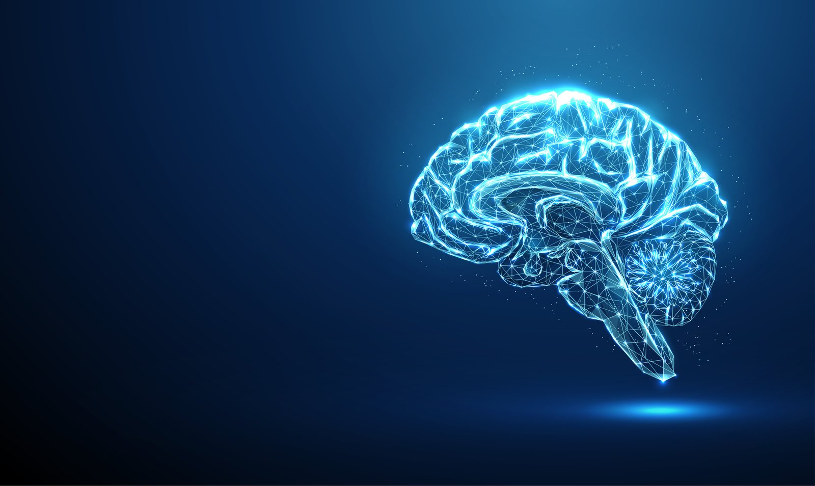
Studying living brains has long been a challenge. Techniques like MRI and CT scans can yield some information, but the details are often lost. Even studies of ex vivo brain tissue are often not as detailed as researchers would prefer, due to the density of tissues impairing image quality, even with fluorescence.
A research team at Kyushu University, led by Takeshi Imai, PhD, a faculty member in the medical sciences department, has been working on developing a technique that not only visually clears brain tissues, but does so in a way that preserves normal brain dynamics. After screening nearly 100 compounds with varying outcomes, the team identified an unlikely winner.
Their work is published in Nature Methods in a paper entitled, “Isotonic and minimally invasive optical clearing media for live cell imaging ex vivo and in vivo.”
Foundationally, effective and clear imaging comes from reducing the opacity of tissues. Light traveling through uncleared tissues can be impeded and scattered when passing through materials with different refractive indices. In tissues, this correlates to the multitude of cellular components including lipids and proteins. Light going through tissues refracts off the cellular components and scatter, resulting in blurred and indistinct images.
Adjusting the refractive index in living tissue is complicated as added materials not only affect opacity of the tissue, but can also change the osmolarity, potentially causing cells to swell, shrink, or behave differently.
The authors described a serendipitous situation that led them to test bovine serum albumin, a common reagent in the lab. They shared that late one evening in the midst of testing a multitude of other clearing candidates, first author Shigenori Inagaki, PhD, assistant professor at Kyushu University, reflected that proteins are polymers and may be an option to clear cells without impacting osmolarity the way that carbohydrates do.
“I tested it three or four times before I believed it,” Inagaki shared. “Of all things, we never expected it would come down to this.”
“Albumin is abundant in blood and highly soluble, which makes it well-suited for clearing,” noted Imai. “It was an accidental discovery, but looking back, it feels almost natural. What evolution has shaped over millions of years is truly impressive.”
The team refined their solution, developing a culture medium that included albumin. This clearing solution, dubbed SeeDB-Live, not only clears the tissue, but does not impact the tissue’s ability to function normally.
“This is the first time tissue clearing has been achieved without altering its biology,” said Imai.
“During the development of SeeDB-Live, we found that neurons are extremely sensitive to ion concentrations, and it took us enormous effort to get the formulation right,” Inagaki added. “Thanks to that fortunate night alone in the lab, I helped myself to an expensive, high-purity BSA I wouldn’t normally dare use.”
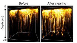
The team tested SeeDB-Live ex vivo in brain slices, immersing them for an hour in the solution. To test the ability to visualize neuronal function, they added a calcium indicator and were able to image neuronal firing deep in the tissue. In living mouse brains the fluorescence is three times brighter following SeeDB-Live application.
“SeeDB-Live minimally affects neuronal electrophysiological properties and sensory responses in vivo, and facilitates fluorescence imaging of deep cortical layers in live animals without detectable toxicity to neurons or behavior,” the authors wrote.
While this clearing solution is a step forward in the researchers’ ability to visualize tissues in more detail and greater depth that previously, there is still more work to fully clarify the applications.
“I feel we have not yet fully materialized its potential,” Inagaki said. Continued work will focus on improving delivery to be less invasive, as the current method for visualizing the brain in vivo requires a surgical window that can cause the animal stress. They also plan to improve solution penetration for deeper visualization into the brain.
“That question came to me about a hundred times, and each time I answered ‘impossible,’” Imai reflects. “But ten years later, here we are. When something seems unachievable, if you keep thinking about it, you may eventually find a way.”
The post Seeing the Brain in a Different Light appeared first on GEN – Genetic Engineering and Biotechnology News.